Competency-based training in clinical ultrasonography requires a clear understanding of skill acquisition trajectories. However, objective data on how residents actually learn in real-time clinical environments remain scarce. This study aims to map the learning pathway of ultrasound residents by analyzing real-time consultation patterns and introducing a novel metric—the skill acquisition half-life—to quantify the rate of skill mastery. We conducted a single-center retrospective observational study in a high-volume outpatient ultrasound department. Over an eight-week period, real-time consultation requests initiated by 45 residents across three postgraduate years (PGY-1 to PGY-3) were logged. Consultations were categorized by examination type (Standard vs. Advanced assignments). Adjusted consultation rates were calculated per clinical session. A negative exponential learning curve model was fitted to derive the skill acquisition half-life (T₁/₂), defined as the time required for the consultation rate to decrease by half. A total of 529 consultations were recorded, with PGY-1 residents accounting for 88.5
This study evaluates the performance of subharmonic-aided pressure estimation (SHAPE) with ultrasound contrast agents using data obtained from calibration procedures. A commercial scanner of ultrasonography (GE Healthcare Logiq E20, Wuxi, China) was used to perform SHAPE on a flowing contrast agent (Sonazoid) in a phantom setup under controlled microbubble stability and flow velocity conditions. Subharmonic time-intensity curves were collected during the SHAPE calibration procedure for analysis. Subharmonic amplitude of diluted contrast agents exhibited a time-dependent decline but was not affected by velocity. SHAPE sensitivity was measured through reciprocal pressurizing and depressurizing sequences to mitigate the effect of subharmonic decline over time. A wide range of mechanical index (MI) levels within the steady growth phase of the calibration curve showed higher SHAPE sensitivity compared to the traditionally recommended “optimal” MI at the maximum slope. The approximate maximum SHAPE sensitivity was −0.04 dB/mm Hg. Subharmonic amplitude was linearly correlated with pressure at a range of MI levels (R2 > 0.9, p < 0.05) but showed significant variations (approximately 2 dB standard deviation) in the time series. The lowered sensitivity compared to previous reports, combined with the substantial variation in subharmonic amplitude, raises concerns about the accuracy and consistency of SHAPE in clinical applications.
Background Differentiated thyroid cancer (DTC) accounts for the majority of thyroid cancers. The preoperative diagnosis of extrathyroidal extension (ETE) in DTC patients is highly important. However, two-dimensional ultrasound (2D-US) has several limitations in diagnosing ETE. This study aimed to evaluate the efficiency of OmniView of three-dimensional ultrasound (3D-OmniView) in assessing the ETE of DTC patients compared with that of 2D-US. Methods Patients who underwent thyroid surgery for nodules adjacent to the thyroid capsule between February 2016 and January 2018 were prospectively enrolled in this study. Both 2D-US and 3D-OmniView were used to evaluate ETE of thyroid nodules. The definition for ETE in ultrasound images was capsule disruption, or capsule disruption and surrounding tissue invasion. Intraoperative and pathological findings of ETE were considered positive. The sensitivity, specificity, positive predictive value (PPV), negative predictive value (NPV), accuracy, and area under the ROC curve (AUC) were calculated. Results A total of 176 DTC nodules from 137 patients were included in this study. ETE was identified in 67.0% of the nodules. The sensitivity, accuracy, NPV and AUC of 3D-OmniView for predicting ETE were significantly greater than those of 2D-US. The sensitivity and specificity of 2D-US and 3D-OmniView were 79.7% and 51.7%, respectively (P < 0.001), and 81.0% and 82.8%, respectively (P = 0.776). Both 2D-US and 3D-OmniView showed better efficacy in evaluating ETE in nodules > 1 cm than in evaluating ETE in nodules ≤ 1 cm. Conclusion 3D-OmniView was more precise in predicting ETE of DTC nodules than 2D-US. 3D-OmniView is recommended for further evaluation of suspicious ETE. ETE was easier to detect by ultrasound for nodules > 1 cm than for nodules ≤ 1 cm.
We report a rare case of Mayer-Rokitansky-Küster-Hauser (MRKH) syndrome accompanied by simultaneous adenomyosis and leiomyoma in rudimentary uterine remnants. A 35-year-old woman presented with progressive lower abdominal pain and underwent diagnostic imaging. The initial ultrasound erroneously suggested an adnexal mass, but subsequent imaging revealed bilateral uterine remnants-one with adenomyotic changes and the other harboring a leiomyoma. Laparoscopic excision confirmed these pathological findings. This case suggests the importance of regular gynecological imaging examinations for MRKH patients and challenges the conventional belief in the non-functional nature of Müllerian remnants. Furthermore, our findings underscore the diagnostic utility of transabdominal ultrasound in accurately identifying complex uterine anomalies.
Unicentric Castleman disease (UCD) is a rare group of non-neoplastic lymphoproliferative disorders. This study aims to summarize the specific ultrasonic manifestations of UCD. This retrospective study included patients who underwent preoperative ultrasound for enlarged lymph nodes and were later diagnosed with UCD between January 2016 and March 2024. Ultrasound features, including lymph node size, cortical characteristics, corticomedullary interface, hyperechoic regions, and Doppler flow signals, were recorded. Pathological types were classified as hyaline vascular (HV), plasma cell (PC), or mixed. The ultrasonic features of each UCD subtype were systematically analyzed. A total of 41 patients were enrolled in the study, comprising 29 with HV-type, 4 with PC-type, and 8 with a mixed type. All patients presented with enlarged lymph nodes (LNs) characterized by a solitary mass, well-defined margins, and increased cortical thickness. Among these, 95.12
Accurate cancer segmentation in PET-CT images is crucial for oncology, yet remains challenging due to lesion diversity, data scarcity, and modality heterogeneity. Existing methods often struggle to effectively fuse cross-modal information and leverage self-supervised learning for improved representation. In this paper, we introduce C-2 MAOT, a Cross-modal Complementary Masked Autoencoder with Optimal Transport framework for PET-CT cancer segmentation. Our method employs a novel modality-complementary masking strategy during pre-training to explicitly encourage cross-modal learning between PET and CT encoders. Furthermore, we integrate an optimal transport loss to guide the alignment of feature distributions across modalities, facilitating robust multi-modal fusion. Experimental results on two datasets demonstrate that C(2)MAOT outperforms existing state-of-the-art methods, achieving significant improvements in segmentation accuracy across five cancer types. These results establish our proposed method as an effective approach for tumor segmentation in PET-CT imaging. Our code is available at https://github.com/hjj194/c2maot.
Transient perivascular inflammation of the carotid artery (TIPIC) syndrome is a relatively rare disease, and ultrasound is the first screening method for initial diagnosis of the disease. Contrast-enhanced ultrasound (CEUS) has unique advantages in the follow-up of patients with TIPIC syndrome. This paper reports a patient with TIPIC syndrome who was treated with acute left neck pain. The inflammation was significantly relieved and subsided after treatment with non-steroidal anti-inflammatory drugs. The ultrasound changes of carotid artery lesions in this patient during follow-up were analyzed, and the application value of CEUS in the follow-up diagnosis of this disease was summarized, in the hope of providing clinical reference.
Multimodal PET-CT segmentation plays a crucial role in medical image analysis, offering vital localization and quantification of tumors and organs. However, automatic segmentation of multimodal medical images remains a significant challenging. In this study, we developed a deep learning-based segmentation model for PET-CT that can simultaneously segment organs and tumor. For the PET and CT, we design dual encoders separately to comprehensively capture the features of both modalities, and then the multimodal features are input to a shared decoder. Additionally, to address the challenge of limited PET-CT data, we developed a model capable of generating PET images from CT scans. This approach allows us to include CT-only datasets in the training process, thereby enhancing the model's generalization and performance. Experimental evaluations on publicly available datasets demonstrate the superiority of our method over benchmark approaches. In addition, we also test the generalization ability of our model on an internal breast cancer dataset. Our code is available at https://github.com/MD7sjh/DuEU-Net.
Papillary thyroid carcinoma (PTC) is a prevalent endocrine malignancy with a propensity for lymph node metastasis (LNM). Extrathyroidal extension (ETE) is a key factor in preoperative LNM prediction. The criteria for ultrasound diagnosis of ETE remain controversial. The aim is to determine if the length of capsule disruption (LCD) on three-dimensional ultrasound (3D-US) can predict cervical LNM in PTC patients. A prospective cohort of 168 patients from Peking Union Medical College Hospital was examined by 3D-US. The LCD was measured using the omniview mode of 3D-US. Statistical analyses included Chi-square tests, T-tests, Mann–Whitney tests, ROC curve analysis, and logistic regression analysis. Of the 126 patients included, 71 had LNM. Younger age, male gender, larger malignant nodules, LCD, echogenic foci, and thyroid capsule invasion were significantly associated with LNM. LCD ≥ 0.42 cm increases LNM risk by 4.097 (p < 0.001). A nomogram was constructed incorporating gender, age, maximum diameter of the largest malignant nodule (MDLM), and LCD to estimate the risk of LNM. The accuracy and AUC of the nomogram were 73.0
IntroductionMedullary thyroid carcinoma (MTC) is aggressive and difficult to distinguish from papillary thyroid carcinoma (PTC) using traditional ultrasound. Objective to establish a standard-based ultrasound imaging model for preoperative differentiation of MTC from PTC.MethodsA retrospective study was conducted on the case data of 213 thyroid cancer patients (82 MTC, 90 lesions; 131 PTC, 135 lesions) from the Department of Diagnostic and Therapeutic Ultrasonography, Tianjin Medical University Cancer Institute and Hospital. We constructed clinical model, radiomics model and comprehensive model by executing machine learning algorithms based on baseline clinical, pathological characteristics and ultrasound image data, respectively.ResultsThe study showed that the comprehensive model observed the highest diagnostic efficacy in differentiating MTC from PTC with AUC, sensitivity, specificity, positive predictive value, negative predictive value and accuracy of 0.93, 0.88, 0.82, 0.77, 0.91, 85.8%. Delong test results showed that the comprehensive model was significantly better than the clinical model (Z=-3.791, P<0.001) and the radiomics model (Z=-2.017, P=0.044). Calibration curves indicated the comprehensive model and the radiomics model exhibited better stability than the clinical model. Decision curves analysis (DCA) demonstrated that the comprehensive model had the highest clinical net benefit.DiscussionsRadiomics model is effective in identifying MTC and PTC preoperatively, and the comprehensive model is better. This approach can aid in identifying the pathologic types of thyroid nodule before clinical operation, supporting personalized medicine in the decision-making process.
BACKGROUND:Corticosteroids have beneficial effects in improving outcomes in hospitalized patients with severe COVID-19 by suppressing excessive immune responses. However, the effect of corticosteroids on the humoral and T-cell responses of survivors of COVID-19 1 year after infection remains uncertain, as it relates to the extent of immediate, antigen-specific defense provided by protective memory. RESEARCH QUESTION:What is the effect of corticosteroids on long-term humoral and T-cell immune responses? STUDY DESIGN AND METHODS:In this retrospective cohort study conducted at a single center, we analyzed data from a cohort who had survived COVID-19 to compare the 1-year seropositivity and titer changes in neutralizing antibodies (NAbs) and SARS-CoV-2-specific antibodies. Additionally, we evaluated the magnitude and rate of SARS-CoV-2-specific T-cell response in individuals who received corticosteroids during hospitalization and those who did not. RESULTS:Our findings indicated that corticosteroids do not statistically influence the kinetics or seropositive rate of NAbs against the Wuhan strain of SARS-CoV-2 from 6 months to 1 year. However, subgroup analysis revealed a numerical increase of NAbs titers, from 20.0 to 28.2, in categories where long-term (> 15 days) and high-dose (> 560 mg) corticosteroids were administered. Similarly, corticosteroids showed no significant effect on nucleoprotein and receptor-binding domain IgG at 1 year, except for spike protein IgG (β, 0.08; 95% CI, 0.04-0.12), which demonstrated a delayed decline of titers. Regarding T-cell immunity, corticosteroids did not affect the rate or magnitude of T-cell responses significantly. However, functional assessment of memory T cells revealed higher interferon-γ responses in CD4 (β, 0.61; 95% CI, 0.10-1.12) and CD8 (β, 0.63; 95% CI, 0.11-1.15) memory T cells in the corticosteroids group at 1 year. INTERPRETATION:Based on our findings, short-term and low-dose corticosteroid therapy during hospitalization does not appear to have a significant effect on long-term humoral kinetics or the magnitude and rate of memory T-cell responses to SARS-CoV-2 antigens. However, the potential harmful effects of long-term and high-dose corticosteroid use on memory immune responses require further investigation.
Objective: This study aimed to investigate the impact of microbubble degradation and flow velocity on Sub-Harmonic Aided Pressure Estimation (SHAPE), and to explore the correlation between subharmonic amplitude and pressure as a single factor. Methods: We develop an open-loop vascular phantom platform system and utilize a commercial ultrasound machine and microbubbles for subharmonic imaging. Subharmonic amplitude was measured continuously at constant pressure and flow velocity to assess the impact of microbubble degradation. Flow velocity was varied within a range of 4-14 cm/s at constant pressure to investigate its relationship to subharmonic amplitude. Furthermore, pressure was varied within a range of 10-110 mm Hg at constant flow velocity to assess its isolated effect on subharmonic amplitude. Results: Under constant pressure and flow velocity, subharmonic amplitude exhibited a continuous decrease at an average rate of 0.221 dB/min, signifying ongoing microbubble degradation during the experimental procedures. Subharmonic amplitude demonstrated a positive correlation with flow velocity, with a variation ratio of 0.423 dB/(cm/s). Under controlled conditions of microbubble degradation and flow velocity, a strong negative linear correlation was observed between pressure and subharmonic amplitude across different Mechanical Index (MI) settings (all R-2 > 0.90). The sensitivity of SHAPE was determined to be 0.025 dB/mmHg at an MI of 0.04. Conclusion: The assessment of SHAPE sensitivity is affected by microbubble degradation and flow velocity. Excluding the aforementioned influencing factors, a strong linear negative correlation between pressure and subharmonic amplitude was still evident, albeit with a sensitivity coefficient lower than previously reported values.
Background Vascular features are not commonly used to evaluate thyroid nodules by conventional ultrasound due to the low sensitivity. Superb Microvascular Imaging (SMI) is a new ultrasonic Doppler technology that specializes in depicting microvessels and low-speed flow. The objective of this study was to explore the value of microflow features on SMI in differentiating malignant from benign thyroid nodules. Methods One hundred and seventy-seven adult patients with thyroid nodules in our center from October 2021 to June 2022 with available histopathological results were recruited, including 125 malignant nodules and 123 benign nodules. Preoperative ultrasound was performed using greyscale, Color Doppler Flow Imaging (CDFI), monochrome SMI (mSMI) and color SMI (cSMI). Vascular features such as flow richness, microflow distribution and microflow patterns of malignant thyroid nodules were compared with those of benign nodules. Results Penetrating vessel ≥ 1 (82.4% in the malignant group vs. 30.9% in the benign group, P < 0.001), the crab claw-like pattern (64.0% vs. 10.6%, P < 0.001) and the root hair-like pattern (8.0% vs. 2.4%, P = 0.049) were common in malignant thyroid nodules, among which the crab claw-like pattern was an independent risk factor for malignant thyroid nodules. The wheel-like pattern (1.6% in the malignant group vs. 33.3% in the benign group, P < 0.001) and the arborescent pattern (0 vs. 19.5%, P < 0.001) were more likely to appear in benign nodules. The diagnostic specificities of the crab claw-like pattern and the root hair-like pattern for malignant thyroid nodules were 0.894, 0.976, and the positive predictive values were 0.860, 0.769. The diagnostic specificities of the wheel-like pattern and the arborescent pattern for benign thyroid nodules were 0.984, 1.000, and the positive predictive values were 0.953, 1.000. Conclusions The crab claw-like pattern and the root hair-like pattern were microflow characteristics of malignant thyroid nodules. The wheel-like pattern and the arborescent pattern could help exclude the diagnosis of thyroid cancer.
BACKGROUND:Thyroid nodules diagnosed in children pose a greater risk of malignancy compared to those in adults. However, there is no ultrasound thyroid nodule evaluation system aimed at children. The objective of this research is to assess the diagnostic performance of the adult-based American College of Radiology Thyroid Imaging Reporting and Data System (ACR-TIRADS) and Chinese Thyroid Imaging Reporting and Data System (C-TIRADS) in pediatric thyroid carcinoma. METHODS:The preoperative ultrasound images of 177 thyroid lesions in 136 pediatric patients aged 18 or younger who underwent thyroid surgery or fine needle aspiration (FNA) at our center from July 2017 to July 2022 were reviewed. The sonographic characteristics of pediatric thyroid carcinoma were compared and analyzed in contrast to benign nodules. All the nodules were evaluated by the ACR-TIRADS and the C-TIRADS respectively. RESULTS:Ultrasound features such as solid composition (94.8%), hypoechogenicity or marked hypoechogenicity (94.8-95.7%) and microcalcification (78.3-84.3%) were more common in pediatric malignant nodules (P-values < 0.05). The areas under receiver operating characteristic curves (AUC) of the ACR-TIRADS and the C-TIRADS in diagnosing pediatric thyroid carcinoma were 0.903-0.906, 0.907-0.909 (P-value > 0.05). The interobserver agreement of both the ACR-TIRADS and the C-TIRADS was strong (weighted Kappa > 0.90). CONCLUSIONS:Both the C-TIRADS and the ACR-TIRADS owned great diagnostic performance and strong interobserver agreement in diagnosing pediatric thyroid carcinoma. However, a more complete and specific ultrasound evaluation system for pediatric thyroid nodules is still needed.
Background: Diffuse sclerosing variant of papillary thyroid carcinoma (DSVPTC) is a rare but high invasive subtype of papillary thyroid carcinoma, which mandates an aggressive clinical strategy. Few studies have focused on the sonographic characteristics of DSVPTC and the role of ultrasound in diagnosis and treatment of this variant remains unknown. This study aimed to identify and understand DSVPTC more accurately under ultrasound in correlation with pathology. Methods: The ultrasound characteristics and histopathologic sections of 10 lesions in 10 DSVPTC patients who underwent thyroid surgery at our center between 2014 and 2020 were reviewed and compared with 184 lesions in 168 classic variant of papillary thyroid carcinoma (cPTC) patients. Results: 6 DSVPTC cases (60%) showed the “snowstorm” pattern on sonogram and 4 cases (40%) presented hypoechoic solid nodules only. Vague borders (100.0% vs. 18.5%, P =0.019) and abundant microcalcifications (66.7% vs. 10.9%, P =0.037) were more common in DSVPTC nodules than in cPTC nodules, corresponding to the infiltrating boundaries and numerous psammoma bodies under the microscope respectively. Most of the DSVPTC cases had a heterogeneous background (80%) and suspicious metastatic cervical lymph nodes (80%) on sonograms. All DSVPTC cases had histopathological metastatic cervical lymph nodes. Conclusion: The sonographic “snowstorm” pattern indicated DSVPTC with whole-lobe occupation. Hypoechoic solid nodules with vague borders and abundant microcalcifications on sonogram suggested DSVPTC lesion with an ongoing invasion. Regardless of which of the two sonograms was shown, the corresponding DSVPTC lesions were aggressive and required the same attention from the surgeons.
e16011 Background: Therapeutic options for patients with previously treated advanced esophageal squamous cell carcinoma (ESCC) are limited. KC1036, a novel oral multi-kinase inhibitor of AXL, VEGFR2 and FLT3, has shown promising antitumor activity in preclinical and phase I studies. Here we report the preliminary outcomes from a phase II study, including the safety of KC1036 in patients with advanced gastrointestinal cancers, and the efficacy of KC1036 as second-line or later treatment in patients with advanced ESCC. Methods: In this multicenter, open label, single-arm phase II study, patients with advanced gastrointestinal cancers refractory to standard treatment were enrolled. For patients with advanced ESCC, those who had received standard first-line PD-1 blockade plus chemotherapy were eligible. KC1036 was administered orally at 60 mg QD, every 21 days as a cycle, until disease progression, death or intolerable toxicities. Primary endpoint was objective response rate (ORR) assessed by investigators per RECIST 1.1. Secondary endpoints included duration of response (DoR), progression-free survival (PFS), and safety. Results: Between June 8, 2022 and December 30, 2022, 58 patients were enrolled and received at least 1 dose of KC1036. The most common tumor type was ESCC (55.2%%, 32/58), followed by gastric cancer (13.8%, 8/58) and colorectal cancer (13.8%, 8/58). The median age was 63.5 (range 55.0-67.0) years. 29 patients (50.0%) had received prior treatment with PD-1 inhibitors and 46 patients (79.3%) had received platinum-containing chemotherapy. Lung metastasis and liver metastasis were observed at baseline in 39.7% (23/58) and 48.3% (28/58) of the patients, respectively. As of the data cut-off date (December 30, 2022), treatment-related adverse events (TRAEs) of any grade occurred in 50 (86.2%, 50/58) patients. 12 patients (20.7%, 12/58) reported grade 3 TRAEs, and the most common grade 3 TRAE was hypertension (5.2%, 3/58). Grade 4-5 TRAEs were reported in 2 patients, including one case of grade 4 esophagomediastinal fistula, and one case of death from unknown cause. 24 out of the 32 patients with advanced ESCC had at least one tumor response evaluation at data cut-off. Among them, 7 had partial response (PR), 13 had stable disease (SD), and 4 had progressive disease (PD) as best response. The ORR for ESCC patients was 29.2% (95% CI: 12.6%, 51.1%), and the DCR was 83.3% (95% CI: 62.6%, 95.3%). The median PFS was 4.2 months (95% CI, 2.20, 5.75), while the median DoR had not been reached. At data cut-off, 11 patients with ESCC were still receiving KC1036 treatment. Conclusions: KC1036 showed a manageable safety profile and promising anti-tumor activity in patients with previously treated advanced ESCC. This study is ongoing with further investigation of KC1036 in ESCC patients. Clinical trial information: NCT05260385 .
Objective:Intravenous leiomyomatosis (IVL) is a rare and aggressive tumor type that has the potential to extend into the inferior vena cava (IVC) and is susceptible to be misdiagnosed and neglected. Despite its clinical significance, there is a paucity of research that has focused on the specific manifestations of IVL on ultrasonography. Therefore, this study aims to systematically analyze the specific ultrasound features of IVL and augment its diagnostic accuracy. Materials and method:Prospective inclusion was granted to patients admitted to our hospital between December 2016 and March 2021 for an IVC-occupying lesion. Multi-modal ultrasonography, encompassing gray-scale and color Doppler, was conducted. Lesions were categorized as IVL or non-IVL based on pathological or follow-up data. Two ultrasound sonographers with over 5 years of experience read and recorded ultrasound data for all lesions, which were subsequently comparatively analyzed to identify specific signs of IVL. Results:A total of 284 patients diagnosed with IVC-occupying lesions were included in the study. The lesion types comprised of IVL (n=67, 23.6%), IVC thrombus (n=135, 47.5%), tumor thrombus of renal carcinoma involving the IVC (n=35, 12.4%), tumor thrombus of liver carcinoma involving the IVC (n=24, 8.5%), leiomyosarcoma of the IVC (n=14, 4.9%), and tumor thrombus of adrenocortical adenocarcinoma (n=9, 4.1%). The presence of "sieve hole" and "multi-track" signs was observed in 20 IVL lesions under the grey-scale modality, while both signs were absent in the non-IVL group (P<0.01). The study found no statistically significant differences in the presentation of "sieve hole" and "multi-track" signs under the grey-scale and color Doppler modalities in cases of intravascular lithotripsy (IVL) (P>0.05). Using these two signs as diagnostic criteria for IVL, the sensitivity, specificity, positive predictive value (PPV), negative predictive value (NPV), miss rate, misdiagnosis rate, and accuracy were determined to be 29.9%, 100%, 100%, 82.2%, 70.1%, 0, and 83.5%, respectively (AUC ROC=0.649; 95%CI: 0.537-0.761). Conclusion:IVL exhibits distinct ultrasound presentations, including "sieve hole" and "multi-track" signs, which demonstrate high specificity and accuracy as diagnostic indicators. Furthermore, these signs are corroborated by pathological evidence and effectively distinguish IVL from other lesions occupying the IVC.
Objective To evaluate extrathyroidal extension (ETE) in papillary thyroid microcarcinoma (PTMC) with three-dimensional tomographic ultrasound imaging (3D-TUI). Methods A total of 97 thyroid nodules of 79 patients with PTMC treated in PUMC Hospital from February 2016 to January 2018 were included in this study.Two ultrasound experts performed independent blinded assessment of the relationship between thyroid nodules and thyroid capsule by two-dimensional ultrasound (2D-US) and 3D-TUI.The results of 2D-US and 3D-TUI in evaluating ETE were compared with intraoperative findings and postoperative histological and pathological results. Results Among the 97 nodules,54 (55.7%) nodules had ETE.The diagnostic sensitivity (68.5% vs.37.0%;χ2=10.737,P=0.002),accuracy (74.5% vs.56.7%;χ2=6.686,P=0.015),and area under the receiver operating characteristic curve[0.761 (95%CI=0.677-0.845) vs.0.592 (95%CI=0.504-0.680);Z=3.500,P<0.001] of 3D-TUI were higher than those of 2D-US.However,3D-TUI and 2D-US showed no significant difference in the specificity (84.1% vs.81.4%;χ2=0.081,P=0.776),negative predictive value (67.9% vs.50.7%;χ2=3.645,P=0.066),or positive predictive value (84.1% vs.71.4%;χ2=1.663,P=0.240). Conclusion Compared with 2D-US,3D-TUI demonstrates increased diagnostic efficiency for ETE of PTMC.
Objective:In recent years, day surgery has gained widespread attention from the medical industry and has been recognized and promoted as a new service model that enhances medical service efficiency and reduces medical expenses. With the rapid increase in the incidence of thyroid tumors, medical institutions are exploring day surgery for thyroid patients in order to meet their needs. This study compared the length of hospital stay, payment methods and costs of thyroid day surgery and general elective surgery, so as to understand the advantages and limitations of day surgery, improve the efficiency of medical resources, and provide reference for other medical institutions to promote day surgery. Methods:In total, 624 day surgery cases and 1,078 inpatient cases underwent the same cataract surgery in the day care surgery and ordinary ward respectively, and were defined as two groups. General information, distribution of surgery, hospitalization time, total costs and especially payment method were analyzed to compare day care versus inpatient. Results:There were no significant difference in these main payment methods and distribution of sex between these two groups(P>0.05).The period of stay in hospital was significantly different (P < 0.05). The total costs were lower for day surgery than for inpatients (P < 0.05). Day surgery patients were younger than inpatients. Patients with day surgery had a higher proportion of unilateral thyroidectomy, and patients with elective operations had a higher proportion of total thyroidectomy.. Conclusion:Day surgery for patients with thyroid tumors is more cost-effective and efficient than inpatient surgery. As a new service model, day surgery not only reduces the burden of patients, but also improves the efficiency of medical insurance funds, helps to relieve the pressure of medical insurance funds, so as to improve social benefits, which should be further promoted with the support of policies.
 Xiao Yang合作论文数21
 Luying Gao合作论文数20
 Jianchu Li (李建初)合作论文数北京协和医院18
 Yuxin Jiang (姜玉新)合作论文数Peking Union Medical College Hospital17
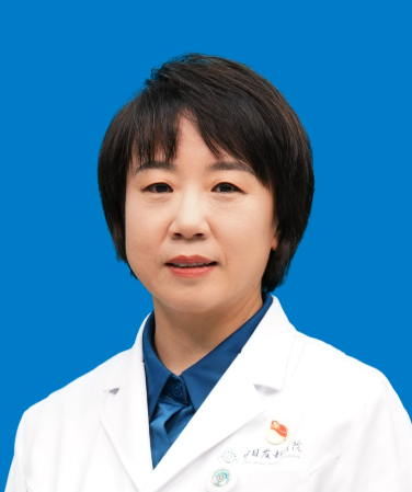 Bo Zhang (张波)合作论文数China-Japan Friendship Hospital17Xingjian Lai合作论文数北京协和医学院(北京协和医院、北京协和医院、北京协和医院),中国北京16Ruina Zhao (赵瑞娜)合作论文数14Xuehua Xi合作论文数13
 Yahong Wang (王亚红)合作论文数Dongzhimen Hospital, Beijing University of Chinese Medicine12Ruyu Liu合作论文数11Xiaoyan Zhang合作论文数11Shenling Zhu合作论文数11Zhitong Ge合作论文数10Sheng Cai (蔡胜)合作论文数6
 Hongyan Wang (王红燕)合作论文数Peking Union Medical College Hospital6Jian-Chu Li合作论文数5Juanjuan Wang合作论文数4Wanying Li合作论文数4Qiong Gao (高琼)合作论文数4
 Ke Lv (吕珂)合作论文数1. Department of Ultrasound, 4Li Zhang合作论文数3Yifan Dong合作论文数3Jia LIU (刘佳)合作论文数3
 Yu Chen (陈钰)合作论文数Peking Union Medical College Hospital3Xinping Tian (田新平)合作论文数2Ruie Feng合作论文数2Yunjiao Yang合作论文数2Jingzhu XU (徐景竹)合作论文数2Bin-yang GAO (高彬洋)合作论文数2Yixuan Zhang (张怡璇)合作论文数2Naili Wang合作论文数2
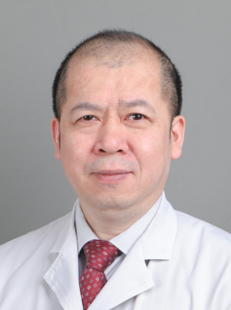 Zhiyong Liang (梁智勇)合作论文数Peking Union Medical College2
 Tao Tan (檀韜)合作论文数Faculty of Applied Sciences, Macao Polytechnic University2Li Tan合作论文数2
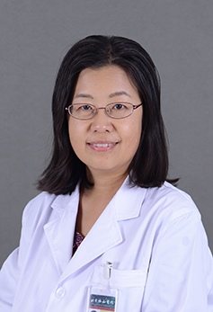 Qingli Zhu (朱庆莉)合作论文数Peking Union Medical College Hospital2
 Wenfeng Song (宋文凤)合作论文数College of Computer Science, Beijing Information Science and Technology University1Fan Yang合作论文数1Jingchuan Zhong合作论文数1Tingxuan Huang合作论文数1Xinglong Liang合作论文数1Gao-Wa Sharen合作论文数1Lixue Huang合作论文数1Xinxin Mao合作论文数1Linfeng Zou合作论文数1
 Guohui Fan合作论文数1Huiting Xu合作论文数1Qing Gao合作论文数1




