Regulation of phospholipid composition is essential for cellular homeostasis. Phosphatidylserine (PS) synthesized in the endoplasmic reticulum (ER) plays critical roles in the plasma membrane and endolysosomal system. Although aberrant PS metabolism is linked to diseases, its cellular effects remain poorly understood. Here, we reveal a conserved role for PS in maintaining Ca2+ homeostasis. PS deficiency in Drosophila leads to mitochondrial damage, which is reversed by reducing inositol 1,4,5-trisphosphate receptor (IP3R)-mediated ER Ca2+ release. Notably, in mammalian cells with pathological PS levels-either deficiency or excess as in Lenz-Majewski syndrome-IP3R activation leads to oscillatory or reduced ER-surface Ca2+ release, contrasting with steady-state conditions. Manipulating phospholipid composition via the phosphatidylethanolamine (PE)-SREBP axis in Drosophila and the phosphatidylcholine (PC)-SREBP axis in mammals normalizes IP3R-mediated Ca2+ release during PS deficiency. These findings establish modulated ER Ca2+ release as a key function of PS and suggest therapeutic strategies for treating lipid metabolic disorders.
Early endosomes are the pivotal sorting station in eukaryotic cells. A longstanding critical question is how the small GTPase Rab5 is precisely targeted to the correct membrane to initiate early endosome formation. Here, we identify Rabex5 and hRME6 as the two guanine-nucleotide exchange factors (GEFs) that together regulate Rab5 recruitment during early endosome formation. Single-molecule imaging of genome-edited cells reveals that Rabex5 and hRME6 are recruited continuously or transiently to nascent uncoated endocytic carriers, respectively. However, in contrast to uncoated endocytic carriers and other intracellular organelles, directing Rabex5 or its GEF domain to clathrin-coated pits or the plasma membrane fails to trigger Rab5 recruitment. Both in vivo and in vitro experiments show that the plasma membrane-enriched phospholipid PI(4,5)P2 prevents Rab5 association with the plasma membrane. Importantly, we found that impaired hydrolysis of PI(4,5)P2 led to reduced early endosome formation in Lowe syndrome cells. Therefore, the spatiotemporal recruitment and activation of Rab5 during early endosome formation are collectively determined by Rabex5/hRME6 recruitment and PI(4,5)P2 depletion during uncoated endocytic carrier formation.
Dynamic protein complex assembly is critical for regulating various biological processes. Proximity labeling (PL), best represented by the ascorbate peroxidase APEX2, allows these molecular events to be captured in living cells in a spatiotemporal manner. However, the hydrogen peroxide (H2O2) dependence of APEX2 has hindered its application in sensitive living systems. Here we introduce ROProx, a radical- and oxygen-driven photoreactive PL technology that leverages the chemically evolved biotin-naphthylamine probe BN2, which has strong binding affinity for APEX2, and the unexpected tyrosyl radicals in APEX2. ROProx labels dynamic cytosolic protein complexes in living cells within seconds, with a range of 10 nm, and is precisely controlled by mild blue light irradiation without H2O2. Additionally, we apply ROProx to explore the phosphotyrosine-dependent GRB2 interactome in living mice by simply injecting BN2 for 5 minutes. ROProx should, therefore, open broad opportunities for PL chemical evolution and applications in other living systems.
In eukaryotes, protein secretion plays essential roles in intercellular communications and extracellular niche-building. Protein secretion generally requires a signal sequence that targets cargos to the canonical secretory pathway consisting of the endoplasmic reticulum (ER), the Golgi apparatus, plasma membrane, and vesicles moving between these compartments. However, cytoplasmic proteins lacking signal sequences (e.g., IL1β, Acb1, FGF2) have been detected, and many have defined functions in the extracellular space, suggesting unconventional protein secretion (UcPS) via alternative pathways. In recent years, scientists have uncovered many new UcPS paradigms, reporting a plethora of mechanisms that collectively form a new field. The inaugural Cold Spring Harbor Asia (CSHA) conference on "Molecular Mechanisms and Physiology of Unconventional Secretion" is the first meeting to bring these researchers together, providing a collegial platform for information sharing at this exciting frontier of cell biology research.
Mammalian cells express seven distinct phosphoinositide species: PI(3)P, PI(4)P, PI(5)P, PI(3,4)P2, PI(3,5)P2, PI(4,5)P2, and PI(3,4,5)P3. With the rapid development of labeling, imaging, and manipulation tools, our understanding of the spatial distribution, functions, and regulation of these phosphoinositides has advanced significantly. Tightly regulated by lipid kinases, phosphatases, and lipid transfer proteins, each phosphoinositide exhibits a unique yet dynamic spatial distribution at both subcellular and suborganelle levels. This distinct spatial organization is critical for controlling membrane trafficking, defining organelle identity and function, mediating signal transduction, and supporting other essential cellular processes. Dysregulation of spatial phosphoinositide signaling has been linked to various human diseases. In this review, we provide a brief overview of current insights into the spatial organization of phosphoinositide signaling, highlighting its key roles in regulating membrane dynamics and signal transduction at the plasma membrane, endosomes and lysosomes, the Golgi apparatus, the ER, and the nucleus.
cTAGE5/MEA6 plays a pivotal role in COPII complex assembly, ER-to-Golgi trafficking, and secretion. However, whether cTAGE5/MEA6 is involved in other cellular functions remains unclear. Here, we show that conditional cTAGE5 knockout results in embryonic lethality during development and premature aging in adult mice. cTAGE5 deficiency leads to abnormal nuclear structure and disturbed cell proliferation in MEF cells. Further mechanistic studies reveal that cTAGE5 localizes not only to the ER exit sites but also to other ER structures, where it interacts with the lamin B receptor (LBR). Loss of cTAGE5 disrupts LBR's localization to the inner nuclear membrane, leading to its retention in the ER and instability. This results in abnormal nuclear (envelope) morphology and cellular senescence, likely driven by activation of the P53/P21 senescence pathway. Thus, our study uncovers cTAGE5's role in maintaining nuclear envelope integrity and highlights its function and potential mechanism in preventing cellular senescence and animal aging.
In plants, genetically encoded probes based on redox-sensitive green fluorescent protein (roGFP) have been used to detect hydrogen peroxide (H2O2) levels by fusing exogenous thiol peroxidases, such as Orp1 and Tsa2. However, the effectiveness of these thiol peroxidases compared to endogenous ones remains unexplored. Here, we develop a H2O2 probe by fusing roGFP2 to an endogenous H2O2 sensor, type II peroxiredoxin (PRXIIB), which displayed enhanced responsiveness and conversion kinetics compared to roGFP2-Orp1 in vitro and superior sensitivity to H2O2 in vivo. The roGFP2-PRXIIB probe allowed robust visualization of H2O2 production in abiotic and biotic stresses, and growing pollen tubes. We further targeted roGFP2-PRXIIB to cytosol, nuclei, mitochondria and chloroplasts to monitor H2O2 accumulation in real time in different subcellular compartments during immune activation, and the analyses revealed different temporal patterns of H2O2 accumulation during pattern- and effector-triggered immune responses in different compartments. Taken together, the work provides an ultra-sensitive probe for H2O2 dynamics in diverse plant biological processes.
Mutations of TRAPPC12 are associated with progressive childhood encephalopathy including abnormal white matter. However, the underlying pathogenesis is still unclear. Here, we found that Trappc12 deficiency in CG4 and oligodendrocyte progenitor cells (OPCs) affects their differentiation and maturation. In addition, TRAPPC12 interacts with Mea6/cTAGE5, and Mea6/cTAGE5 ablation in OPCs affects their proliferation and differentiation, leading to marked hypomyelination, compromised synaptic functionality, and aberrant behaviors in mice. We reveal that TRAPPC12 is associated with COPII components at ER exit site, and Mea6/cTAGE5 cKO disrupts the trafficking pathway by affecting the distribution and/or expression of TRAPPC12, SEC13, SEC31A, and SAR1. Moreover, we observed marked disturbances in the secretion of pleiotrophin (PTN) in Mea6-deficient OPCs. Notably, exogenous PTN supplementation ameliorated the differentiation deficits of these OPCs. Collectively, our findings indicate that the association between TRAPPC12 and MEA6 is important for cargo trafficking and white matter development.
The spatiotemporal transition of small GTPase Rab5 to Rab7 is crucial for early-to-late endosome maturation, yet the precise mechanism governing Rab5-to-Rab7 switching remains elusive. USP8, a ubiquitin-specific protease, plays a prominent role in the endosomal sorting of a wide range of transmembrane receptors and is a promising target in cancer therapy. Here, we identified that USP8 is recruited to Rab5-positive carriers by Rabex5, a guanine nucleotide exchange factor (GEF) for Rab5. The recruitment of USP8 dissociates Rabex5 from early endosomes (EEs) and meanwhile promotes the recruitment of the Rab7 GEF SAND-1/Mon1. In USP8-deficient cells, the level of active Rab5 is increased, while the Rab7 signal is decreased. As a result, enlarged EEs with abundant intraluminal vesicles accumulate and digestive lysosomes are rudimentary. Together, our results reveal an important and unexpected role of a deubiquitinating enzyme in endosome maturation.
The class I phosphatidylinositol 3-kinase (PI3K)-AKT signaling pathway is a key regulator of cell survival, growth, and proliferation and is among the most frequently mutated pathways in cancer. However, where and how PI3K-AKT signaling is spatially activated and organized in mammalian cells remains poorly understood. Here, we identify focal adhesions (FAs) as subcellular signaling hubs organizing the activation of PI3K-PI(3,4,5)P3-AKT signaling in human cancer cells containing p110α mutations under basal conditions. We find that class IA PI3Ks are preferentially recruited to FAs for activation, resulting in localized production of PI(3,4,5)P3 around FAs. As the effector protein of PI(3,4,5)P3, AKT1 molecules are dynamically recruited around FAs for activation. The spatial recruitment/activation of the PI3K-PI(3,4,5)P3-AKT cascade is regulated by activated FA kinase (FAK). Furthermore, combined inhibition of p110α and FAK results in a more potent inhibitory effect on cancer cells. Thus, our results unveil a growth-factor independent, compartmentalized organization mechanism for PI3K-PI(3,4,5)P3-AKT signaling.
BACKGROUND:beta-adrenergic receptor (beta-AR) overactivation is a major pathological cue associated with cardiac injury and diseases. AMPK (AMP-activated protein kinase), a conserved energy sensor, regulates energy metabolism and is cardioprotective. However, whether AMPK exerts cardioprotective effects via regulating the signaling pathway downstream of beta-AR remains unclear.METHODS:Using immunoprecipitation, mass spectrometry, site-specific mutation, in vitro kinase assay, and in vivo animal studies, we determined whether AMPK phosphorylates beta-arrestin-1 at serine (Ser) 330. Wild-type mice and mice with site-specific mutagenesis (S330A knock-in [KI]/S330D KI) were subcutaneously injected with the beta-AR agonist isoproterenol (5 mg/kg) to evaluate the causality between beta-adrenergic insult and beta-arrestin-1 Ser330 phosphorylation. Cardiac transcriptomics was used to identify changes in gene expression from beta-arrestin-1-S330A/S330D mutation and beta-adrenergic insult.RESULTS:Metformin could decrease cAMP/PKA (protein kinase A) signaling induced by isoproterenol. AMPK bound to beta-arrestin-1 and phosphorylated Ser330 with the highest phosphorylated mass spectrometry score. AMPK activation promoted beta-arrestin-1 Ser330 phosphorylation in vitro and in vivo. Neonatal mouse cardiomyocytes overexpressing beta-arrestin-1-S330D (active form) inhibited the beta-AR/cAMP/PKA axis by increasing PDE (phosphodiesterase) 4 expression and activity. Cardiac transcriptomics revealed that the differentially expressed genes between isoproterenol-treated S330A KI and S330D KI mice were mainly involved in immune processes and inflammatory response. beta-arrestin-1 Ser330 phosphorylation inhibited isoproterenol-induced reactive oxygen species production and NLRP3 (NOD-like receptor protein 3) inflammasome activation in neonatal mouse cardiomyocytes. In S330D KI mice, the beta-AR-activated cAMP/PKA pathways were attenuated, leading to repressed inflammasome activation, reduced expression of proinflammatory cytokines, and mitigated macrophage infiltration. Compared with S330A KI mice, S330D KI mice showed diminished cardiac fibrosis and improved cardiac function upon isoproterenol exposure. However, the cardiac protection exerted by AMPK was abolished in S330A KI mice.CONCLUSIONS:AMPK phosphorylation of beta-arrestin-1 Ser330 potentiated PDE4 expression and activity, thereby inhibiting beta-AR/cAMP/PKA activation. Subsequently, beta-arrestin-1 Ser330 phosphorylation blocks beta-AR-induced cardiac inflammasome activation and remodeling.
Endocytosis and recycling control the uptake and retrieval of various materials, including membrane proteins and lipids, in all eukaryotic cells. These processes are crucial for cell growth, organization, function and environmental communication. However, the mechanisms underlying efficient, fast endocytic recycling remain poorly understood. Here, by utilizing a biosensor and imaging-based screening, we uncover a recycling mechanism that couples endocytosis and fast recycling, which we name the clathrin-associated fast endosomal recycling pathway (CARP). Clathrin-associated tubulovesicular carriers containing clathrin, AP1, Arf1, Rab1 and Rab11, while lacking the multimeric retrieval complexes, are generated at subdomains of early endosomes and then transported along actin to cell surfaces. Unexpectedly, the clathrin-associated recycling carriers undergo partial fusion with the plasma membrane. Subsequently, they are released from the membrane by dynamin and re-enter cells. Multiple receptors utilize and modulate CARP for fast recycling following endocytosis. Thus, CARP represents a previously unrecognized endocytic recycling mechanism with kiss-and-run membrane fusion. Xu, Liang, Li, Dang et al. delineate the clathrin-associated fast endosomal recycling pathway, which involves clathrin-associated carriers derived from early endosomes partially fusing with the plasma membrane before release from the membrane.
Distinct phospholipid species display specific distribution patterns across cellular membranes, which are important for their structural and signaling roles and for preserving the integrity and functionality of the plasma membrane and organelles. Recent advancements in lipid biosensor technology and imaging modalities now allow for direct observation of phospholipid distribution, trafficking, and dynamics in living cells. These innovations have markedly advanced our understanding of phospholipid function and regulation at both cellular and subcellular levels. Herein, we summarize the latest developments in phospholipid biosensor design and application, emphasizing the contribution of cutting-edge imaging techniques to elucidating phospholipid dynamics and distribution with unparalleled spatiotemporal precision.
Environmental osmolarity plays a crucial role in regulating the functions and behaviors of both host cells and pathogens. However, it remains unclear whether and how environmental osmotic stimuli modulate bacterial-host interfacial adhesion. Using single-cell force spectroscopy, we revealed that the interfacial adhesion force depended nonlinearly on the osmotic prestimulation of host cells but not bacteria. Quantitatively, the adhesion force increased dramatically from 25.98 nN under isotonic conditions to 112.45 or 93.10 nN after the host cells were treated with the hypotonic or hypertonic solution. There was a strong correlation between the adhesion force and the number of host cells harboring adherent/internalized bacteria. We further revealed that enhanced overexpression levels of collagen XV and II were responsible for the increases in interfacial adhesion under hypotonic and hypertonic conditions, respectively. This work provides new opportunities for developing host-directed antibacterial strategies related to interfacial adhesion from a mechanobiological perspective.
Upon endoplasmic reticulum (ER) stress, activation of the ER-resident transmembrane protein kinase/endoribonuclease inositol-requiring enzyme 1 (IRE1) initiates a key branch of the unfolded protein response (UPR) through unconventional splicing generation of the transcription factor X-box-binding protein 1 (XBP1s). Activated IRE1 can form large clusters/foci, whose exact dynamic architectures and functional properties remain largely elusive. Here we report that, in mammalian cells, formation of IRE1 alpha clusters is an ER membrane-bound phase separation event that is coupled to the assembly of stress granules (SGs). In response to different stressors, IRE1 alpha clusters are dynamically tethered to SGs at the ER. The cytosolic linker portion of IRE1 alpha possesses intrinsically disordered regions and is essential for its condensation with SGs. Furthermore, disruption of SG assembly abolishes IRE1 alpha clustering and compromises XBP1 mRNA splicing, and such IRE1 alpha-SG coalescence engenders enrichment of the biochemical components of the pro-survival IRE1 alpha-XBP1 pathway during ER stress. Our findings unravel a phase transition mechanism for the spatiotemporal assembly of IRE1 alpha-SG condensates to establish a more efficient IRE1 alpha machinery, thus enabling higher stress-handling capacity. Liu, Zhang, Yao et al. report that IRE1 alpha clustering, known to be part of the unfolded protein response, is membrane-bound phase separation and that IRE1 can coalesce with the phase-separated stress granules.
HER2 belongs to the human epidermal growth factor receptor tyrosine kinase family. Its overexpression or hyperactivation is a leading cause for multiple types of cancers. HER2 functions mainly through dimerization with other family members, such as EGFR. However, the molecular details for heterodimer assembly have not been completely understood. Here, we report cryo-EM structures of the EGF- and epiregulin-bound EGFR/HER2 ectodomain complexes at resolutions of 3.3 Å and 4.5 Å, respectively. Together with the functional analyses, we demonstrate that only the dimerization arm of HER2, but not that of EGFR, is essential for their heterodimer formation and signal transduction. Moreover, we analyze the differential membrane dynamics and transient interactions of endogenous EGFR and HER2 molecules in genome-edited cells using single-molecule live-cell imaging. Furthermore, we show that the interaction with HER2 could allow EGFR to resist endocytosis. Together, this work deepens our understanding of the unique structural properties and dynamics of the EGFR/HER2 complex.
Salmonella enterica, the etiological agent of gastrointestinal and systemic diseases, translocates a plethora of virulence factors through its type III secretion systems to host cells during infection. Among them, SpvB has been reported to harbor an ADP-ribosyltransferase domain in its C terminus, which destabilizes host cytoskeleton by modifying actin. However, whether this effector targets other host factors as well as the function of its N terminus still remains to be determined. Here, we found that SpvB targets clathrin and its adaptor AP-1 (adaptor protein 1) via interactions with its N-terminal domain. Notably, our data suggest that SpvB-clathrin/AP-1 associations disrupt clathrin-mediated endocytosis and protein secretion pathway as well. In addition, knocking down of AP-1 promotes Salmonella intracellular survival and proliferation in host cells.
Adrenergic receptors (ARs) and catalytic receptors (CRs), two major classes of cell-surface receptors, play essential roles in a wide range of physiological and pathological processes. Studies over the years have revealed that ARs and CRs, along with their associated signaling transduction pathways, are not isolated in the cells. Instead, there exists functional crosstalk, involving either activation or inhibition, among specific members of ARs and CRs. Although the dynamics and mechanism of individual receptors within each family have been extensively studied, we have just begun to understand the spatiotemporal dynamics, functional consequences, and underlying mechanisms of the crosstalk between ARs and CRs. In this review, we will provide a concise overview of recent progress in identifying and elucidating the crosstalk, either unidirectional or bidirectional, between ARs and CRs.
 Song Dang合作论文数15
 Xiaohong Fang (方晓红)合作论文数Key Laboratory of Molecular Nanostructure and Nanotechnology, Chinese Academy of Sciences15
 Youyi Zhang (张幼怡)合作论文数Peking University Third Hospital14Yongtao Du合作论文数9
 Nan Li合作论文数9
 Zijian Li (李子健)合作论文数Peking University Third Hospital;Department of Pharmacy Administration and Clinical Pharmacy, School of Pharmaceutical Sciences, Peking University;International Research Center for Medicinal Administration, Peking University8
 Wouter Boomsma合作论文数Department of Computer Science, Faculty of Science, University of Copenhagen7ZhiZhen Lv (吕志珍)合作论文数7
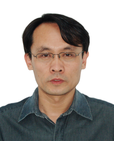 Jinghe Yuan (袁景和)合作论文数Institute of Chemistry, Chinese Academy of Sciences;University of Chinese Academy of Sciences6
 Han Xiao (肖晗)合作论文数Peking University Third Hospital;Shihezi University School of Medicine6Jimin Wu (吴济民)合作论文数5Wesley Skillern合作论文数4Xuejie Zhang合作论文数4
 Gokul Upadhyayula合作论文数Cell & Developmental Biology Faculty, Department of Molecular and Cell Biology, University of California4
 Mei Ding (丁梅)合作论文数School of Basic Medical Sciences, Faculty of Medical Sciences, Wuhan University;Taikang Center for Life and Medical Sciences, Wuhan University4Gaudin Raphaël合作论文数CNRS4
 Ming Xu (徐明)合作论文数Graduate School, Peking University Health Science Center;Peking University Third Hospital4Yue Miao合作论文数3Xinyi Zhang合作论文数3Baolei Wang合作论文数3Fei Liu (刘飞)合作论文数3
 Liang Jingjing合作论文数3
 Jiachao Xu合作论文数3Yi-ying Chou合作论文数Departments of Microbiology and3
 Jianyong Huang (黄建永)合作论文数School of Mechanics and Engineering Science, Peking University3
 Dong Li (李栋)合作论文数School of Life Sciences, Tsinghua University3Minghe Ma合作论文数3Siying Qin合作论文数2Xiaocen Duan合作论文数2
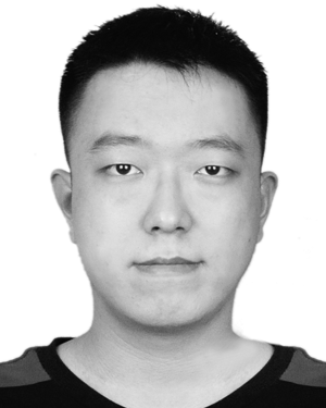 Jiahao Liu合作论文数2Hongwei Xu合作论文数2Yiming Han合作论文数2Yuting Feng合作论文数2
 Wangxi Luo合作论文数Temple Univ, Dept Biol, Philadelphia, PA 19122 USA2jianghui zhang (张江晖)合作论文数2Shijia Huang合作论文数Beijing Advanced Innovation Center for Structural Biology, Tsinghua-Peking Joint Center for Life Sciences, Center for Plant Biology, School of Life Sciences, Tsinghua University2
 Zhe Zhang (张哲)合作论文数School of Life Sciences, Peking University;Peking University-Tsinghua University Joint Center for Life Sciences2
 Yuting Guo合作论文数Chinese Acad Sci, CAS Ctr Excellence Biomacromol, Inst Biophys, Natl Lab Biomacromol, Beijing, Peoples R China2
 Xiaohua Yan (严晓华)合作论文数School of Basic Medical Sciences, Nanchang University2
 Lei Shi (石磊)合作论文数Institute of Materia Medica, Chinese Academy of Medical Science & Peking Union Medical College2
 Zhiheng Xu (许执恒)合作论文数Institute of Genetics and Developmental Biology, Chinese Academy of Sciences;University of Chinese Academy of Sciences2
 Xiaoyun Liu (刘小云)合作论文数School of Basic Medical Sciences, Peking University2Aiju Tian合作论文数Beijing Geriatric Hospital;Department of Cardiology, Fuwai Hospital, Chinese Academy of Medical Sciences & Peking Union Medical College2
 Tie Xia (夏铁)合作论文数School of Medicine, Tsinghua University2
 Eli Song (宋婀莉)合作论文数Institute of Biophysics, Chinese Academy Sciences2
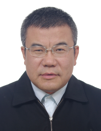 Chunru Wang (王春儒)合作论文数Key Laboratory of Molecular Nanostructure and Nanotechnology, Chinese Academy of Sciences2Tsuyoshi Sakai合作论文数2Lei Qin合作论文数2
 Jianmin Zhou (周俭民)合作论文数Yazhouwan National Laboratory;University of Chinese Academy of Sciences;Institute of Genetics and Developmental Biology, Chinese Academy of Sciences2




