HuFceRI transfected RBL or LAD2 cells were incubated with IgE ± MEDI4212. Bound IgE was cross-linked using a commercial anti-IgE and cell activation detected using calcium signalling or β-hexosaminidase release. Effect of MEDI4212 on IgE/CD23 interaction was assessed by incubation of IL-4 stimulated IM9 cells with IgE ± MEDI4212 and bound IgE detected by flow cytometry. Effects on IgE production in human B cells (differentiated with APC + IL-4, ± MEDI4212) was evaluated by flow cytometry. Serum free-IgE was measured using ELISA.
Disclaimer/Complaints regulations If you believe that digital publication of certain material infringes any of your rights or (privacy) interests, please let the Library know, stating your reasons. In case of a legitimate complaint, the Library will make the material inaccessible and/or remove it from the website. Please Ask the Library: http://uba.uva.nl/en/contact, or a letter to: Library of the University of Amsterdam, Secretariat, Singel 425, 1012 WP Amsterdam, The Netherlands. You will be contacted as soon as possible.
Disclaimer/Complaints regulations If you believe that digital publication of certain material infringes any of your rights or (privacy) interests, please let the Library know, stating your reasons. In case of a legitimate complaint, the Library will make the material inaccessible and/or remove it from the website. Please Ask the Library: https://uba.uva.nl/en/contact, or a letter to: Library of the University of Amsterdam, Secretariat, Singel 425, 1012 WP Amsterdam, The Netherlands. You will be contacted as soon as possible.
Although T cells infiltrate many types of murine and human neoplasms, in many instances tumor-specific cytotoxicity is not observed. Strategies to stimulate CTL-mediated antitumor immunity have included in vitro stimulation and/or genetic engineering of T cells, followed by adoptive transfer into tumor-bearing hosts. In this model of B cell lymphoma in SJL/J mice, we used Tim-3+ T-bet+ Th1 cells to facilitate the development of tumor-specific CTL. Tumor-specific Th1 cell lines were polarized with IL-12 during in vitro stimulation and long term maintenance. As few as 5 million Tim-3+ T-bet+ Th1 cells enabled recipients to resist growth of malignant transplantable cells. In addition, similar numbers of Th1 cells injected into 2- to 3-mo-old mice inhibited development of the spontaneous primary lymphomas, which normally arise in 90% of aging mice. CFSE+ Th1 cells colocalized with injected tumor cells in vivo and formed conjugates with the tumor cells within follicles, whereas in nontumor-challenged recipients the CFSE+ Th1 cells localized only within the T cell zones of the spleen. These results provide evidence that adoptive immunotherapy with Tim-3+ T-bet+ tumor-specific Th1 cells can be used to induce host cytotoxic responses that inhibit the development and growth of neoplastic cells.
Isolated peripheral blood CD4 cells from allergic individuals express CC chemokine receptor (CCR)3 and CCR4 after expansion in vitro. In addition, human T helper type 2 (Th2) cells polarized in vitro selectively express CCR3 and CCR4 at certain stages of activation/differentiation and respond preferentially to the ligands eotaxin and monocyte-derived chemokine (MDC). However, controversy arises when the in vivo significance of this distinct expression is discussed. To address the functional role of CCR3/eotaxin and CCR4/MDC during the in vivo recruitment of Th2 cells, we have transferred effector Th cells into naive mice to induce allergic airway disease. Tracking of these cells after repeated antigen challenge has established that both CCR3/eotaxin and CCR4/MDC axes contribute to the recruitment of Th2 cells to the lung, demonstrating the in vivo relevance of the expression of these receptors on Th2 cells. We have shown that involvement of the CCR3/eotaxin pathway is confined to early stages of the response in vivo, whereas repeated antigen stimulation results in the predominant use of the CCR4/MDC pathway. We propose that effector Th2 cells respond to both CCR3/eotaxin and CCR4/MDC pathways initially, but that a progressive increase in CCR4-positive cells results in the predominance of the CCR4/MDC axis in the long-term recruitment of Th2 cells in vivo.
 J. Bertin合作论文数3Jose-Carlos Gutierrez-Ramos合作论文数3
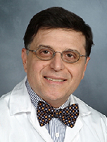 Giorgio Inghirami合作论文数Department of Pathology and Laboratory Medicine, Weill Cornell Medical College, Cornell University;NewYork-Presbyterian Hospital3K Sjoerdsma合作论文数2james w metzger合作论文数2E. Wierenga合作论文数2L. Franchi合作论文数2
 Nesrin Ozoren合作论文数Center for Life Sciences and Technologies, Bogazici University2
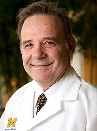 Gabriel Nunez合作论文数Department of Pathology, University of Michigan Medical School2Nicholas M. Ponzio合作论文数2
 Byung S Kim合作论文数Department of Microbiology-Immunology;Medical School, Northwestern University;Medical School, Northwestern University2
 Mathilde Body-Malapel合作论文数Amphi J & K, Université Lille Nord de France2Mani Mohindru合作论文数2Lisa Brown合作论文数2Gillian A. Kingsbury合作论文数2C TOUVAY合作论文数2Lianne Wassink合作论文数2KR Taylor合作论文数1C.P. Page合作论文数1M Koneru合作论文数1T-D Kanneganti合作论文数1A. L. Cogen合作论文数1S. Oakley合作论文数1
 Trang Nguyen合作论文数1D. R. Gaya合作论文数1M. R. Staempfli合作论文数1Claire L Dobson合作论文数1Rosemarie H DeKruyff合作论文数1sofia persdotter合作论文数1Wj Simmons合作论文数1K B Nguyen合作论文数1Mythili Koneru合作论文数1
 Hal Hoffman合作论文数Department of Pediatrics, University of California, San Diego;Division of Rheumatology, Allergy & Immunology, Department of Medicine, School of Medicine, University of California1A Cahill合作论文数1P. Shen合作论文数1
 Ismail Erturk合作论文数Department of Medical Oncology, Gülhane Training and Research Hospital1
 Naohiro Inohara合作论文数Department of Pathology, Medical School, University of Michigan-Ann Arbor1Anderson Ian合作论文数1
 Mathieu Morissette合作论文数Département De Médecine, Faculté De Médecine, Université Laval;Centre De Recherche De L'institut Universitaire De Cardiologie Et De Pneumonie De Québec;Centre De Recherche Sur Le Cancer, Université Laval1
 Richard Gallo合作论文数Department of Dermatology, School of Medicine, University of California, San Diego1Phillip Monk合作论文数1Benson G合作论文数1
 Clare Lloyd合作论文数National Heart & Lung Institute, Faculty of Medicine, Imperial College London1Nikota J K合作论文数1
 Parul Singh合作论文数1Jagirdar Rajesh合作论文数1Carlsson Mats合作论文数1
 Cohen E Suzanne合作论文数MedImmune Ltd;Cambridge;MedImmune Ltd1Christine A. Biron合作论文数1




