Normal diploid somatic mammalian cell division generates 2 daughter cells as a result of a strict and well-controlled mitotic process. However, some defects during the progression of that process could generate an unbalanced distribution of chromosomes, aneuploidy and eventually, a malignant phenotype. Previous observations using a transgenic mouse model with diminished DNA repair capacity revealed the presence of nuclear buds (NBs) induced in vitro by the nucleoside analog zidovudine (Retrovir®, 3′-azido-3′-deoxythymidine, AZT). Here we used bone marrow mesenchymal cells, taken from mice with the Xpa–/–Trp53+/– genotype, that were cultured and exposed to 0 and 100 µM AZT for 24 hours. Fixed and denatured cells were processed by fluorescence in situ hybridization (FISH) with whole chromosome painting probes used to identify chromosomes in cells growing on glass chamber slides (2 probes/slide). A variety of sizes and shapes of NBs were observed. Some NBs had a large connection with the main nucleus (>¼ of the NB diameter), others hada smaller connection (<¼ of the NB diameter), some were circular and positioned close to the nucleus, while some resided in the cytoplasm separated from the nucleus or connected by a thin chromatin strand. We had hypothesized that NBs would progress in the process of budding until separation occurred, but this was not proven by time-lapse photography studies performed for 20 hours. From 1,126 cells scored in the unexposed cultures, 10.39 % of cells carried NBs, while from 1,108 cells scored in the AZT-exposed cultures 29.16% of cells carried NBs (p = 0.001). In AZT-exposed cells there were a total of 322 NBs scored; 46.6% or 150 NBs contained positive signals for one or both probes used, while 53% or 172 NBs had no probe signal. In addition, FISH analysis showed no preferential localization of any chromosome within the NBs. Among the NBs that carried no probe signal, the presence of positive signals with inversion of DAPI imaging demonstrated centromeric content. It has been hypothesized that NBs occur as a result of expulsion of amplified DNA from the main nucleus; however, this data demonstrates that NBs may contain any chromosome, suggesting that NBs do not consist of just amplified DNA.
Schizophrenia is a debilitating brain disorder characterized by hallucinations, delusions, disordered thinking, diminished emotion, and cognitive impairment. Age of onset of schizophrenia is typically in late adolescence and early adulthood. Given a worldwide prevalence of approximately 1%, schizophrenia is the fourth leading cause of disability and major public health burden. Family, twin and adoption studies have demonstrated a large genetic component, with estimates of heritability around 81%.(1) Childhood onset schizophrenia is rare, with the prevalence estimated to be approximately 1/300th the rate of the more typical adult onset form. As has been the case for other complex disorders such as breast cancer and Alzheimer’s disease, the study of extreme early onset cases might facilitate the discovery of disease genes.(2) Since 1990, we have recruited and rigorously characterized 92 patients with COS.(3) None of these patients had notable dysmorphologies indicative of an obvious chromosomal anomaly. However, there was an increased rate of early pre-psychotic neurodevelopmental disorders relative to that seen in adult onset patients.(4) Such disorders are seen in many genetic syndromes of pediatric onset;(5) the mean IQ of this group was 80, somewhat lower than that of adult onset patients. Follow-up every 2 years for reevaluation confirms the stability of the diagnosis of schizophrenia and has shown continuity with the more common adult onset form of the disorder (AOS).(3, 6) Previously we reported that high resolution karyotyping and fluorescent in situ hybridization (FISH) revealed 4 cases with the typical 3 Mb 22q11 deletion,(7) 1 case with atypical Turner’s syndrome (46,X,del(X)(q24-ter),(8) and 1 case with an inherited 1;7 balanced translocation.(9) The rate of 4 out of 92 cases for 22q11 deletion is significantly higher than that seen in samples of unselected patients with AOS.(7) In addition, one case with 5q32-ter segmental uniparental isodisomy (35 Mb) was observed through loss of heterozygosity (LOH) on the Affymetrix NspI 250K SNP array and confirmed with genotyping of microsatellite markers.(10) Numerous case reports for Turner syndrome and trisomy X provide evidence for the increased risk of psychotic disorders and relevant psychopathological manifestations among adults with X-chromosome anomalies.(11–13) Unrelated studies report rates of atypical Turner syndrome in the general and adult psychiatric population to be 0.2% and 0.17%, respectively.(13, 14) Women with Turner syndrome also show impairments in emotional and visual-spatial processing, decreased full-scale IQ, and share similarities with schizophrenia patients in structural and functional brain abnormalities.(15, 16) Less is known about the association between trisomy X and psychosis; however DeLisi et al. reported the prevalence of trisomy X to be 0.63% in studies of adult onset schizophrenia compared to the 0.39% in the general population.(12, 17) Here we report 2 additional cases with X chromosome anomalies in the NIMH COS cohort: one mosaic Turner syndrome (46,X,i(X)(q10)(22%)/45,X(78%)), and one trisomy X (47, XXX), bringing the total prevalence to 3/38 (7.9%) among the females in this cohort. The prevalence of these X chromosome anomalies in the NIMH COS population is significantly greater than the reported rates in either the general population (0.2%) or adult onset schizophrenic populations (0.4%, p=0.001) (Table I). Table 1 Rates of specific sex chromosome anomalies in childhood onset schizophrenia (COS), adult onset schizophrenia (AOS), and general population samples Further, in accordance with Kaplan & Cotton’s (1968) hypothesis of “the possibility that other and more subtle genetic instabilities may be associated with the schizophrenic disorders,” we hypothesized that X-chromosome anomalies may be associated with previously unidentified micro-deletions and/or duplications that predispose to schizophrenia.(18) To test this hypothesis, we completed whole genome high density SNP and CGH arrays to ascertain novel structural variants (see details in Walsh et al 2008).(19) Briefly, Affymetrix Mapping 500K SNP arrays and Agilent 185K/244K oligo arrayCGH were completed according to manufacturer protocols (www.affymetrix.com, www.agilent.com) and only variants that were identified through both methods were considered for analysis. Results did not reveal any further de novo chromosomal events among the probands with X-chromosome anomalies that could be implicated in the etiology of schizophrenia. Nor did these cases tend to have a higher rate of rare structural variants as compared to karyotypically normal female COS probands (results not shown). However, the one case with atypical Turner syndrome who was missing part of one Xq arm was found to have also a 17 Mb duplication of chromosome 16q22.2-ter. Spectral karyotyping revealed that the extra copy of this segment of chromosome 16 was attached to the missing arm of the X chromosome. The duplicated region of chromosome 16 contains approximately 100 known genes, but presents an interesting case since it is not clear what the functionality of the derived chromosome X;16 would be. It is conceivable that the entire chromosome would be subject to X-inactivation, though functional studies would be warranted for this case. To summarize, this report confirms that subsyndromal sex chromosome anomalies in COS are significantly higher than that found in the community or in AOS. The role of these anomalies in schizophrenia is unclear, though dysregulation of emotional processing may be one component. While we had hypothesized that these cases may have other, more subtle submicroscopic structural variants that could be involved in schizophrenia etiology, this was not confirmed. To date, the overall rate of rate of large chromosomal abnormalities and 22q11 deletions among this COS cohort is 10%.
Therapeutic irradiation for head and neck cancer, and the autoimmune disease Sjogren's syndrome, lead to loss of salivary parenchyma. They are the two main causes of irreversible salivary gland hypofunction. Such patients cannot produce adequate levels of saliva, leading to considerable morbidity. We are working to develop an artificial salivary gland for such patients. A major problem in this endeavor has been the difficulty in obtaining a suitable autologous cellular component. This article describes a method of culturing and expanding primary salivary cells obtained from human submandibular glands ( huSMGs) that is serum free and yields cells that are epithelial in nature. These include morphological ( light and transmission electron microscopy [TEM]), protein expression (immunologically positive for ZO-1, claudin-1, and E-cadherin), and functional evidence. Under confocal microscopy, huSMG cells show polarization and appropriately localize tight junction proteins. TEM micrographs show an absence of dense core granules, but confirm the presence of tight and intermediate junctions and desmosomes between the cells. Functional assays showed that huSMG cells have high transepithelial electrical resistance and low rates of paracellular fluid movement. Additionally, huSMG cells show a normal karyotype without any morphological or numerical abnormalities, and most closely resemble striated and excretory duct cells in appearance. We conclude that this culture method for obtaining autologous human salivary cells should be useful in developing an artificial salivary gland.
Acute myeloid leukemia subtype M4 with eosinophilia is associated with a chromosome 16 inversion that creates a fusion gene CBFB-MYH11. We have previously shown that CBFB-MYH11 is necessary but not sufficient for leukemogenesis. Here, we report the identification of genes that specifically cooperate with CBFB-MYH11 in leukemogenesis. Neonatal injection of Cbfb-MYH11 knock-in chimeric mice with retrovirus 4070A led to the development of acute myeloid leukemia in 2-5 months. Each leukemia sample contained one or a few viral insertions, suggesting that alteration of one gene could be sufficient to synergize with Cbfb-MYH11. The chromosomal position of 67 independent retroviral insertion sites (RISs) was determined, and 90% of the RISs mapped within 10 kb of a flanking gene. In total, 54 candidate genes were identified; six of them were common insertion sites (CISs). CIS genes included members of a zinc finger transcription factors family, Plag1 and Plagl2, with eight and two independent insertions, respectively. CIS genes also included Runx2, Myb, H2T24, and D6Mm5e. Comparison of the remaining 48 genes with single insertion sites with known leukemia-associated RISs indicated that 18 coincide with known RISs. To our knowledge, this retroviral genetic screen is the first to identify genes that cooperate with a fusion gene important for human myeloid leukemia.
Summary: C57BL/6 is a well‐characterized mouse strain that is used extensively for immunological and neurological research. The establishment of C57BL/6 ES cell lines has facilitated the study of gene‐altered mice in a pure genetic background—however, relatively few such lines exist. Using a defined media supplement, knockout serum replacement (KSR) with knockout DMEM (KSR‐KDMEM), we find that we can readily establish ES cell lines from blastocysts of C57BL/6J mice. Six lines were established, all of which were karyotypically normal and could be maintained in the undifferentiated state on mouse embryonic fibroblast (MEF) feeders. One line was further tested and found to be karyotypically stable and germline competent, both prior to manipulation and after gene targeting. For this cell line, efficiencies of cell cloning and chimera generation were greater when maintained in KSR‐KDMEM. Our work suggests that the use of defined serum‐free media may facilitate the generation of ES cells from inbred mouse strains. genesis 39:100–104, 2004. Published 2004 Wiley‐Liss, Inc.
We report a 19‐year‐old, non‐Amish Caucasian female patient with primary amenorrhea caused by complete lack of Müllerian fusion with vaginal agenesis or Müllerian aplasia (MA), postaxial polydactyly (PAP), and tetralogy of Fallot. The genital tract anomaly of MA with and without renal or skeletal anomalies comprises Mayer–Rokitansky–Kuster–Hauser syndrome, which has not been reported with tetralogy of Fallot. The phenotypic triad of anomalies most closely resembled McKusick–Kaufman syndrome (MKS; OMIM 236700), a rare multiple congenital anomaly syndrome comprised of hydrometrocolpos (HMC), PAP, and congenital heart malformation that is inherited in an autosomal recessive pattern. While upper reproductive tract anomalies have not been reported with MKS, they have been reported with Bardet–Biedl syndrome (BBS), a syndrome that significantly overlaps with MKS. Both MKS and BBS can be caused by mutations in the MKKS or BBS6 gene on chromosome 20p12 and BBS is also associated with mutations in other genes ( BBS1 , BBS2 , BBS4 , and BBS7 ). To address this heterogenity, we sequenced the causative genes in MKS and BBS but no mutations in these five genes were identified. Fluorescence in situ hybridization (FISH) excluded large deletions of chromosome 20p12 and microsatellite marker studies confirmed biparental inheritance for all of the known BBS loci. The dual midline fusion defects of tetralogy of Fallot and MA suggests that either this patient has a unique syndrome with a distinct genetic etiology or that she has a genetically heterogeneous or variant form of MKS. Published 2004 Wiley‐Liss, Inc.
(11:00–11:15 AM) 1. Triplication of Alpha-Synuclein is Causal of Neuropathologically Confirmed Parkinson’s Disease, Diffuse Lewy Body Disease, and Multiple System Atrophy in a Single Kindred K. Gwinn-Hardy, M. Farrer, D. Dickson, L.-W. Jin, J. Johnson, A. Singleton, S. Hague, J. Kachergus, M. Hulihan, T. Peuralinna, A. Dutra, S. Lincoln, A. Crawley, M. Hanson, M. Cookson, M. Muenter, M. Baptista, D. Miller, R. Nussbaum, J. Biancato, D. Maragnore, C. Adler, D. Murphy, J. Hardy, and A. Singleton; Bethesda, MD, Seattle, WA, Jacksonville, FL, Scottsdale, AZ, Washington, DC, and Rochester, MN
Impaired Autophagic-Lysosomal Fusion in Parkinson's Patient Midbrain Neurons Occurs through Loss of ykt6 and Is Rescued by Farnesyltransferase Inhibition,
The reported draft human genome sequence includes many contigs that are separated by gaps of unknown sequence. These gaps may be due to chromosomal regions that are not present in the Escherichia coli libraries used for DNA sequencing because they cannot be cloned efficiently, if at all, in bacteria. Using a yeast artificial chromosome (YAC)/ bacterial artificial chromosome (BAC) library generated in yeast, we found that approximately 6% of human DNA sequences tested transformed E. coli cells less efficiently than yeast cells, and were less stable in E. coli than in yeast. When the ends of several YAC/BAC isolates cloned in yeast were sequenced and compared with the reported draft sequence, major inconsistencies were found with the sequences of those YAC/BAC isolates that transformed E. coli cells inefficiently. Two human genomic fragments were re-isolated from human DNA by transformation-associated recombination (TAR) cloning. Re-sequencing of these regions showed that the errors in the draft are the results of both missassembly and loss of specific DNA sequences during cloning in E. coli. These results show that TAR cloning might be a valuable method that could be widely used during the final stages of the Human Genome Project.
BACKGROUND:Adult bone marrow-derived (BMD) cells could be used to repair damaged organs and tissues, but the intrinsic plasticity of these cells has been questioned by results of in-vitro studies suggesting that such cells might fuse with other cells giving the appearance of differentiation. We aimed to determine whether fusion events are important in vivo.METHODS:To test whether BMD cells can colonise an epithelial tissue and differentiate there without fusion, we did in-situ hybridisation with Y and X chromosome probes labelled with 35-sulphur or digoxigenin, or labelled fluorescently. We did immunohistochemistry with anticytokeratin 13 along with fluorescence in-situ hybridisation to identify Y-chromosome positive buccal epithelial cells in cheek scrapings obtained from five females who had received either a bone-marrow transplant or an allogeneic mobilised peripheral-blood progenitor-cell transplant (enriched in CD34+ cells) from male donors.FINDINGS:When examined 4-6 years after male-to-female marrow-cell transplantation, all female recipients had Y-chromosome-positive buccal cells (0.8-12.7%). In more than 9700 cells studied, we detected only one XXXY-positive cell (0.01%) and one XXY cell (0.01%), both of which could have arisen when an XY cell fused with an XX cell.INTERPRETATION:Male BMD cells migrate into the cheek and differentiate into epithelial cells, an occurrence that does not depend on fusion of BMD cells to recipient cells. This finding might be an example of transdifferentiation of haemopoietic or stromal progenitor cells. Plasticity of BMD cells could be useful in regenerative medicine.
The genes encoding basic helix-loop-helix (bHLH) transcription factors have been implicated in many aspects of neural development, including cell growth, differentiation, and cell migration. Using both genomic and cDNA mouse and human clones encoding a neural-specific bHLH protein, human BHLHB5 was cloned and mapped to a region on chromosome 8q13 that segregates with Duane syndrome. Genomic sequence analysis of human BHLHB5 and mouse Bhlhb5 revealed that they contain a single exon encoding 381- and 355-amino-acid bHLH proteins, respectively. Multiple amino acid sequence alignments of the Bhlhb5 family members revealed several conserved motifs and an identical 147-amino-acid carboxy-terminal region that contains a 60-amino-acid bHLH domain. A 27-bp trinucleotide repeat (CAG)(9) encoding polyserine was found in human BHLHB5, but only one CAG was found at the corresponding position in the mouse Bhlhb5 and hamster BETA3 genes. Northern blot analysis of human BHLHB5 revealed brain-specific expression with the highest abundance in the cerebellum. Mouse Bhlhb5 can strongly repress a human PAX6 promoter.
In the Chapel Hill colony of factor VIII-deficient dogs, abnormal sequence (ch8, for canine hemophilia 8, GenBank no. ) follows exons 1-22 in the factor VIII transcript in place of exons 23-26. The canine hemophilia 8 locus (ch8) sequence was found in a 140-kb normal dog genomic DNA bacterial artificial chromosome (BAC) clone that was completely outside the factor VIII gene, but not in BAC clones containing the factor VIII gene. The BAC clone that contained ch8 also contained a homologue of F8A (factor 8 associated) sequence, which participates in a common inversion that causes severe hemophilia A in humans. Fluorescence in situ hybridization analysis indicated that exons 1-26 normally proceed sequentially from telomere to centromere at Xq28, and ch8 is telomeric to the factor VIII gene. The appearance of an "upstream" genomic sequence element (ch8) at the end of the aberrant factor VIII transcript suggested that an inversion of genomic DNA replaced factor VIII exons 22-26 with ch8. The F8A sequence appeared also in overlapping normal BAC clones containing factor VIII sequence. We hypothesized that homologous recombination between copies of canine F8A inside and outside the factor VIII gene had occurred, as in human hemophilia A. High-resolution fluorescent in situ hybridization on hemophilia A dog DNA revealed a pattern consistent with this inversion mechanism. We also identified a HindIII restriction fragment length polymorphism of F8A fragments that distinguished hemophilia A, carrier, and normal dogs' DNA. The Chapel Hill hemophilia A dog colony therefore replicates the factor VIII gene inversion commonly seen in humans with severe hemophilia A.
Holoprosencephaly (HPE) is the most common congenital malformation of the brain and face in humans. In this study we report the analysis of SIL (SCL interrupting locus) as a candidate gene for HPE. Fluorescent in situ hybridization (FISH) analysis using a BAC 246e16 confirmed the assignment of SIL to 1p32. Computational analysis of SIL at the protein level revealed a 73% overall identity between the human and murine proteins. Denaturing high performance liquid chromatography (dHPLC) techniques were used to screen for mutations and these studies identified several common polymorphisms but no disease-associated mutations, suggesting that SIL is not a common factor in HPE pathogenesis in humans.
 E Pak合作论文数8M Stoll合作论文数5H. Cardoso合作论文数5M Muenke合作论文数4Erich Roessler合作论文数4J Johnson合作论文数3A Crawley合作论文数3A Peceli合作论文数3A Dehejia合作论文数3S Hague合作论文数3J Kachergus合作论文数3M Hulihan合作论文数3T Peuralinna合作论文数3K. Gwinn-Hardy合作论文数3M Hanson合作论文数3D Miller合作论文数3
 Mark R. Cookson合作论文数Laboratory of Neurogenetics, National Institute on Aging, National Institutes of Health3Polymeropoulos M H合作论文数3
 Andrew B Singleton合作论文数Global Parkinson’s Genetics Program3MJ Baptista合作论文数3
 Robert Nussbaum合作论文数School of Medicine, University of California, San Francisco3
 John Hardy (约翰·哈迪)合作论文数Department of Neurodegenerative Disease, Queen Square Institute of Neurology, Faculty of Brain Sciences, University College London3
 Sarah J. Lincoln合作论文数Mayo Clinic3Manfred D. Muenter合作论文数3Elisabeth Leroy合作论文数3Christian N. Lavedan合作论文数3
 Matthew Farrer合作论文数Department of Neurology, College of Medicine, University of Florida3J Rubenstein合作论文数2B. Pike合作论文数2R.L. Nussbaum合作论文数2Jn Lozier合作论文数2Da Bellinger合作论文数2JD Karkera合作论文数2nan zhou合作论文数2R Nussbaum合作论文数2
 Thomas Haaf合作论文数Institute of Human Genetics, Julius-Maximilians-Universitat Wurzburg2
 Ilan Kirsch合作论文数Adaptive Biotechnologies, Inc2Bruce J. Baum合作论文数2Charles H. Adler合作论文数2
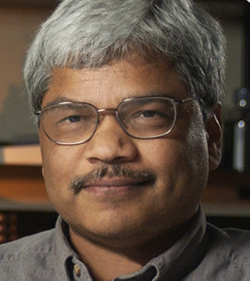 Settara Chandrasekharappa合作论文数Cancer Genomics Unit, National Human Genome Research Institute, National Institutes of Health2
 Simon Tran合作论文数Faculty of Dental Medicine and Oral Health Sciences, McGill University2
 Zhili Zheng合作论文数National Institutes of Health Surgery Branch, Center for Cancer Research, National Cancer Institute 10 Center Drive, Building 10/Rm 3-5940 Bethesda MD 20892 USA2
 Paul P. Liu合作论文数National Human Genome Research Institute, National Institutes of Health2
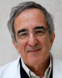 Shai Izraeli合作论文数Department of Human Molecular Genetics & Biochemistry, Sackler Faculty of Medicine, Tel-Aviv University;Division of Pediatric Hematology and Oncology, Schneider Children's Medical Center2Holly Root合作论文数2
 Tim Nichols合作论文数Division of Cardiology, Department of Medicine, School of Medicine, The University of North Carolina at Chapel Hill;Francis Owen Blood Research Laboratory, The University of North Carolina at Chapel Hill2Andrei Glinka合作论文数2
 Jan Blancato合作论文数Institute for Molecular and Human Genetics, Georgetown University Medical Center2A. Schumacher-Schuh合作论文数1




