Nanoparticles govern early (chemical-controlled) and late (diffusion-controlled) stages of epoxy crosslinking, respectively. Surface functionalization of nanoparticles with macromolecules was recognized as an effective route to compensate for inadequate diffusion-controlled crosslinking. However, the effect of bulk composition amended with cations having catalytic effect towards epoxy ring opening has remained hitherto undescribed. In this work, cathodic electrochemical method was applied to obtain naked iron oxide nanoparticles (IONPs), polyethylene glycol (PEG) capped IONPs (PEG-IONPs) and zinc-doped IONPs (Zn-doped PEG-IONPs) for evaluation of the effects of surface (alone) and surface-bulk (concurrent) modification of IONPs. Various techniques including FTIR, XRD, VSM, and FE-SEM were employed to probe into changes in the surface and bulk of nanoparticles. Cure Index calculated based on nonisothermal differential scanning calorimetry (DSC) allowed for labeling epoxy nanocomposites as Poor (Red) or Good (Blue) cured networks. Partial substitution of Fe2+ cations by Zn2+ in the bulk of Zn-doped PEG-IONPs with respect to PEG-IONPs labeled their corresponding epoxy nanocomposites in Blue and Red. Although at low and high heating rates epoxy/PEG-IONPs was labeled Red, epoxy/Zn-doped PEG-IONPs nanocomposites was unconditionally labeled Blue thanks to a more significant catalytic effect springing from Zn2+ cations in Zn-doped IONPs compared to hydroxyl groups alone in PEG-IONPs.
The enteric microbiota is characterised by a balance and composition that is unique to the host. It is important to understand the mechanisms through which the host can maintain the composition of the gut microbiota. MicroRNAs (miRNA) are implicated in intercellular communication and have been isolated from bodily fluids including stool. Recent findings suggest that miRNA produced by the host's intestinal epithelial cells (IECs) participate in shaping the microbiota. To investigate whether miRNA expression was influenced by the gut microbiota we measured the expression of miRNAs expressed by intestinal epithelial cells in faeces. Specifically, we measured miRNA expression in faeces from germ-free (GF) and conventional mice and similarly in a rat model of antibiotic-mediated depletion of the gut microbiota control rats. In adult male GF and conventional mice and adult Sprague Dawley (SD) rats were treated with a combination of antibiotics for 8 weeks; total RNA was extracted from faecal pellets taken at week 0, 2, 4, 6 week 8 and the expression of let-7b-3p, miR-141-3p, miR-200a-3p and miR-1224-5p (miRNAs known to be expressed in IECs) were measured relative to U6 at each time point using qRT-PCR. In GF animals the expression of let-7b, miR-141 and miR-200a in faeces was lower compared to conventional mice. Following antibiotic-mediated depletion of gut microbiota, rats showed two divergent profiles of miRNA expression. Following two weeks of antibiotic treatment, the expression of let-7b and miR-1224 dropped significantly and remained low for the remainder of the study. The expression of miR-200a and miR-141 was significantly higher at week 2 than before antibiotic treatment commenced. Subsequently, the expression of miR-200a and miR-141 decreased at week 4 and continued to decrease at week 6. This data demonstrates that miRNAs can be used as an independent, non-invasive marker of microbial fluctuations along with gut pathology in the intestine.
The amygdala is a key brain region that is critically involved in the processing and expression of anxiety and fear-related signals. In parallel, a growing number of preclinical and human studies have implicated the microbiome-gut-brain in regulating anxiety and stress-related responses. However, the role of the microbiome in fear-related behaviours is unclear. To this end we investigated the importance of the host microbiome on amygdala-dependent behavioural readouts using the cued fear conditioning paradigm. We also assessed changes in neuronal transcription and post-transcriptional regulation in the amygdala of naive and stimulated germ-free (GF) mice, using a genome-wide transcriptome profiling approach. Our results reveal that GF mice display reduced freezing during the cued memory retention test. Moreover, we demonstrate that under baseline conditions, GF mice display altered transcriptional profile with a marked increase in immediate-early genes (for example, Fos, Egr2, Fosb, Arc) as well as genes implicated in neural activity, synaptic transmission and nervous system development. We also found a predicted interaction between mRNA and specific microRNAs that are differentially regulated in GF mice. Interestingly, colonized GF mice (ex-GF) were behaviourally comparable to conventionally raised (CON) mice. Together, our data demonstrates a unique transcriptional response in GF animals, likely because of already elevated levels of immediate-early gene expression and the potentially underlying neuronal hyperactivity that in turn primes the amygdala for a different transcriptional response. Thus, we demonstrate for what is to our knowledge the first time that the presence of the host microbiome is crucial for the appropriate behavioural response during amygdala-dependent memory retention.
Detection of Clostridium difficile infection is important for clinical laboratories, owing to debilitating disease, severe outcomes, patient awareness, and public reporting of hospital data. This study evaluated the performance of 4 nucleic acid amplification test (NAAT) assays as part of a 2-step algorithm that involves reflexive NAAT following enzyme immunoassay (EIA) testing that is indeterminate for glutamate dehydrogenase (GDH) antigen and toxin A/B (GDH+/toxin− or GDH−/toxin+). A total of 500 stool specimens from consecutive patients were tested by each of the 5 methods and also evaluated as part of a 2-step algorithm. A specimen was considered positive for presence of C. difficile if it tested positive by 3 of 4 molecular methods or toxigenic culture. The sensitivity and specificity of the GDH-EIA method were each 93%. The toxin EIA had only 48% sensitivity, but it had 99% specificity. Sensitivity and specificity of 2-step algorithmic testing ranged from 88% to 93% and 99% to 100%, respectively, offering similar performance to stand-alone NAAT testing.
Gut microbiota colonization is a key event for host physiology that occurs early in life. Disruption of this process leads to altered brain development which ultimately manifests as changes in brain function and behaviour in adulthood. Studies using germ-free (GF) mice highlight the extreme impact on brain health that results from life without commensal microbes. However, the impact of microbiota disturbances occurring in adulthood is less studied. To this end, we depleted the gut microbiota of 10-week-old male SpragueDawley rats via chronic antibiotic treatment. Following this marked, sustained depletion of the gut bacteria, we investigated behavioural and molecular hallmarks of gut-brain communication. Our results reveal that depletion of the gut microbiota during adulthood results in deficits in spatial memory as tested by Morris water maze, decreased visceral sensitivity and a greater display of depressive-like behaviours in the forced swim test. In tandem with these clear behavioural alterations we found changes in altered CNS serotonin concentration along with changes in the mRNA levels of corticotrophin releasing hormone receptor 1 and glucocorticoid receptor. Additionally, we found changes in the expression of brain derived neurotrophic factor (BDNF), a hallmark of altered microbiota-gut-brain axis signalling. In summary, this model of antibiotic-induced depletion of the gut microbiota can be used for future studies interested in the impact of the gut microbiota on host health without the confounding developmental influence of early-life microbial alterations.
The prefrontal cortex (PFC) is a key region implicated in a range of neuropsychiatric disorders such as depression, schizophrenia and autism. In parallel, the role of the gut microbiota in contributing to these disorders is emerging. Germ-free (GF) animals, microbiota-deficient throughout life, have been instrumental in elucidating the role of the microbiota in many aspects of physiology, especially the role of the microbiota in anxiety-related behaviours, impaired social cognition and stress responsivity. Here we aim to further elucidate the mechanisms of the microbial influence by investigating changes in the homeostatic regulation of neuronal transcription of GF mice within the PFC using a genome-wide transcriptome profiling approach. Our results reveal a marked, concerted upregulation of genes linked to myelination and myelin plasticity. This coincided with upregulation of neural activity-induced pathways, potentially driving myelin plasticity. Subsequent investigation at the ultrastructural level demonstrated the presence of hypermyelinated axons within the PFC of GF mice. Notably, these changes in myelin and activity-related gene expression could be reversed by colonization with a conventional microbiota following weaning. In summary, we believe we demonstrate for the first time that the microbiome is necessary for appropriate and dynamic regulation of myelin-related genes with clear implications for cortical myelination at an ultrastructural level. The microbiota is therefore a potential therapeutic target for psychiatric disorders involving dynamic myelination in the PFC.
Severe mental illness is characterised by a 15-year mortality gap driven by cardiometabolic disease. Antipsychotic treatment leads to increased appetite and rapid weight gain. The 12-week lifestyle pilot intervention improved dietary intake and prevented antipsychotic-induced weight gain. Here we report two-year outcomes.Participants were exposed to an extended program. Weight and waist circumference were measured, and food frequency questionnaire completed.Diet quality was higher, and discretionary food intake was 40% lower, at two-years compared to baseline. Weight and waist-circumference did not increase.This pilot study demonstrated sustained effectiveness of a dietetic intervention in youth with first-episode psychosis with improvements in diet quality and no increase in weight secondary to antipsychotic medication initiation.
Event Abstract Back to Event Regulation of microRNAs in the Amygdala by the Gut Microbiota: Implications for Brain and Behaviour Alan E. Hoban1* 1 Alimentary Pharmabiotic Centre/Department of Anatomy and Neuroscience University College Cork, Department of Anatomy and Neuroscience University College Cork, Ireland Background: The ability of the gut microbiota to influence brain and behaviour is a relatively new area of research. One of the most consistent findings is in relation to anxiety-like behaviours and the stress response. However, the molecular mechanisms underpinning this remains poorly understood but may well be due to alterations in gene expression. It is unknown if the gut microbiota also recruits microRNA machinery to wield this influence. The aim of this experiment was to establish if germ-free animals have altered microRNA expression patterns in the amygdala, a key brain region for anxiety and fear. Methods: Using Next Generation Sequencing, we assessed alterations in microRNA expression in the amygdala of conventional, germ-free and colonized germ-free mice. Results: The microbiota-deficient germ-free animals display altered expression of 54 miRNAs in the amygdala compared to conventional animals. However, colonisation of the germ-free animals post weaning normalises the expression of 6 miRNAs, suggesting partial reversibility of the cumulative molecular changes. Within these, miR-182 and mir-183 have been previously linked to amygdala-dependent stress-related outputs in preclinical models Conclusion: This is, to our knowledge, the first demonstration that the gut microbiota can regulate miRNA expression in the amygdala. Further studies are required to verify the exact contribution of these miRNAs to amygdala-dependent anxiety-related behaviours. The results from this study will be essential in increasing our understanding of the molecular mechanisms underpinning the impact of microbiota-gut-brain axis communication on both brain and behaviour. Further analysis of mRNA targets may reveal important molecular pathways for microbiota-gut-brain axis signalling. Acknowledgements The Alimentary Pharmabiotic Centre is a research centre funded by Science Foundation Ireland (SFI), through the Irish Government’s National Development Plan. The authors and their work were supported by SFI (grant numbers SFI/12/RC/2273, 02/CE/B124 and 07/CE/B1368) and by the Health Research Board (HRB) through Health Research Awards (grant no HRA_POR/2011/23; TGD, JFC and GC and HRA_POR/2012/32; JFC, TGD). JFC is also funded by the European Community's Seventh Framework Programme (grant no.: FP7/2007–2013, grant agreement 201 714). The Centre was previously funded by GlaxoSmithKline. GC is supported by a NARSAD Young Investigator Grant from the Brain and Behavior Research Foundation (Grant Number 20771). The authors report no conflict of interest. Keywords: Anxiety, MicroRNAs, Amygdala, germ-free, microbiota–gut–brain axis Conference: Neuroscience Ireland Young Neuroscientists Symposium 2014 , Dublin, Ireland, 20 Sep - 20 Sep, 2014. Presentation Type: Poster Presentation Topic: Early Career Neuroscience Citation: Hoban AE (2014). Regulation of microRNAs in the Amygdala by the Gut Microbiota: Implications for Brain and Behaviour. Front. Neurosci. Conference Abstract: Neuroscience Ireland Young Neuroscientists Symposium 2014 . doi: 10.3389/conf.fnins.2014.87.00023 Copyright: The abstracts in this collection have not been subject to any Frontiers peer review or checks, and are not endorsed by Frontiers. They are made available through the Frontiers publishing platform as a service to conference organizers and presenters. The copyright in the individual abstracts is owned by the author of each abstract or his/her employer unless otherwise stated. Each abstract, as well as the collection of abstracts, are published under a Creative Commons CC-BY 4.0 (attribution) licence (https://creativecommons.org/licenses/by/4.0/) and may thus be reproduced, translated, adapted and be the subject of derivative works provided the authors and Frontiers are attributed. For Frontiers’ terms and conditions please see https://www.frontiersin.org/legal/terms-and-conditions. Received: 13 Sep 2014; Published Online: 14 Sep 2014. * Correspondence: Mr. Alan E Hoban, Alimentary Pharmabiotic Centre/Department of Anatomy and Neuroscience University College Cork, Department of Anatomy and Neuroscience University College Cork, Cork, Ireland, alanhoban@live.ie Login Required This action requires you to be registered with Frontiers and logged in. To register or login click here. Abstract Info Abstract The Authors in Frontiers Alan E Hoban Google Alan E Hoban Google Scholar Alan E Hoban PubMed Alan E Hoban Related Article in Frontiers Google Scholar PubMed Abstract Close Back to top Javascript is disabled. Please enable Javascript in your browser settings in order to see all the content on this page.
 John Cryan合作论文数Department of Anatomy and Neuroscience, University College Cork;School of Pharmacy, University College Cork9
 Timothy Dinan合作论文数Alimentary Pharmabiotic Centre, University College Cork;Department of Psychiatry, University College Cork7
 Gerard Clarke合作论文数Department of Psychiatry and Neurobehavioural Science, University College Cork, Ireland6
 Catherine Stanton合作论文数Food Biosciences Department, Teagasc;Moorepark Food Research Centre, Teagasc;APC Microbiome Institute, University College Cork4
 Roman Manuel Stilling合作论文数Department of Psychiatry and Psychotherapy, University Medical Center4
 Patterson Elaine合作论文数4
 Gerard Moloney合作论文数Department of Anatomy and Neuroscience, University College Cork4Moloney Rachel D合作论文数Biosciences Institute, University College Cork3
 Anna V Golubeva合作论文数3
 Orla O’Sullivan合作论文数Department of Biochemistry, University College3
 Fergus Shanahan合作论文数Department of Medicine, University College Cork, National University of Ireland3S. Beers合作论文数2
 El Aidy Sahar合作论文数Top Inst Food & Nutr2
 Karen Anne McVey Neufeld合作论文数Brain-Body Institute;St. Joseph's Healthcare;McMaster University;Hamilton;Brain-Body Institute, McMaster University2Borre Yuliya E合作论文数2
 Paul Ross合作论文数APC Microbiome Institute, University College Cork2Y.M. Nolan合作论文数1A. Moya-Pérez合作论文数1T. Dinan合作论文数1
 Jennifer Deane合作论文数1Ciarán O’Brien合作论文数1
 Patrick Fitzgerald合作论文数APC Microbiome Ireland1Maria Francesca Viola合作论文数1
 Feargal J Ryan合作论文数Precis Med Theme, South Australian Hlth & Med Res Inst1
 Kieran Rea合作论文数APC Microbiome Ireland, Univ Coll Cork1
 O'Leary Olivia F合作论文数Department of Anatomy and Neuroscience, University College Cork1
 Kennedy Paul J合作论文数Alimentary Pharmabiotic Centre, University College Cork1
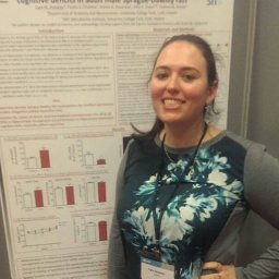 Hueston Cara M合作论文数Department of Psychology, Binghamton University1
 Karen Scott合作论文数The Institute of Medical Sciences, School of Medicine, Medical Sciences and Nutrition, University of Aberdeen1
 Danka Kozareva合作论文数1
 James D. O’leary Mbbch合作论文数Hospital for Sick Children, University of Toronto1
 John R. Kelly合作论文数1
 Paul D. Cotter合作论文数Food Biosciences Department, Teagasc;SeqBiome1Lucinda V. Scott合作论文数1Lieve Desbonnet合作论文数1
 Lívia H. Morais合作论文数Centro Politécnico, Universidade Federal do Paraná1
 Marcus J. Claesson合作论文数Alimentary Pharmabiotic Centre, University College Cork1




