DIII-D physics research addresses critical challenges for the operation of ITER and the next generation of fusion energy devices. This is done through a focus on innovations to provide solutions for high performance long pulse operation, coupled with fundamental plasma physics understanding and model validation, to drive scenario development by integrating high performance core and boundary plasmas. Substantial increases in off-axis current drive efficiency from an innovative top launch system for EC power, and in pressure broadening for Alfven eigenmode control from a co-/counter- I p steerable off-axis neutral beam, all improve the prospects for optimization of future long pulse/steady state high performance tokamak operation. Fundamental studies into the modes that drive the evolution of the pedestal pressure profile and electron vs ion heat flux validate predictive models of pedestal recovery after ELMs. Understanding the physics mechanisms of ELM control and density pumpout by 3D magnetic perturbation fields leads to confident predictions for ITER and future devices. Validated modeling of high- Z shattered pellet injection for disruption mitigation, runaway electron dissipation, and techniques for disruption prediction and avoidance including machine learning, give confidence in handling disruptivity for future devices. For the non-nuclear phase of ITER, two actuators are identified to lower the L–H threshold power in hydrogen plasmas. With this physics understanding and suite of capabilities, a high poloidal beta optimized-core scenario with an internal transport barrier that projects nearly to Q = 10 in ITER at ∼ 8 MA was coupled to a detached divertor, and a near super H-mode optimized-pedestal scenario with co- I p beam injection was coupled to a radiative divertor. The hybrid core scenario was achieved directly, without the need for anomalous current diffusion, using off-axis current drive actuators. Also, a controller to assess proximity to stability limits and regulate β N in the ITER baseline scenario, based on plasma response to probing 3D fields, was demonstrated. Finally, innovative tokamak operation using a negative triangularity shape showed many attractive features for future pilot plant operation.
The use of hybrid PET/MRI for clinical staging is growing in several cancer forms and, consequently, PET/MRI has also gained interest in the assessment of non-small cell lung cancer (NSCLC) and lung lesions. However, lung evaluation with PET/MRI is associated with challenges related to technical issues and diagnostic image quality. We, therefore, investigated the published literature on PET/MRI for clinical staging in NSCLC or lung nodule detection specifically addressing diagnostic accuracy and technical issues. The data originates from a systematic search performed in PubMed/MEDLINE, Embase, and Cochrane Library on hybrid PET/MRI in patients with cancer for a scoping review published earlier ( https://doi.org/10.1007/s00259-019-04402-8 ). Studies in English and German evaluating the diagnostic performance of hybrid PET/MRI for NSCLC or lung nodule detection in cancer patients were selected. Data reported in peer-reviewed journals without restrictions to year of publication were included. A total of 3138 publications were identified from which 116 published 2012–2018 were included. Of these, nine studies addressed PET/MRI in NSCLC (4) or lung nodule detection (5). Overall, PET/MRI did not provide advantages in preoperative T- and N-staging in NSCLC compared to PET/CT. The data on M-staging were too few for conclusions to be drawn. The lung nodule detection rate of PET/MRI was comparable to that of PET/CT for FDG-avid nodules larger than 10 mm, but the sensitivity of PET/MRI for detection of non-FDG-avid nodules smaller than 5 mm was low. PET/MRI did not provide advantages in T- and N-staging of NSCLC compared to PET/CT. PET/MRI had a comparable sensitivity for detection of FDG-avid lung nodules and nodules over 10 mm, but PET/CT yielded a higher detection rate in non FDG-avid lung nodules under 5 mm. With PET/MRI, the overall detection rate for lung nodules in various cancer types remains inferior to that of PET/CT due to the lower diagnostic performance of MRI than CT in the lungs.
"Comments on ‘Search for tumor-specific frequencies of amplitude modulated 27 MHz electromagnetic fields in mice with hepatocarcinoma xenografted tumors’." International Journal of Radiation Biology, 96(7), pp. 845–846
PET/MRI was introduced for clinical use in 2011 and is now an established modality for the imaging of brain and certain pelvic cancers, whereas clinical use for the imaging of other forms of cancer is not yet widespread. We therefore systematically investigated what has been published on the use of PET/MRI compared to PET/CT in the imaging of cancers outside the brain, focusing on clinical areas of application related to diagnosis, staging and restaging. A systematic search of PubMed/MEDLINE, Embase and the Cochrane Library was performed. Studies evaluating the diagnostic performance of simultaneous PET/MRI in cancer patients were chosen. A total of 3,138 publications were identified and 116 published during the period 2012–2018 were included and were grouped according to the major cancer forms: 13 head and neck (HNC), 9 breast (BC), 21 prostate (PC), 14 gynaecological, 13 gastrointestinal (GIC), and 46 various cancers. Data from studies comparing PET/MRI and PET/CT for staging/restaging suggested the superiority of 18F-FDG PET/MRI for the detection of tumour extension and retropharyngeal lymph node metastases in nasopharyngeal cancer, and for the detection of liver metastases and possibly bone marrow metastases in high-risk BC. FDG PET/MRI tended to be inferior for the detection of lung metastases in HNC and BC. 68Ga-PSMA-11 PET/MRI was superior to PET/CT for the detection of local PC recurrence. FDG PET/MRI was superior to FDG PET/CT for the detection of local tumour invasion in cervical cancer and had higher accuracy for the detection of liver metastases in colorectal cancer. The scoping review methodology resulted in the identification of a huge number of records, of which less than 5% were suitable for inclusion and only a limited number allowed conclusions on the advantages/disadvantages of PET/MRI compared to PET/CT in the oncological setting. There was evidence to support the use of FDG PET/MRI in staging of nasopharyngeal cancer and high-risk BC. Preliminary data indicate the superiority of PET/MRI for the detection of local recurrence in PC, local tumour invasion in cervical cancer, and liver metastases in colorectal cancer. These conclusions are based on small datasets and need to be further explored.
Combined myocardial flow reserve (MFR) by PET and CT coronary angiography (CTA) is a promising tool for assessment of coronary artery disease. Prior analyses of MFR/CTA has been performed as side-by-side interpretation, not as volume rendered, full hybrid analysis, with fused MFR/CTA. We aimed to: (i) establish a method for full hybrid analysis of MFR/CTA, (ii) validate the inter- and intra-observer reproducibility of MFR values, and (iii) determine the diagnostic value of side-by-side versus full hybrid MFR/CTA with 15-O-water PET. Forty-four outpatients scheduled for invasive coronary angiography (ICA) were enrolled prospectively. All underwent rest/stress 15-O-water PET/CTA with ICA as reference. Within two observers of different experience, the Pearson r at global and territorial level exceeded 0.953 for rest, stress, and MFR values, as determined by Carimas software. Within and between observers, the mean differences between rest, stress, and MFR values were close to zero and the confidence intervals for 95% limits of agreement were narrow. The diagnostic performance of full hybrid PET/CTA did not outperform the side-by-side approach, but performed better than MFR without CTA at vessel level: specificity 93% (95% confidence limits: 89–97%) versus 76% (64–88%), p = 0.0004; positive predictive value 71% (55–86%) versus 51% (37–65%), p = 0.0001; accuracy 90% (84–95%) versus 77% (69–84%), p = 0.0009. MFR showed high reproducibility within and between observers of different experience. The full hybrid model was not superior to side-by-side interpretation of MFR/CTA, but proved better than MFR alone at vessel level with regard to specificity, positive predictive value, and accuracy.
ISS Fro Un D Ho De D pa OBJECTIVES This study sought to compare the per-patient diagnostic performance of coronary computed tomography angiography (CTA)-derived fractional flow reserve (FFRCT) with that of single-photon emission computed tomography (SPECT), using a fractional flow reserve (FFR) value of #0.80 as the reference for diagnosing at least 1 hemodynamically significant stenosis in a head-to-head comparison of patients with intermediate coronary stenosis as determined by coronary CTA. BACKGROUND No previous study has prospectively compared the diagnostic performance of FFRCT and myocardial perfusion imaging by SPECT in symptomatic patients with intermediate range coronary artery disease (CAD). METHODS This study was conducted at a single-center as a prospective study in patients with stable angina pectoris (N 1⁄4 143). FFRCT and SPECT analyses were performed by core laboratories and were blinded for the personnel responsible for downstream patient management. FFRCT #0.80 distally in at least 1 coronary artery with a diameter$2 mm classified patients as having ischemia. Ischemia by SPECT was encountered if a reversible perfusion defect (summed difference score $2) or transitory ischemic dilation of the left ventricle (ratio >1.19) were found. RESULTS The per-patient diagnostic performance for identifying ischemia (95% confidence interval [CI]), FFRCT versus SPECT, were sensitivity of 91% (95% CI: 81% to 97%) versus 41% (95% CI: 29% to 55%; p < 0.001); specificity of 55% (95% CI: 44% to 66%) versus 86% (95% CI: 77% to 93%; p < 0.001); negative predictive value of 90% (95% CI: 82% to 98%) versus 68% (95% CI: 59% to 77%; p 1⁄4 0.001); positive predictive value of 58% (95% CI: 48% to 68%) versus 67% (95% CI: 51% to 82%; p 1⁄4 NS); and accuracy of 70% (95% CI: 62% to 77%) versus 68% (95% CI: 60% to 75%; p 1⁄4 NS) respectively. CONCLUSIONS In patients with stable chest pain and CAD as determined by coronary CTA, the overall diagnostic accuracy levels of FFRCT and SPECT were identical in assessing hemodynamically significant stenosis. However, FFRCT demonstrated a significantly higher diagnostic sensitivity than SPECT. (J Am Coll Cardiol Img 2018;11:1640–50) © 2018 The Authors. Published by Elsevier on behalf of the American College of Cardiology Foundation. This is an open access article under the CC BY-NC-ND license (http://creativecommons.org/licenses/by-nc-nd/4.0/). N 1936-878X https://doi.org/10.1016/j.jcmg.2018.05.004 m the Department of Cardiology, Hospital of Southwest Denmark, Esbjerg, Denmark; Institute of Regional Health Research, iversity of Southern Denmark, Odense, Denmark; Department of Cardiology, Odense University Hospital, Odense, Denmark; epartment of Nuclear Medicine, Aalborg University Hospital, Aalborg, Denmark; Department of Cardiology, Aarhus University spital, Skejby, Aarhus, Denmark; Department of Epidemiology, Biostatistics and Bioinformatics, University of Southern nmark, Odense, Denmark; Department of Nuclear Medicine, Odense University Hospital, Odense, Denmark; and the epartment of Radiology and Nuclear Medicine, Hospital of Southwest Denmark, Esbjerg, Denmark. Supported by rticipating departments. No external funding was used. Dr. Nørgaard has received institutional research grants from Siemens, ABBR EV I A T I ON S
OBJECTIVES:In stable coronary artery disease (CAD), coronary revascularisation may reduce mortality of patients with a certain amount of left ventricular myocardial ischaemia. However, revascularisation does not always follow the guidance suggested by ischaemia testing. We compared outcomes in patients without ischaemia who had either revascularisation or medical treatment.DESIGN AND POPULATION:Based on registries, 1327 consecutive patients with normal myocardial perfusion scintigraphy (MPS) and 278 with fixed perfusion defects were followed for a median of 6.1 years. Most patients received medical therapy alone (Med), but 26 (2%) with a normal MPS and 15 (5%) with fixed perfusion defects underwent revascularisation (Revasc).OUTCOME MEASURES:Incidence rates of all-cause death (ACD) and rates of cardiac death/myocardial infarction (CD/MI).RESULTS:With a normal MPS, the ACD rate was 6.2%/year in the Revasc group versus 1.9%/year in the Med group (p=0.01); the CD/MI rates were 6.9%/year and 0.6%/year, respectively (p<0.00001). Results persisted after adjustment for predictors of revascularisation, in particular angina score, and in comparisons of matched Revasc and Med patients. With fixed defects, the ACD rate was 9.1%/year in the Revasc group and 6.7%/year in the Med group (p=0.44); the CD/MI rate was 5.0%/year versus 4.2%/year, respectively (p=0.69). If adjusted for angiographic variables or analysed in matched subsets, differences remained insignificant.CONCLUSIONS:With normal MPS, revascularisation conferred a higher risk, even after adjustment for predictors of revascularisation. With fixed defects, the Revascversus Med difference was close to equipoise. Hence, in patients with stable CAD without ischaemia, we could not find evidence to justify exceptional revascularisation.
Objective Patients with normal stress perfusion have an excellent prognosis. Prospective studies on the diagnostic accuracy of stress-only scans with contemporary, independent examinations as gold standards are lacking. Patients and methods A total of 109 patients with typical angina and no previous coronary artery disease underwent a 2-day stress (exercise)/rest, gated, and attenuation-corrected (AC), 99m-technetium-sestamibi perfusion study, followed by invasive coronary angiography. The stress datasets were evaluated twice by four physicians with two different training levels (expert and novice): familiar and unfamiliar with AC. The two experts also made a consensus reading of the integrated stress-rest datasets. The consensus reading and quantitative data from the invasive coronary angiography were applied as reference methods. Results The sensitivity/specificity were 0.92-1.00/0.73-0.90 (reference: expert consensus reading), 0.93-0.96/0.63-0.82 (reference: >= 1 stenosis>70%), and 0.75-0.88/0.70-0.88 (reference: >= 1 stenosis>50%). The four readers showed a high and fairly equal sensitivity independent of their familiarity with AC. The expert familiar with AC had the highest specificity independent of the reference method. The intraobserver and interobserver agreements on the stress-only readings were good (readers without AC experience) to excellent (readers with AC experience). Conclusion AC stress-only images yielded a high sensitivity independent of the training level and experience with AC of the nuclear physician, whereas the specificity correlated positively with both. Interobserver and intraobserver agreements tended to be the best for physicians with AC experience. Copyright (c) 2017 Wolters Kluwer Health, Inc. All rights reserved.
OBJECTIVE:While nuclear myocardial perfusion imaging (MPI) offers many benefits to patients with known or suspected cardiovascular disease, concerns exist regarding radiation-associated health effects. Little is known regarding MPI practice in Africa. We sought to characterise radiation doses and the use of MPI best practices that could minimise radiation in African nuclear cardiology laboratories, and compare these to practice worldwide.METHODS:Demographics and clinical characteristics were collected for a consecutive sample of 348 patients from 12 laboratories in six African countries over a one-week period from March to April 2013. Radiation effective dose (ED) was estimated for each patient. A quality index (QI) enumerating adherence to eight best practices, identified a priori by an IAEA expert panel, was calculated for each laboratory. We compared these metrics with those from 7 563 patients from 296 laboratories outside Africa.RESULTS:Median (interquartile range) patient ED in Africa was similar to that of the rest of the world [9.1 (5.1-15.6) vs 10.3 mSv (6.8-12.6), p = 0.14], although a larger proportion of African patients received a low ED, ≤ 9 mSv targeted in societal recommendations (49.7 vs 38.2%, p < 0.001). Bestpractice adherence was higher among African laboratories (QI score: 6.3 ± 1.2 vs 5.4 ± 1.3, p = 0.013). However, median ED varied significantly among African laboratories (range: 2.0-16.3 mSv; p < 0.0001) and QI range was 4-8.CONCLUSION:Patient radiation dose from MPI in Africa was similar to that in the rest of the world, and adherence to best practices was relatively high in African laboratories. Nevertheless there remain opportunities to further reduce radiation exposure to African patients from MPI.
Purpose The Region of Southern Denmark (RSD), covering 1.2 of Denmark's 5.6 million inhabitants, established a task force to (1) retrieve literature evidence for the clinical use of positron emission tomography (PET)/CT and provide consequent recommendations and further to (2) compare the actual use of PET/CT in the RSD with these recommendations. This article summarizes the results.Methods A Work Group appointed a professional Subgroup which made Clinician Groups conduct literature reviews on six selected cancers responsible for 5,768 (62.6 %) of 9,213 PET/CT scans in the RSD in 2012. Rapid Evidence Assessment was applied, using the methodology of systematic reviews with predefined limitations to search PubMed, Embase and the Cochrane Library for articles published in English/Danish/Swedish/Norwegian since 2002. PICO questions were defined, data recorded and quality appraised and rated with regard to strength and evidence level. Consequent recommendations for applications of PET/CT were established. The actual use of PET/CT was compared with these, where grades A and B indicated "established" and "useful" and grades C and D "potentially useful" and "non-recommendable" indications, respectively.Results Of 11,729 citations, 1,729 were considered for review, and 204 were included. The evidence suggested usefulness of PET/CT in lung, lymphoma, melanoma, head and neck, and colorectal cancers, whereas evidence was sparse in gynaecological cancers. The agreement between actual use of PET/CT and literature-based recommendations was high in the first five mentioned cancers in that 96.2 % of scans were made for grade A or B indications versus only 22.2 % in gynaecological cancers.Conclusion Evidence-based usefulness was reported in five of six selected cancers; evidence was sparse in the sixth, gynaecological cancers. Actual use of PET/CT agreed well with recommendations.
Mathew Mercuri, PhD; Thomas N. B. Pascual, MD; John J. Mahmarian, MD; Leslee J. Shaw, PhD; Maurizio Dondi, MD; Diana Paez, MD; Andrew J. Einstein, MD, PhD; for the INCAPS Investigators Group
AIMS Our aim was to address the combined influence of myocardial perfusion defects and left ventricular ejection fraction (LVEF) on outcome with coronary revascularisation in stable CAD patients. METHODS AND RESULTS Of 527 patients with ischaemia by myocardial perfusion scintigraphy, 343 had medical therapy (Med) and 184 revascularisation (Revasc). During 5.3 years of follow-up, there was no intergroup difference in rates of death/myocardial infarction. Propensity score adjustment demonstrated a benefit of Revasc over Med with large defects (>14% of the myocardium), marked ischaemia (>10% of the myocardium), or LVEF <50%. However, defect size, ischaemia, and LVEF were correlated. In multivariate models, the Med versus Revasc hazard ratio (HR) was 4.06 times larger for LVEF <50% than for LVEF ≥50% (p=0.04) and 3.01 times larger for marked compared to mild/moderate ischaemia (p=0.11), whereas the effect of large compared to small/moderate defects vanished when adjusted for LVEF and ischaemia (HR=1.01, p=0.99). Considering the outcome difference as a function of both LVEF and ischaemia, we found no advantage or even a disadvantage of revascularisation in patients with mild/moderate ischaemia and preserved LVEF. CONCLUSIONS A benefit of revascularisation was found only in case of marked ischaemia or LVEF <50%. For treatment triage, both perfusion parameters and LVEF should be considered.
Patients presenting with cystic lesions in the neck without obvious signs of malignancy constitute a diagnostic challenge since fine needle aspiration is often insufficient and a diagnosis may not be reached until surgical resection/biopsy is performed. The differential diagnosis of a cystic cervical mass comprises a variety of benign conditions, but malignancy must be ruled out. We examined the diagnostic performance of fluorine-18 fluorodeoxyglucose (18F-FDG) PET/CT to identify malignancy.
AIMSReporting of quantitative myocardial blood flow (MBF) is typically performed in standard coronary territories. However, coronary anatomy and myocardial vascular territories vary among individuals, and a coronary artery may erroneously be deemed stenosed or not if territorial demarcation is incorrect. So far, the diagnostic consequences of calculating individually vs. standardly assessed MBF values have not been reported. We examined whether individual reassignment of vascular territories would improve the diagnostic accuracy of MBF with regard to the detection of significant coronary artery disease (CAD).METHODS AND RESULTSForty-four patients with suspected CAD were included prospectively and underwent coronary CT-angiography and quantitative MBF assessment with O-15-water PET followed by invasive, quantitative coronary angiography, which served as reference. MBF was calculated in the vascular territories during adenosine stress according to a standardized 17-segment American Heart Association model and an individualized model, using CT-angiography to adjust the coronary territories to their feeding vessels. Individually defined territories deviated from standard territories in 52% of patients. However, MBF in the three coronary territories defined by standard and individualized models did not differ significantly, except in one patient, in whom the MBF of an individualized coronary territory deviated sufficiently as to change the test from a false positive to a true negative result in this particular territory.CONCLUSIONDisparity between standardized and individualized vascular territories was present in half of the patients, but had little clinical impact. Still, caution should be taken not always to rely on standard territories, as this may at times cause misinterpretation.
This study estimates current rates of stress-only imaging in cardiology patients in the United States and worldwide, as well as the potential effect of changes in this rate on the radiation burden to the US population.
OBJECTIVE:To evaluate the Bone Scan Index (BSI) for prediction of castration resistance and prostate cancer-specific survival (PCSS). In this retrospective study, we used novel computer-assisted software for automated detection/quantification of bone metastases by BSI. Patients with prostate cancer are M-staged by whole-body bone scintigraphy (WBS) and categorised as M0 or M1. Within the M1 group, there is a wide range of clinical outcomes. The BSI was introduced a decade ago providing quantification of bone metastases by estimating the percentage of bone involvement. Being too time consuming, it never gained widespread clinical use.PATIENTS AND METHODS:In all, 88 patients with prostate cancer awaiting initiation of androgen-deprivation therapy due to metastases were included. WBS was performed using a two-headed γ-camera. BSI was obtained using the automated platform EXINI bone (EXINI Diagnostics AB, Lund, Sweden). In Cox proportional hazard models, time to castration-resistant prostate cancer (CRPC) and PCSS were modelled as the dependent variables, whereas prostate-specific antigen (PSA) level, Gleason score and BSI were used as explanatory factors. For Kaplan-Meier estimates, BSI groups were dichotomously split into: BSI <1 and BSI ≥1. Discrimination between prognostic models was explored using the concordance index (C-index).RESULTS:The mean (range) age of the patients was 72 (52-92) years, the median (range) PSA level was 73 (4-5 740) ng/mL, the mean (range) Gleason score was 7.7 (2-10), and the mean (range) BSI was 1.0 (0-9.2). During a mean (range) follow-up of 26 (8-49) months, 48 patients became castration resistant and 15 had died; most (13) from prostate cancer. In multivariate analysis including PSA level, Gleason score and BSI, only prediction by BSI was statistically significant. This was true both for time to CRPC (hazard ratio [HR] 1.45, 95% confidence interval [CI] 1.22-1.74; C-index increase from 0.49 to 0.69) and for PCSS (HR 1.34, 95% CI 1.07-1.67; C-index increase from 0.76 to 0.95).CONCLUSION:BSI obtained using a novel automated computer-assisted algorithm appears to be a useful predictor of outcome for time to CRPC and PCSS in patients with hormone-sensitive metastatic prostate cancer.
Aims To characterize patient radiation doses from nuclear myocardial perfusion imaging (MPI) and the use of radiation-optimizing ‘best practices’ worldwide, and to evaluate the relationship between laboratory use of best practices and patient radiation dose. Methods and results We conducted an observational cross-sectional study of protocols used for all 7911 MPI studies performed in 308 nuclear cardiology laboratories in 65 countries for a single week in March–April 2013. Eight ‘best practices’ relating to radiation exposure were identified a priori by an expert committee, and a radiation-related quality index (QI) devised indicating the number of best practices used by a laboratory. Patient radiation effective dose (ED) ranged between 0.8 and 35.6 mSv (median 10.0 mSv). Average laboratory ED ranged from 2.2 to 24.4 mSv (median 10.4 mSv); only 91 (30%) laboratories achieved the median ED ≤ 9 mSv recommended by guidelines. Laboratory QIs ranged from 2 to 8 (median 5). Both ED and QI differed significantly between laboratories, countries, and world regions. The lowest median ED (8.0 mSv), in Europe, coincided with high best-practice adherence (mean laboratory QI 6.2). The highest doses (median 12.1 mSv) and low QI (4.9) occurred in Latin America. In hierarchical regression modelling, patients undergoing MPI at laboratories following more ‘best practices’ had lower EDs. Conclusion Marked worldwide variation exists in radiation safety practices pertaining to MPI, with targeted EDs currently achieved in a minority of laboratories. The significant relationship between best-practice implementation and lower doses indicates numerous opportunities to reduce radiation exposure from MPI globally.
 Torben Haghfelt合作论文数31
 Oke Gerke合作论文数22P. F. Hoilund-Carlsen合作论文数21Poul Flemming Høilund-Carlsen合作论文数20Werner Vach合作论文数19
 Hans Mickley合作论文数Department of Cardiology, Odense University Hospital18
 Henrik Wulff Christensen合作论文数17Henrik Petersen合作论文数Odense14Per Thayssen合作论文数11
 Søren Hess合作论文数Department of Nuclear Medicine, Odense University Hospital11
 Lisette Okkels Jensen合作论文数Odense University Hospital;Research unit of Cardiology, Department of Clinical Research, The Faculty of Health Sciences, University of Southern Denmark10
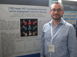 Anders Thomassen合作论文数Department of Nuclear Medicine, Odense University Hospital10Mikael K. Poulsen合作论文数9Annegrete Veje合作论文数8
 Axel Cosmus Pyndt Diederichsen合作论文数Odense University Hospital8Poul Fl. Høilund-Carlsen合作论文数7Mette Moldrup合作论文数7
 Peter Grupe合作论文数7M. Moldrup合作论文数7
 Henriksen Jan Erik合作论文数Dept Endocrinol, Odense Univ Hosp6Braad Poul-Erik合作论文数Department of Nuclear Medicine, Odense University Hospital6
 Henning Beck-Nielsen合作论文数6
 Jordi S Dahl合作论文数Research Unit of Cardiology, Department of Clinical Research, Syddansk Universitet;Department of Cardiology, Odense University Hospital;Mayo Clinic in Rochester6Jane Angel Simonsen合作论文数6A Veje合作论文数6F. P. Esteves合作论文数5Al Gaster合作论文数5
 Joao V. Vitola合作论文数5
 Henry Gewirtz合作论文数5Oliver Lindner合作论文数5
 Lars Edenbrandt合作论文数Department of Molecular and Clinical Medicine, University of Gothenburg5S. Saranovic合作论文数4R. Peace合作论文数4K Churchwell合作论文数4S. Unger合作论文数4I. Duncan合作论文数4K. Thamnirat合作论文数4B. Adams合作论文数4O. Lang合作论文数4J. B. Meeks合作论文数4R. Yudistiro合作论文数4C. Bavelaar合作论文数4W. H. Moore合作论文数4A. Vieni合作论文数4E. Etherton合作论文数4H. Golan合作论文数4M. Ratniece合作论文数4J. L. Lopez合作论文数4J. Canessa合作论文数4




