Handball is one of the top four athletic games with highest injury risks. The jump shot is the most accomplished goal shot technique and the lower extremities are mostly injured. As a basis for ankle sprain simulation, the aim of this study was to extend the ankle region of an existing musculoskeletal full-body model through incorporation of three prominent lateral ankle ligaments: ligamentum fibulotalare anterius (LFTA), ligamentum fibulotalare posterius (LFTP), ligamentum fibulocalcaneare (LFC). The specific objective was to calculate and visualise ligament force scenarios during the jumping and landing phases of controlled jump shots. Recorded kinematic data of performed jump shots and the corresponding ground reaction forces were used to perform inverse dynamics. The calculated peak force of the LFTA (107 N) was found at maximum plantarflexion and of the LFTP (150 N) at maximum dorsiflexion. The peak force of the LFC (190 N) was observed at maximum dorsiflexion combined with maximum eversion. Within the performed jump shots, the LFTA showed a peak force (59 N to 69 N) during maximum plantarflexion in the final moment of the lift off. During landing, the force developed by the LFTA reached its peak value (61 N to 70 N) at the first contact with the floor. After that, the LFTP developed a peak force (70 N to 118 N). This model allows the calculation of forces in lateral ankle ligaments. The information obtained in this study can serve as a basis for future research on ankle sprain and ankle sprain simulation.
REASONS FOR PERFORMING STUDY:Skeletal muscle activity can be concentric or eccentric, anisometric or isometric and correlation of the equine splenius muscle activity with the movement of its effector joints at walk and trot has not yet been fully characterised.OBJECTIVE:Investigating activity of the splenius muscle together with kinematics of head and cranial neck at walk and trot.MATERIALS AND METHODS:Kinematics and surface electromyography were measured in 6 horses (8-20-years-old, 450-700 kg) without signs of neck pain. Markers were placed on left and right crista facialis, and on left and right cervical vertebrae 1 and 3. Head and neck angle was calculated in sagittal and horizontal planes. Electrodes were placed over both splenius muscles at the level of C2. Left and right muscle activity was compared using Student t test for paired samples and correlations calculated using Pearson correlation coefficient. Significance was set at P < 0.05.RESULTS:In all horses, maximum surface electromyography (sEMG) values at the trot were higher than at the walk. The intraindividual differences between maximum and minimum values of the EMG ranged from 45-127 mV in walk and from 154-524 mV in trot. Flexion-extension C1 angle changed by 43° in walk and 27° in trot. For each motion cycle, 2 EMG maxima were found in both gaits, occurring just prior to maximum extension of the C1 angle. Lateral bending at C1 angle changed by 16° in walk and 17° in trot and EMG reached maximum values bilaterally during maximum lateral bending at walk.CONCLUSIONS:The splenius muscle reaches maximum activity at the beginning of the forelimb stance phases in trot, indicating functional stabilisation against flexion of the head and neck. Unilateral activity of the splenius muscle representing stabilisation against lateral movement was not found.
REASON FOR PERFORMING STUDY Weight boots are commonly used for Icelandic horses to increase the height of the flight arc of the forelimbs in toelt. OBJECTIVE To show the influence of weights and toelting speed on the height of the swing phase. MATERIALS AND METHODS Eight Icelandic horses (mean ± s.d. 12 ± 3 years old, 369 ± 46 kg) were used. Reflecting makers were placed on the dorsal side of each hoof. The motion was collected with a kinematic system (10 cameras, 120 Hz sample rate, 1.3 Mpixels resolution). The horses were ridden in toelt by 2 experienced riders on a treadmill at 2 different speeds (2.96 m/s ± 0.30 and 4.10 m/s ± 0.32). At each speed the horses were measured wearing no boots, light boots (170 g) and heavy boots (280 g) on both fore hooves. The measurement sequence was varied between horses. A Kolmogorov-Smirnov test was carried out to test for normal distribution of data and ANOVA for repeated measurements were used to compare differences (P < 0.05). RESULTS The weight as well as the speed of toelt had a significant influence on the height of the flight arc. At the lower speed, the mean ± s.d. height was 163 ± 55 mm, whereas at the higher speed the mean height was 228 ± 60 mm. The heavy weights increased the mean height at the lower speed from 152 ± 38 to 169 ± 48 mm and at the higher speed from 214 ± 60 to 245 ± 60 mm. CONCLUSIONS This investigation shows that Icelandic horses can be expected to show a better toelt in competitions with weights, and ridden at a higher speed. For muscle adaptation to occur, weights should therefore be used during competitions and training.
REASONS FOR PERFORMING STUDY Identifying the underlying problem of equine back pain and diseases of the spine are significant problems in veterinary orthopaedics. A study to validate a preliminary biomechanical model of the equine back based on CT images including longissimus dorsi (LD) muscle is therefore important. OBJECTIVES Validation of the back model by comparing the shortening of LD muscles in the model with integrated EMG (IEMG) at stance during induced lateral flexion of the spine. METHODS Longissimus dorsi muscle activity at stance has been used for validation. EMG electrodes were placed laterally at the level of T12, T16 and L3. Reflective markers have been attached on top of the spinous processes T5, T12, T16, L1 and the sacral bone (OS1, OS2) for motion tracking analysis. A virtual model of the equine's back (T1-S5) was built with inclusion of a simplified LD muscle by 2 separate contours left and right of the spine, starting at tuber coxae laterally and attaching to the spinous process T5 medially. Shortening of LD during induced lateral flexion caused by the kinematic data (input) was compared to the 3 EMG signals (T12, T16 and L3) on the active side via correlation. RESULTS Pearson correlation coefficient between IEMG and shortening length of LD in the model was (mean ± s.d.) 0.95 ± 0.07 for the left side and 0.91 ± 0.07 for the right side of LD. CONCLUSIONS Activity of the LD muscles is mainly responsible for stabilisation of the vertebral column with isometric muscle contraction against dynamic forces in walk and trot. This validation requires muscle shortening in the back, like induced lateral flexion at stance. The length of the shortening muscle model and the IEMG show a linear relationship. These findings will help to model the LD for forward simulations, e.g. from force to motion.
REASONS FOR PERFORMING STUDY:Equine ataxia is routinely evaluated subjectively by clinicians; however, objective measurements of the movement and coordination of ataxic horses have not been reported.OBJECTIVES:To document the movement pattern of ataxic horses and compare the results to the movement of neurologically sound horses with, and without, sedation.METHODS:Seventeen ataxic horses were evaluated walking and trotting on a treadmill using a 3D high speed video system. From the horizontal movement of hoof markers the autocorrelation function (ACF) of the left forelimb and the cross correlation function (CCF) between the left forelimb and other 3 limbs (CCF foreleft/hindright [flhr], foreleft/ hindleft [flhl], foreleft/foreright [flfr]) was calculated. This resulted in a value of motion cycle consistency. The results were compared to data of 17 neurologically normal horses with, and without, sedation with detomidine. For statistical analysis the t test for independent samples was used.RESULTS:Comparing normal (NO) and ataxic (AT) horses at the walk, highly significant differences for the ACF and for all the CCFs were documented. At the trot, ACF and CCFs flhr and flfr were significantly different in the NO and AT groups. Comparing sedated and ataxic horses, only the CCF flfr at walk and at trot were significantly different.CONCLUSION AND POTENTIAL RELEVANCE:Ataxia is best documented and examined at the walk. At trot pendulum effect may make coordination easier for horses. The results of this study serve as basic data for evaluating questionably ataxic horses.
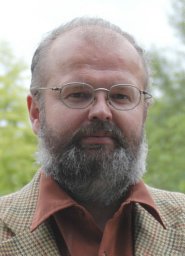 Christian Peham合作论文数Clinic of Orthopaedics in Ungulates, University of the Veterinary Medicine Vienna5
 Theresia Licka合作论文数Royal Dick Sch Vet Studies, Univ Edinburgh4
 M Groesel合作论文数3
 Rebeka R Zsoldos合作论文数3C Kicker合作论文数1A B Kotschwar合作论文数1D Girtler合作论文数1Lindner M合作论文数1Riha A合作论文数1Rumpler B合作论文数1
 Margit Gfoehler合作论文数1Ian G Mayhew合作论文数1




