Medical phrase grounding (MPG) maps textual descriptions of radiological findings to corresponding image regions. These grounded reports are easier to interpret, especially for non-experts. Existing MPG systems mostly follow the referring expression comprehension (REC) paradigm and return exactly one bounding box per phrase. Real reports often violate this assumption. They contain multi-region findings, non-diagnostic text, and non-groundable phrases, such as negations or descriptions of normal anatomy. Motivated by this, we reformulate the task as generalised medical phrase grounding (GMPG), where each sentence is mapped to zero, one, or multiple scored regions. To realise this formulation, we introduce the first GMPG model: MedGrounder. We adopted a two-stage training regime: pre-training on report sentence-anatomy box alignment datasets and fine-tuning on report sentence-human annotated box datasets. Experiments on PadChest-GR and MS-CXR show that MedGrounder achieves strong zero-shot transfer, state-of-the-art overall performance on PadChest-GR, competitive results on MS-CXR, and the largest gains on multi-region phrases, while using far fewer human box annotations. Finally, we show that MedGrounder can be composed with existing report generators to produce grounded reports without retraining the generator.
Accurate interpretation of chest radiograph images and generation of narrative reports is essential for patient care but places a heavy burden on radiologists and clinical experts. While AI models for automated report generation show promise, standardized evaluation frameworks remain limited. Here we present the ReXrank Challenge V1.0, a competition in the generation of chest radiograph reports utilizing ReXGradient, the largest test dataset consisting of 10,000 studies across 67 sites. The challenge attracted diverse participants from academic institutions, industry, and independent research teams, resulting in 8 new submissions alongside 16 state-of-the-art models previously benchmarked. Through comprehensive evaluation using multiple metrics, we analyzed model performance across various dimensions: differences between normal and abnormal studies, generalization capabilities across healthcare sites, and error rates in identifying clinical findings. This benchmark reveals that automated chest X-ray report generation remains fundamentally unsolved, with significant performance gaps between normal and abnormal studies, where even top-performing models achieve less than 45% error-free reporting on abnormal cases, and substantial variability across healthcare institutions, indicating that robust, clinically-ready systems require continued development before widespread deployment.
Diabetic foot osteomyelitis (DFO) is a leading cause of lower-extremity complications in individuals with diabetes, and timely, accurate screening is critical to prevent severe outcomes such as limb amputation. Although conventional radiography remains the most accessible imaging modality, the subtle and heterogeneous appearance of DFO often results in delayed or missed detection. Despite the rich morphological information encoded in foot radiographs, current deep learning methods tend to underperform in capturing localized pathological patterns due to architectural limitations. In this work, we propose Dual Backbone with Gated Fusion and Transformer encoder (DualBack-GFT), a deep learning framework for automated detection and localization of DFO in plain radiographs. The model leverages two complementary backbones, EfficientNet-B6 and ResNet-50, fused via a gated mechanism that adaptively combines image-specific features. These fused representations are further refined using transformer encoders, which effectively model long-range dependencies. The architecture operates in two stages: binary classification followed by confidence-weighted bounding-box localization. We evaluate DualBack-GFT on a curated, expert-annotated baseline dataset of diabetic foot X-rays with both diagnostic and bounding-box labels. The model achieves an AUC of 0.9683 and an average ground truth coverage of 62.71%, outperforming established baselines. These results underscore the potential of dual-stage, attention-enhanced models for interpretable and robust DFO assessment in clinical radiographs.
Chest X-ray (CXR) radiology report generation (RRG) models have shown rapid progress on automated metrics, yet their clinical utility remains uncertain due to limited qualitative evaluation by radiologists. We present CXRMate-2, a state-of-the-art CXR RRG model that enables tractable reinforcement learning (RL) through structured multimodal temporal embeddings and high-resolution visual feature compression, for efficient, unified conditioning of an LLM decoder on visual, textual, and temporal context from a study and its prior. This enables group relative policy optimisation (GRPO), where a proposed reward function is used to improve semantic alignment with radiologist reports. Across the MIMIC-CXR, CheXpert Plus, and ReXgradient datasets, CXRMate-2 achieves statistically significant improvements over strong benchmarks, including gains of 11.2 To directly compare CXRMate-2 against radiologist reporting, we conduct a blinded, randomised qualitative retrospective evaluation. Three consultant radiologists compare generated and radiologist reports across 120 studies from the MIMIC-CXR test set. Generated reports were deemed acceptable (defined as preferred or rated equally to radiologist reports) in 45 Together, these results define a clear pathway to clinically acceptable CXR RRG. Improving recall and the detection of subtle findings represents the primary remaining barrier to non-inferiority with radiologist reporting, positioning CXR RRG for prospective evaluation in assistive, radiologist-led workflows.
Biomedical texts, such as research articles and clinical reports, are often written in highly technical language, making them difficult for patients and the general public to understand. The BioLaySumm 2025 Shared Task addresses this challenge by promoting the development of models that generate lay summarisations of biomedical content. This paper focuses on Subtask 2.1: Radiology Report Generation with Layman's Terms. In this work, we evaluate two large language model (LLM) architectures, T5-large (700M parameter encoder-decoder model) and LLaMA-3.2-3B (3B parameter decoder-only model). Both models are trained under fully-supervised conditions using the task's multi-source dataset. Our results show that T5-large consistently outperforms LLaMA-3.2-3B across nine out of ten metrics, including relevance, readability, and clinical accuracy, despite having only a quarter of the parameters. Our T5-based model achieved the top rank in both the open-source and close-source tracks of the subtask 2.1.
Medical Phrase Grounding (MPG) maps radiological findings described in medical reports to specific regions in medical images. The primary obstacle hindering progress in MPG is the scarcity of annotated data available for training and validation. We propose anatomical grounding as an in-domain pre-training task that aligns anatomical terms with corresponding regions in medical images, leveraging large-scale datasets such as Chest ImaGenome. Our empirical evaluation on MS-CXR demonstrates that anatomical grounding pre-training significantly improves performance in both a zero-shot learning and fine-tuning setting, outperforming state-of-the-art MPG models. Our fine-tuned model achieved state-of-the-art performance on MS-CXR with an mIoU of 61.2, demonstrating the effectiveness of anatomical grounding pre-training for MPG.
This study investigates the integration of diverse patient data sources into multimodal language models for automated chest X-ray (CXR) report generation. Traditionally, CXR report generation relies solely on CXR images and limited radiology data, overlooking valuable information from patient health records, particularly from emergency departments. Utilising the MIMIC-CXR and MIMIC-IV-ED datasets, we incorporate detailed patient information such as vital signs, medicines, and clinical history to enhance diagnostic accuracy. We introduce a novel approach to transform these heterogeneous data sources into embeddings that prompt a multimodal language model; this significantly enhances the diagnostic accuracy of generated radiology reports. Our comprehensive evaluation demonstrates the benefits of using a broader set of patient data, underscoring the potential for enhanced diagnostic capabilities and better patient outcomes through the integration of multimodal data in CXR report generation.
Radiation therapy plays a crucial role in cancer treatment, necessitating precise delivery of radiation to tumors while sparing healthy tissues over multiple days. Computed tomography (CT) is integral for treatment planning, offering electron density data crucial for accurate dose calculations. However, accurately representing patient anatomy is challenging, especially in adaptive radiotherapy, where CT is not acquired daily. Magnetic resonance imaging (MRI) provides superior soft-tissue contrast. Still, it lacks electron density information, while cone beam CT (CBCT) lacks direct electron density calibration and is mainly used for patient positioning. Adopting MRI-only or CBCT-based adaptive radiotherapy eliminates the need for CT planning but presents challenges. Synthetic CT (sCT) generation techniques aim to address these challenges by using image synthesis to bridge the gap between MRI, CBCT, and CT. The SynthRAD2023 challenge was organized to compare synthetic CT generation methods using multi-center ground truth data from 1080 patients, divided into two tasks: (1) MRI-to-CT and (2) CBCT-to-CT. The evaluation included image similarity and dose-based metrics from proton and photon plans. The challenge attracted significant participation, with 617 registrations and 22/17 valid submissions for tasks 1/2. Top-performing teams achieved high structural similarity indices (≥0.87/0.90) and gamma pass rates for photon (≥98.1%/99.0%) and proton (≥97.3%/97.0%) plans. However, no significant correlation was found between image similarity metrics and dose accuracy, emphasizing the need for dose evaluation when assessing the clinical applicability of sCT. SynthRAD2023 facilitated the investigation and benchmarking of sCT generation techniques, providing insights for developing MRI-only and CBCT-based adaptive radiotherapy. It showcased the growing capacity of deep learning to produce high-quality sCT, reducing reliance on conventional CT for treatment planning.
Clinical documentation is an important aspect of clinicians' daily work and often demands a significant amount of time. The BioNLP 2024 Shared Task on Streamlining Discharge Documentation (Discharge Me!) aims to alleviate this documentation burden by automatically generating discharge summary sections, including brief hospital course and discharge instruction, which are often time-consuming to synthesize and write manually. We approach the generation task by fine-tuning multiple open-sourced language models (LMs), including both decoder-only and encoder-decoder LMs, with various configurations on input context. We also examine different setups for decoding algorithms, model ensembling or merging, and model specialization. Our results show that conditioning on the content of discharge summary prior to the target sections is effective for the generation task. Furthermore, we find that smaller encoder-decoder LMs can work as well or even slightly better than larger decoder based LMs fine-tuned through LoRA. The model checkpoints from our team (aehrc) are openly available.
The Shared Task on Large-Scale Radiology Report Generation (RRG24) aims to expedite the development of assistive systems for interpreting and reporting on chest X-ray (CXR) images. This task challenges participants to develop models that generate the findings and impression sections of radiology reports from CXRs from a patient's study, using five different datasets. This paper outlines the e-Health CSIRO team's approach, which achieved multiple first-place finishes in RRG24. The core novelty of our approach lies in the addition of entropy regularisation to self-critical sequence training, to maintain a higher entropy in the token distribution. This prevents overfitting to common phrases and ensures a broader exploration of the vocabulary during training, essential for handling the diversity of the radiology reports in the RRG24 datasets. Our model is available on Hugging Face https://huggingface.co/aehrc/cxrmate-rrg24.
Radiologists face high burnout rates, partially due to the increasing volume of Chest X-rays (CXRs) requiring interpretation and reporting. Automated CXR report generation holds promise for reducing this burden and improving patient care. While current models show potential, their diagnostic accuracy is limited. Our proposed CXR report generator integrates elements of the radiologist workflow and introduces a novel reward for reinforcement learning. Our approach leverages longitudinal data from a patient’s prior CXR study and effectively handles cases where no prior study exists, thus mirroring the radiologist’s workflow. In contrast, existing models typically lack this flexibility, often requiring prior studies for the model to function optimally. Our approach also incorporates all CXRs from a patient’s study and distinguishes between report sections through section embeddings. Our reward for reinforcement learning leverages CXR-BERT, which forces our model to learn the clinical semantics of radiology reporting. We conduct experiments on publicly available datasets — MIMIC-CXR and Open-i IU X-ray — with metrics shown to more closely correlate with radiologists’ assessment of reporting. Results from our study demonstrate that the proposed model generates reports that are more aligned with radiologists’ reports than state-of-the-art models, such as those utilising large language models, reinforcement learning, and multi-task learning. The proposed model improves the diagnostic accuracy of CXR report generation, which could one day reduce radiologists’ workload and enhance patient care. Our Hugging Face checkpoint (https://huggingface.co/aehrc/cxrmate) and code (https://github.com/aehrc/cxrmate) are publicly available.
We describe the participation of team e-Health CSIRO in the BioNLP RadSum task of 2023. This task aims to develop automatic summarisation methods for radiology. The subtask that we participated in was multimodal; the impression section of a report was to be summarised from a given findings section and set of Chest X-rays (CXRs) of a subject’s study. For our method, we adapted an encoder-to-decoder model for CXR report generation to the subtask. e-Health CSIRO placed seventh amongst the participating teams with a RadGraph ER F1 score of 23.9.
Automatically generating a report from a patient’s Chest X-rays (CXRs) is a promising solution to reducing clinical workload and improving patient care. However, current CXR report generators—which are predominantly encoder-to-decoder models—lack the diagnostic accuracy to be deployed in a clinical setting. To improve CXR report generation, we investigate warm starting the encoder and decoder with recent open-source computer vision and natural language processing checkpoints, such as the Vision Transformer (ViT) and PubMedBERT. To this end, each checkpoint is evaluated on the MIMIC-CXR and IU X-ray datasets. Our experimental investigation demonstrates that the Convolutional vision Transformer (CvT) ImageNet-21K and the Distilled Generative Pre-trained Transformer 2 (DistilGPT2) checkpoints are best for warm starting the encoder and decoder, respectively. Compared to the state-of-the-art (M2 Transformer Progressive), CvT2DistilGPT2 attained an improvement of 8.3% for CE F-1, 1.8% for BLEU-4, 1.6% for ROUGE-L, and 1.0% for METEOR. The reports generated by CvT2DistilGPT2 have a higher similarity to radiologist reports than previous approaches. This indicates that leveraging warm starting improves CXR report generation. Code and checkpoints for CvT2DistilGPT2 are available at https://github.com/aehrc/cvt2distilgpt2.
Speech enhancement plays an essential role in a wide range of speech processing applications. Recent studies on speech enhancement tend to investigate how to effectively capture the long-term contextual dependencies of speech signals to boost performance. However, these studies generally neglect the time-frequency (T-F) distribution information of speech spectral components, which is equally important for speech enhancement. In this paper, we propose a simple yet very effective network module, which we term the T-F attention (TFA) module, that uses two parallel attention branches, i.e., time-frame attention and frequency-channel attention, to explicitly exploit position information to generate a 2-D attention map to characterise the salient T-F speech distribution. We validate our TFA module as part of two widely used backbone networks (residual temporal convolution network and Transformer) and conduct speech enhancement with four most popular training objectives. Our extensive experiments demonstrate that our proposed TFA module consistently leads to substantial enhancement performance improvements in terms of the five most widely used objective metrics, with negligible parameter overheads. In addition, we further evaluate the efficacy of speech enhancement as a front-end for a downstream speech recognition task. Our evaluation results show that the TFA module significantly improves the robustness of the system to noisy conditions.
Deep learning models are powerful tools for addressing challenging medical imaging problems. However, for an ever-growing range of applications, interpreting a model’s prediction remains non-trivial. Understanding decisions made by black-box algorithms is critical, and assessing their fairness and susceptibility to bias is a key step towards healthcare deployment. In this paper, we propose DBCE (Deformation Based Counterfactual Explainability). We optimise a diffeomorphic transformation that deforms a given input image to change the prediction of the model. This provides anatomically meaningful saliency maps indicating tissue atrophy and expansion, which can be easily interpreted by clinicians. In our test case, DBCE replicates the transition of a patient from healthy control (HC) to Alzheimer’s disease (AD). We benchmark DBCE against three commonly used saliency methods. We show that it provides more meaningful saliency maps when applied to one subject and disease-consistent atrophy patterns when used over a larger cohort. In addition, our method fulfils a recent sanity check and is repeatable for different model initialisations in contrast to classical sensitivity-based methods.
Deep learning has achieved substantial improvement on single-channel speech enhancement tasks. However, the performance of multi-layer perceptions (MLPs)-based methods is limited by the ability to capture the long-term effective history information. The recurrent neural networks (RNNs), e.g., long short-term memory (LSTM) model, are able to capture the long-term temporal dependencies, but come with the issues of the high latency and the complexity of training.To address these issues, the temporal convolutional network (TCN) was proposed to replace the RNNs in various sequence modeling tasks. In this paper we propose a novel TCN model that employs multi-branch structure, called multi-branch TCN (MB-TCN), for monaural speech enhancement.The MB-TCN exploits split-transform-aggregate design, which is expected to obtain strong representational power at a low computational complexity.Inspired by the TCN, the MB-TCN model incorporates one dimensional causal dilated CNN and residual learning to expand receptive fields for capturing long-term temporal contextual information.Our extensive experimental investigation suggests that the MB-TCNs outperform the residual long short-term memory networks (ResLSTMs), temporal convolutional networks (TCNs), and the CNN networks that employ dense aggregations in terms of speech intelligibility and quality, while providing superior parameter efficiency. Furthermore, our experimental results demonstrate that our proposed MB-TCN model is able to outperform multiple state-of-the-art deep learning-based speech enhancement methods in terms of five widely used objective metrics.
The performance of speech coding, speech recognition, and speech enhancement largely depends upon the accuracy of the linear prediction coefficient (LPC) of clean speech and noise in practice. Formulation of speech and noise LPC estimation as a supervised learning problem has shown considerable promise. In its simplest form, a supervised technique, typically a deep neural network (DNN) is trained to learn a mapping from noisy speech features to clean speech and noise LPCs. Training targets for DNN to clean speech and noise LPC estimation fall into four categories: line spectrum frequency (LSF), LPC power spectrum (LPC-PS), power spectrum (PS), and magnitude spectrum (MS). The choice of appropriate training target as well as the DNN method can have a significant impact on LPC estimation in practice. Motivated by this, we perform a comprehensive study on the training targets using two state-of-the-art DNN methods--- residual network and temporal convolutional network (ResNet-TCN) and multi-head attention network (MHANet). This study aims to determine which training target as well as DNN method produces more accurate LPCs in practice. We train the ResNet-TCN and MHANet for each training target with a large data set. Experiments on the NOIZEUS corpus demonstrate that the LPC-PS training target with MHANet produces a lower spectral distortion (SD) level in the estimated speech LPCs in real-life noise conditions. We also construct the AKF with the estimated speech and noise LPC parameters from each training target using ResNet-TCN and MHANet. Subjective AB listening tests and seven different objective quality and intelligibility evaluation measures (CSIG, CBAK, COVL, PESQ, STOI, SegSNR, and SI-SDR) on the NOIZEUS corpus demonstrate that the AKF constructed with MHANet-LPC-PS driven speech and noise LPC parameters produced enhanced speech with higher quality and intelligibility than competing methods.
Tuberculosis (TB) is one of the leading causes of death worldwide. Automated detection of lung caverns associated with TB in Computed Tomography (CT) could help clinicians optimise treatment. However, caverns detection on 3D CT data is challenging due to the curse of dimensionality, thus requiring larger training data and more computational resource. Our team (AEHRC CSIRO) participated in ImageCLEFmed TB caverns detection 2022 to address this challenge, by developing a 2D YOLO-based model (TBdet-2D), and an efficient 3D Retina-U-Net-based model (TBdet-3D). Both networks were trained on 559 CT data with data augmentation and tested on 140 data provided by the challenge. The results show that TBdet-3D (mAP_IoU 0.504) outperformed TBdet-2D model (mAP_IoU 0.308) on testing data, indicating that employing a 3D approach instead of a 2D approach is more appropriate for the task. Our team placed first among the participating teams in this challenge. An overview of ImageCLEFmed Tuberculosis 2022 is available at: https://www.imageclef.org/2022/medical/tuberculosis.
As part of Best of Labs, we have been invited to conduct further investigation on the ImageCLEFmed Caption task of 2021. The task required participants to automatically compose coherent captions for a set of medical images. The most popular means of doing this is with an encoder-to-decoder model. In this work, we investigate a set of choices with regards to aspects of an encoder-to-decoder model. Such choices include what pre-training data should be used, what architecture should be used for the encoder, whether a natural language understanding (e.g., BERT) or generation (e.g., GPT2) checkpoint should be used to initialise the parameters of the decoder, and what formatting should be applied to the ground truth captions during training. For each of these choices, we first made assumptions about what should be used for each choice and why. Our empirical evaluation then either proved or disproved these assumptions—with the aim to inform others in the field. Our most important finding was that the formatting applied to the ground truth captions of the training set had the greatest impact on the scores of the task’s official metric. In addition, we discuss a number of inconsistencies in the results that others may experience when developing a medical image captioning system.
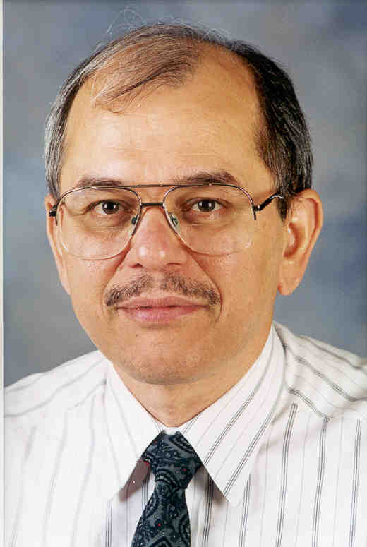 Kuldip Paliwal合作论文数Signal Processing Laboratory, School of Engineering and Built Environment, Griffith University18
 Jason A Dowling合作论文数15
 Bevan R. Koopman合作论文数The University of Queensland;Health Search Team, Australian e-Health Research Centre, Commonwealth Scientific and Industrial Research Organisation13
 Qiquan Zhang合作论文数Harbin Inst Technol, Sch Elect & Informat Engn, Shenzhen, Shenzhen, Peoples R China6
 Sujan Kumar Roy合作论文数Dept. of Computer Science and Engineering, The University of Rajshahi5
 Min Hang合作论文数Ingham Inst Appl Med Res3
 Haizhou Li (李海洲)合作论文数School of Data Science, Chinese University of Hong Kong, Shenzhen;Department of Electrical and Computer Engineering, National University of Singapore;Machine Listening Lab, University of Bremen3
 Gregg Belous合作论文数3
 Mingjiang Wang (王明江)合作论文数School of Integrated Circuits, Harbin Institute of Technology, Shenzhen3Léo Lebrat合作论文数CSIRO Health and Biosecurity, The Australian eHealth Research Centre2
 Jinghui Liu合作论文数Australian e-Health Research Centre, CSIRO2
 Qi Song合作论文数2Timothy Roberts合作论文数Griffith Univ, Signal Proc Lab, 170 Kessels Rd, Nathan, Qld 4111, Australia2
 Ashley Gillman合作论文数CSIRO, Australian E Hlth Res Ctr, Brisbane, Qld, Australia2
 Rodrigo Santa Cruz合作论文数Australian Centre for Robotic Vision, Australian National University2
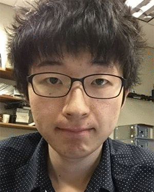 Zhaoheng Ni合作论文数2
 Chenxu Wang (王晨旭)合作论文数College of Information Science and Engineering, Harbin Institute of Technology, Weihai2
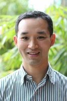 Anthony Nguyen合作论文数The Australian e-Health Research Centre, Commonwealth Scientific and Industrial Research Organisation2
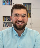 Ibrahim Hamamci合作论文数ETH AI Center;Universitätsspital Zürich;Department of Quantitative Biomedicine, Universität Zürich1f rogez合作论文数1
 Olivier Salvado合作论文数1
 Vincent Dore合作论文数1Jurgen Mejan-Fripp合作论文数1Shaury Srivastav合作论文数1
 Fanhua Shang (尚凡华)合作论文数College of Intelligence and Computing, Tianjin University1Ashu Gupta合作论文数1Satoshi Kondo合作论文数1
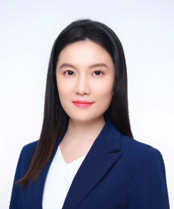 Xinyuan Qian (钱馨园)合作论文数School of Computer & Communication Engineering, University of Science and Technology Beijing1
 Yongsheng Gao合作论文数School of Engineering and Built Environment, Griffith University;Institute for Integrated and Intelligent Systems, Griffith University1Arthur Jr Galapon合作论文数1Kenza Bouzid合作论文数1Mercy Ranjit合作论文数1Joshua Peters合作论文数1Evi M. C. Huijben合作论文数1Bulat Ibragimov合作论文数1Satoshi Kasai合作论文数1
 Suraj Pai合作论文数GROW School for Oncology and Reproduction, Maastricht University Medical Centre+1Zijie Chen合作论文数1
 Maarten Terpstra合作论文数Computational Imaging Group for MR Diagnostics & Therapy, University Medical Center Utrecht1Juhyung Ha合作论文数1Adrian Thummerer合作论文数1
 Han Liu合作论文数Vanderbilt University1Salamata Konate合作论文数1
 Firas Khader合作论文数Department of Diagnostic and Interventional Radiology, University Hospital RWTH Aachen1y gim合作论文数1
 Delwyn Moller合作论文数1
 Pengxin Yu (于朋鑫)合作论文数1
 Bowen Xin合作论文数Univ Sydney, Sch Comp Sci, Sydney, NSW, Australia1Tao Song合作论文数1




