Signal-to-Noise (SNR) ratio is a limiting factor for blood flow imaging. It is known that coded excitation transmission methods can generally improve blood flow imaging outcomes. However, to our knowledge, the impact of coded excitation on modern spatiotemporal tissue filtering techniques has not been performed. Here, we evaluate this impact using 13-bit Barker codes, which theroretically increase SNR by 11.14 dB. We analyze singular value decomposition subspaces from matched coded and uncoded data to quantify differences in the tissue, blood, and noise subspaces. We show that the coded blood subspace contains over 30 more singular vectors, on average, than the uncoded, indicating that coded excitation induces broadening of the blood subspace that requires adjustments to filtering parameters to fully capture the blood flow signal.
Power Doppler imaging is an invaluable tool for blood flow assessment. However, its quality can be degraded in scenarios where the signal-to-noise ratio (SNR) is low. To address this, coded excitation schemes have been proposed to use longer transmit pulses without a reduction in axial resolution. One particular method that can generate arbitrarily long pulses is the use of compound Barker codes, which can provide significant improvements in SNR in the context of power Doppler imaging. However, the combination of these methods has yet to be implemented in real time. Therefore, in this work, we leverage the parallel processing of GPUs to exhibit the first real-time demonstration of power Doppler imaging using compound Barker code excitation.
Functional ultrasound has previously been demonstrated in small animal models and in adult humans without the skull. More recently, transcranial functional ultrasound in adults was demonstrated in central regions of the brain where larger, higher-velocity vessels are clustered. This was enabled in part because of coded transmit waveforms that boost SNR. Here, we demonstrate that functional assessment of more peripheral regions of the brain are possible with better clutter filtering that better isolates slow flow in high clutter and noise environments. To accomplish this, we utilize an adaptive normalized cross-correlation singular value selection technique to increase the effective field-of-view to include both the mid-brain and peripheral regions. We show correlation between the functional ultrasound signal and concurrently-acquired oxygen saturation data (0.744 +/- 0.0423) to confirm functional response.
Functional ultrasound was initially demonstrated in rodent models, but more recently, transcranial functional ultrasound has been demosntrated in humans in applications where the skull can be bypassed. This includes scenarios like surgery where the skull is removed or in neonates where the skull has not yet fused. These are important demonstrations of the technology, but they are insufficient for widespread clinical or scientific investigation in humans. Here, we demonstrate transcranial functional ultrasound in 13 subjects. To do this, we use a breath hold task. To accomplish transcranial functional ultrasound, we mostly use conventional methods, but we introduce direct axial motion correction and compound Barker coded excitation. Across 13 subjects we achieve a correlation of 0.53 +/- 0.08 between the functional ultrasound signal and the block design. This level of correlation matches similar fMRI studies.
Transcranial functional ultrasound is a relatively new technology for assessing functional responses in the brain. So far, transcranial functional ultrasound without contrast agents has only been applied to human imaging during surgery with the skull removed or in imaging neonates. Here, we demonstrate the feasibility of transcranial functional ultrasound through the adult skull. To overcome the severe attenuation from the skull we use a previously developed a compound Barker coded excitation method. For clutter filtering, we use an adaptive demodulation motion correction method with singular value decomposition (SVD) filtering. As a demonstration, we assess cerebrovascular reactivity induced by a breath hold. We measured the power Doppler signal in the vasculature surrounding the midbrain in a healthy adult volunteer during a five minute task of alternating free breathing and breath holding periods. We showed that the power Doppler signal in the blood vessels was highly correlated to the breath hold task (ρ=0.53) and the oxygen saturation (ρ=0.61) as measured by finger pulse oximeter. We also observed a delayed vasodilatory response in the power Doppler signal that reflected the delayed drop in oxygen saturation from the breath hold. These results demonstrate that contrast-free transcranial functional imaging in adults is possible using coded excitation to increase SNR and blood flow sensitivity and with appropriate motion compensation and clutter filtering techniques.
Background: Fifteen million Americans suffer from chronic obstructive pulmonary disease (COPD). After failed optimal medical therapy, the only definitive treatment is lung transplantation, which is limited due to donor scarcity. Extracorporeal membrane oxygenation (ECMO) can be used in acute exacerbations of COPD or as a bridge to transplant for waitlisted COPD patients, but it is not currently suited for long-term respiratory support. Existing venovenous (VV) ECMO configurations often utilize a dual lumen cannula that is inserted into the jugular vein and is designed for short term use. While effective, dual lumen cannulas are associated with high resistance and malposition which limits blood flow, promotes cavitation, and increases blood trauma. Furthermore, patient ambulation is cumbersome because of the difficulty of securing the dual lumen cannula safely to the patient. A novel ECMO system that provides durable respiratory support could reduce morbidity and mortality in patients and facilitate ambulatory use. To this end, a dual lumen cannula that is attached to the heart and tunneled to a more ergonomic position could provide more durable, long term ECMO support. Here, we describe our initial efforts to develop a durable VV ECMO cannula with reduced resistance. Methods: A novel cannula for long-term VV ECMO support was developed using computer aided design. The cannula is designed for surgical attachment to the right atrium with the cannula’s drainage tip at the cavoatrial junction and the reinfusion port directed towards the tricuspid valve to minimize recirculation. Pressure drop was evaluated for the novel cannula, 28Fr, and 32Fr Crescent cannulas (MC3, Dexter, MI) for comparison (N≥5) in circuits primed with deionized water at ambient temperature. A Rotaflow (Maquet, Wayne, NJ) blood pump was operated at 2700-3625 RPM, and pressure drop was assessed for flow rates between 0.1 – 5 LPM. Porcine hearts were harvested from healthy pigs and utilized for ex vivo cannula fit studies. Water flow through the cannula into the right atrium was evaluated using videography. Results: The novel cannula drainage pressure drop (-12±1.1mmHg) was similar to the 28Fr Crescent (-13± 0.3mmHg) and 32Fr Crescent (-9±0.3mmHg) at 2LPM (Figure 1). Reinfusion pressure drop was similar between the novel cannula (22±1.2mmHg) and the 32Fr Crescent (21±0.4mmHg), and higher for the 28Fr Crescent (43±0.9mmHg) at 2LPM. Total pressure drop was similar between the novel cannula (33.4±1.2mmHg) and 32Fr Crescent (29.4±0.5mmHg) and higher for the 28Fr Crescent (57±1.0mmHg) at 2LPM. In ex-vivo heart studies flow through the novel cannula was directed towards the tricuspid valve and entered the right ventricle. Conclusions: Our novel cannula possesses an excellent pressure-flow profile, comparable to the largest-bore commercially available dual lumen cannulas. This novel cannula will enhance durability and ease of use for patients requiring durable mechanical respiratory support.Figure 1. Pressure Flow Curve for Novel VV ECMO Cannula
Functional ultrasound imaging (fUSI) is a promising new modality for neuroimaging that measures the power Doppler signal as a proxy for neural activation. However, clinical translation of noninvasive, transcranial fUSI (tfUSI) is hindered by the low signal-to-noise ratio (SNR) from the skull which greatly limits blood flow sensitivity. To overcome this issue, we demonstrate a coded excitation approach to increase SNR in transcranial power Doppler imaging and thereby increase blood flow sensitivity. In three healthy adult subjects we showed an average SNR gain of 17.77 $\pm 1.05\ \mathbf{dB}$ , contrast-to-noise ratio gain of 4.97 $\pm 4.18\ \mathbf{dB}$ , and contrast ratio gain of $22.53 \pm 11.34\ \text{dB}$ using a 65 bit code. These promising results indicate that tfUSI may be feasible using our coded excitation approach.
Evaluating blood flow in the liver has clinical implications for functional and oncological assessment. Here, we explore visualization of temporally varying power Doppler signals in the 3 vasculature systems in the liver using coded excitation acquisitions and block-wise SVD filtering that mitigate the limitations of poor SNR, spectral overlap, and shorter ensembles. We acquired 2 seconds of data with matched ECG traces, and then evaluated 100 sample ensembles across the ECG trace to assess optimal cardiac phases for forming images. We show that highest CNR and SNR values are obtained from images formed during diastole.
Spatial block-wise filtering architectures improve noise stationarity locally, making SVD-based filtering more robust (Song et al., 2017). Temporal block-wise filtering has been proposed to augment spatial block-wise methods for 3D Doppler data acquired via mechanical translation (Chen et al., 2021) and compensate for bulk motion in cardiac power Doppler imaging (Zhang et al., 2021). We combine spatial and temporal block-wise architectures to suppress noise through depth and better capture time-varying signals with power Doppler. We show that a combination of spatial and temporal block-wise methods provides improved CNR (3.9 dB gain), SNR (0.9 dB gain), and CR (11.7 dB gain) over spatial block-wise alone and propose two methods for selecting temporal block sizes. First, we select temporal ensembles from systole, early-, and late-diastole from matched ECG data. Second, we derive a temporal SVD cutoff between the blood and noise subspaces for the entire ensemble. We use that cutoff as a proxy for correlation length and select ensembles accordingly. We collected 2 s of liver data from a healthy volunteer (nine angled plane waves at 600 Hz PRF and 4.1667 MHz center frequency) and applied spatial and temporal block-wise SVD filtering with 100-sample ensembles and 50-sample temporal block sizes overlapped at 20%.
Non-contrast ultrasound blood flow imaging is difficult at slow blood flow rates. Singular value decomposition (SVD) and independent component analysis (ICA) are useful for separating tissue, blood, and noise sources for Doppler filtering. In addition, it has been shown that applying SVD and ICA in a block-wise manner further improves source separation; noise within a small block is theoretically more stationary, and thus easier to separate. Yet, there is much discussion on how to select independent components; several methods have been introduced with some success. We present a novel, adaptive hierarchical clustering approach for selecting appropriate independent components for blood flow image filtering that utilizes Kurtosis and Normalized Cross Correlation. Components are clustered based on the Kurtosis and NCC of each component; an optimal number of clusters is chosen using the Silhouette Method. Appropriate clusters are selected based on the Autocorrelation Function of each cluster. Our method was tested on 1 mm/s and 5 mm/s flowrate phantoms containing a 0.6 mm vessel and resulted in average SNR and CNR increases of 6.2 dB and 3.7 dB, respectively, for 1 mm/s blood flow velocities. We demonstrate that our method improves tissue and noise suppression throughout the field of view while maintaining blood flow information.
Brown adipose tissue (BAT) is a highly vascularized tissue that uptakes and oxidizes fatty acids from the circulation in response to a cold stimulus, resulting in thermogenesis. Tissue perfusion has been proposed as an approach to understand BAT metabolism, but current imaging techniques require invasive contrast or ionizing radiation. Power Doppler ultrasound imaging enables sensitive, high temporal resolution measures of the movement of blood as it perfuses a tissue without the need for contrast injections. The purpose of this study was to explore the utility of non‐contrast ultrasound perfusion imaging of human BAT during personalized cooling. Five healthy subjects [4 men; age: 31.8 ± 5.6 yrs.; body mass index: 22.5 ± 3.3 kg/m2; total body fat % (dual‐energy x‐ray absorptiometry scan): 22.9 ±7.0 %] underwent an individualized, perception‐based cooling protocol to stimulate BAT. Infrared thermography, a surrogate measure of BAT activity, and power Doppler ultrasound images were acquired over the right and left supraclavicular space, respectively, every five minutes in thermoneutrality (TN; duration: 15 min) and during cold exposure (CE) to the participant’s shiver threshold (duration: 58.3 ± 10.8 min; cooling dose: 319.7 ± 140.4 °C*min). Ultrasound images were post‐processed with a block‐wise, independent component analysis filter to analyze signal changes related to perfusion. BAT regions of interest were defined, and TN and CE conditions were compared as the mean ± standard deviation of the difference in the 95th percentile skin temperatures (infrared thermography) and the mean power Doppler signal (ultrasound). Supraclavicular skin temperature increased by 0.56 ± −0.21 °C (95% bootstrap confidence interval (CI): −0.57 to 1.72 °C), indicating a potential thermogenic response of BAT to individualized cooling (TN: 33.8 ± 1.1 °C vs. CE: 34.3 ± 0.9 °C). Similarly, the mean power Doppler signal increased by 11.6 ± 3.8 dB (95% CI: 3.2 to 19.4 dB) following cold exposure (TN: 46.8 ± 5.3 dB vs. CE: 58.3 ± 9.1 dB). These preliminary data demonstrate the feasibility of non‐contrast ultrasound perfusion imaging to detect the microvascular response of BAT to a cold stimulus in healthy adults. Power Doppler ultrasound imaging could prove useful when combined with existing noninvasive modalities (e.g. magnetic resonance imaging) to assess both the perfusion and metabolic substrate uptake response of BAT to potential obesity‐targeted therapies.Support or Funding InformationNIDDK/NIH R01‐DK‐105371, NCATS/NIH UL1‐TR000445
Non-contrast ultrasound blood flow imaging is difficult at slow blood flow rates. For slow flow rates, tissue signal overpowers blood signal more than at faster velocities, masking blood flow, resulting in low CR and SNR values. Singular value decomposition (SVD) and independent component analysis (ICA) are useful for separating tissue, blood, and noise sources for Doppler filtering. In addition, it has been shown that applying SVD and ICA in a block-wise manner further improves source separation; noise within a small block is theoretically more stationary, and thus easier to separate. Here, we evaluate and compare the efficacy of SVD and ICA component sorting/classification methods by analyzing the sensitivity and specificity of each method, as well as introduce a novel Hierarchical Clustering method for component sorting.
Slow blood flow imaging has proven to be a difficult clinical problem. Imaging modalities, such as MR Angiography and Contrast-Enhanced Ultrasound, attempting to solve this problem are expensive and time-consuming. Both eigen-based filters and spatial filters have been proposed to improve ultrasound power Doppler blood flow images. Block-wise methods and Independent Component Analysis (ICA) filters have each individually been previously shown to improve tissue clutter and noise suppression. Here, we aim to develop a Block-wise implementation of ICA to evaluate blood flow in ultrasound blood flow imaging. We show through phantom studies that by applying ICA in a block-wise manner, we see qualitative improvements as compared to other eigen-based filters applied in global and block-wise manners.
 Brett Byram合作论文数Department of Biomedical Engineering, Duke University10
 Emelina Vienneau合作论文数Vanderbilt University5
 Victoria L Morgan合作论文数Department of Radiology, Vanderbilt University Medical Center;Vanderbilt University Institute of Imaging Science2
 Kaitlyn M. Tracy合作论文数1
 Sean Francois合作论文数1William Tucker合作论文数1Michael Cortelli合作论文数1Caitlin T. Demarest合作论文数1Mark K. George合作论文数1Carl A Johnson合作论文数1
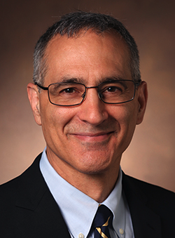 Matthew D. Bacchetta合作论文数Department of Cardiac Surgery, Vanderbilt University Medical Center;Vanderbilt University Adult Hospital1
 Jaime Tierney合作论文数Vanderbilt Univ, Dept Biomed Engn, Nashville, TN 37235 USA1
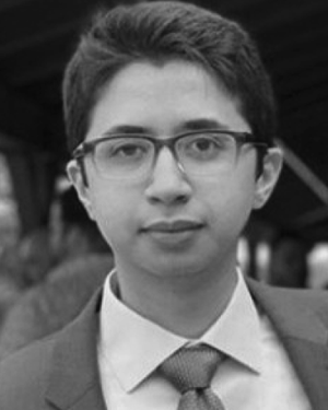 Christopher Khan合作论文数1
 Rei Ukita合作论文数Department of Cardiac Surgery, Vanderbilt University Medical Center1
 Greg Walker合作论文数Dept Mech Engn, Vanderbilt Univ1Pasrija Chetan合作论文数Division of Cardiac Surgery, University of Maryland School of Medicine1
 Coolbaugh Crystal L合作论文数Biomedical Engineering Graduate Group, University of California Davis1Skoog David J合作论文数1
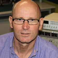 Stephen Wilson合作论文数MD Learning Hub, Faculty of Medicine, University of Queensland1
 Keith E. Cook合作论文数Department of Biomedical Engineering, College of Engineering, Carnegie Mellon University1
 Bruce Damon合作论文数Department of Bioengineering, The Grainger College of Engineering, University of Illinois Urbana-Champaign;Stephens Family Clinical Research Institute, Carle Health1




