Mammalian fertility depends on the production of an oocyte capable of fertilization and supporting early embryo development. This requires both cytoplasmic and nuclear, i.e. chromosomal, competence, processes that were initiated decades prior to ovulation. Current demographic changes with delayed motherhood are increasingly in conflict with these biological processes. This brief review highlights the key stages in oocyte development, as well as recent findings that continue to inform on how the oocyte is able to maintain function over such a prolonged period. These include minimizing oocyte damage caused by the production of reactive oxygen species, the importance of intercellular communication with the surrounding somatic cells, and the molecular mechanisms that underpin the fidelity of chromosome cohesion and then separation at the resumption of meiosis. Some of these are already approaching clinical testing and interventions, with new approaches in the coming years potentially being able to 'put back the clock' to improve oocyte quality.
Meiosis generates gametes through a specialised cell cycle that reduces the genome by half. Homologous chromosomes are segregated in meiosis I and sister chromatids are segregated in meiosis II. Centromeres and kinetochores play central roles in instructing this specialised chromosome segregation pattern. Accordingly, kinetochores acquire meiosis-specific modifications. Here we contextualise recent highlights in our understanding of how centromeres and kinetochores direct the sorting of chromosomes into gametes via meiosis.
The Target of Rapamycin Complex 1 (TORC1) integrates cellular cues and adapts cell growth and metabolism through substrate-specific phosphorylation. A genetic screen for suppressors of a conditional mutant of the cohesin loader Mis4 identified hypomorphic mutants of TORC1. Downregulation of TORC1 enhanced the binding of cohesin and its loader to their regular sites on chromosomes. In the context of impaired cohesin loading, TORC1 downregulation rescued chromosome segregation whereas upregulation had the opposite effect, suggesting environmental cues impinge on the robustness of chromosome segregation. TORC1 co-purified with cohesin from cellular extracts and the phosphorylation level of specific residues on Mis4 and cohesin were reduced in TORC1 mutants. Cohesin mutations mimicking the non-phosphorylated state mirrored the effects of TORC1 downregulation. Challenging cells with various conditions revealed that Mis4 and TORC1 regulate a common set of genes involved in the response to environmental changes. These genes are preferentially located far from centromeres and close to telomeres. We propose that cohesin is an effector of TORC1, orchestrating alterations in chromosome structure that facilitate cellular adaptation to environmental changes. ### Competing Interest Statement The authors have declared no competing interest.
In brief:Chromosome missegregation and declining energy metabolism are considered to be unrelated features of oocyte ageing that contribute to poor reproductive outcomes. Given the bioenergetic cost of chromosome segregation, we propose here that altered energy metabolism during ageing may be an underlying cause of age-related chromosome missegregation and aneuploidy. Abstract:Advanced reproductive age in women is a major cause of infertility, miscarriage and congenital abnormalities. This is principally caused by a decrease in oocyte quality and developmental competence with age. Oocyte ageing is characterised by an increase in chromosome missegregation and aneuploidy. However, the underlying mechanisms of age-related aneuploidy have not been fully elucidated and are still under active investigation. In addition to chromosome missegregation, oocyte ageing is also accompanied by metabolic dysfunction. In this review, we integrate old and new perspectives on oocyte ageing, chromosome segregation and metabolism in mammalian oocytes and make direct links between these processes. We consider age-related alterations to chromosome segregation machinery, including the loss of cohesion, microtubule stability and the integrity of the spindle assembly checkpoint. We focus on how metabolic dysfunction in the ageing oocyte disrupts chromosome segregation machinery to contribute to and exacerbate age-related aneuploidy. More specifically, we discuss how mitochondrial function, ATP production and the generation of free radicals are altered during ageing. We also explore recent developments in oocyte metabolic ageing, including altered redox reactions (NAD+ metabolism) and the interactions between oocytes and their somatic nurse cells. Throughout the review, we integrate the mechanisms by which changes in oocyte metabolism influence age-related chromosome missegregation.
SummaryGametes are produced via meiosis, a specialized cell division associated with frequent errors which cause birth defects and infertility. Uniquely in meiosis I, homologous chromosomes segregate to opposite poles, usually requiring their linkage by chiasmata, the products of crossover recombination1. The spindle checkpoint delays cell cycle progression until all chromosomes are properly attached to microtubules2but the steps leading to the capture and alignment of chromosomes on the meiosis I spindle remain poorly understood. In budding yeast meiosis I, Mad2 and Mad3BUBR1are equally important for spindle checkpoint delay, but biorientation of homologs on the meiosis I spindle requires Mad2, but not Mad3BUBR13,4. Here we show that Mad3BUBR1promotes accurate meiosis I homolog segregation outside its canonical checkpoint role, independently of Mad2. We find that Mad3BUBR1associates with the TOGL1 domain of Stu1CLASP, a conserved plus-end microtubule protein which is important for chromosome capture onto the spindle. Homologous chromosome pairs that are proficient in crossover formation, but which fail to biorient, rely on Mad3BUBR1-Stu1CLASPto ensure their efficient attachment to microtubules and segregation during meiosis I. Furthermore, we show that Mad3BUBR1-Stu1CLASPare essential to rescue the segregation of mini-chromosomes lacking crossovers. Our findings define a new pathway ensuring microtubule-dependent chromosome capture and demonstrate that spindle checkpoint proteins safeguard the fidelity of chromosome segregation both by actively promoting chromosome alignment and delaying cell cycle progression until this has occurred.
Abstract The cell cycle is ordered by a controlled network of kinases and phosphatases. To generate gametes via meiosis, two distinct and sequential chromosome segregation events occur without an intervening S phase. How canonical cell cycle controls are modified for meiosis is not well understood. Here, using highly synchronous budding yeast populations, we reveal how the global proteome and phosphoproteome change during the meiotic divisions. While protein abundance changes are limited to key cell cycle regulators, dynamic phosphorylation changes are pervasive. Our data indicate that two waves of cyclin-dependent kinase (Cdc28Cdk1) and Polo (Cdc5Polo) kinase activity drive successive meiotic divisions. These two distinct phases of phosphorylation are ensured by the meiosis-specific Spo13 protein, which rewires the phosphoproteome. Spo13 binds to Cdc5Polo to promote phosphorylation in meiosis I, particularly of substrates containing a variant of the canonical Cdc5Polo motif. Overall, our findings reveal that a master regulator of meiosis directs the activity of a kinase to change the phosphorylation landscape and elicit a developmental cascade.
Aneuploid human eggs (oocytes) are a major cause of infertility, miscarriage, and chromosomal disorders. Such aneuploidies increase greatly as women age, with defective linkages between sister chromatids (cohesion) in meiosis as a common cause. We found that loss of a specific pool of the cohesin protector protein, shugoshin 2 (SGO2), may contribute to this phenomenon. Our data indicate that SGO2 preserves sister chromatid cohesion in meiosis by protecting a "cohesin bridge" between sister chromatids. In human oocytes, SGO2 localizes to both sub-centromere cups and the pericentromeric bridge, which spans the sister chromatid junction. SGO2 normally colocalizes with cohesin; however, in meiosis II oocytes from older women, SGO2 is frequently lost from the pericentromeric bridge and sister chromatid cohesion is weakened. MPS1 and BUB1 kinase activities maintain SGO2 at sub-centromeres and the pericentromeric bridge. Removal of SGO2 throughout meiosis I by MPS1 inhibition reduces cohesion protection, increasing the incidence of single chromatids at meiosis II. Therefore, SGO2 deficiency in human oocytes can exacerbate the effects of maternal age by rendering residual cohesin at pericentromeres vulnerable to loss in anaphase I. Our data show that impaired SGO2 localization weakens cohesion integrity and may contribute to the increased incidence of aneuploidy observed in human oocytes with advanced maternal age.
Genomes are organised into DNA loops by the Structural Maintenance of Chromosomes (SMC) proteins. SMCs establish functional chromosomal sub-domains for DNA repair, gene expression and chromosome segregation, but how SMC activity is specifically targeted is unclear. Here, we define the molecular mechanism targeting the condensin SMC complex to specific chromosomal regions in budding yeast. A conserved pocket on the condensin HAWK subunit Ycg1 binds to chromosomal receptors carrying a related motif, CR1. In early mitosis, CR1 motifs in receptors Sgo1 and Lrs4 recruit condensin to pericentromeres and rDNA, to facilitate sister kinetochore biorientation and rDNA condensation, respectively. We additionally find that chromosome arm condensation begins as sister kinetochores come under tension, in a manner dependent on the Ycg1 pocket. We propose that multiple CR1-containing proteins recruit condensin to chromosomes and identify several additional candidates based on their sequence. Overall, we uncover the molecular mechanism that targets condensin to functionalise chromosomal domains to achieve accurate chromosome segregation during mitosis.
Full text Figures and data Side by side Abstract Editor's evaluation Introduction Results Discussion Materials and methods Data availability References Decision letter Author response Article and author information Metrics Abstract Small RNAs target their complementary chromatin regions for gene silencing through nascent long non-coding RNAs (lncRNAs). In the ciliated protozoan Tetrahymena, the interaction between Piwi-associated small RNAs (scnRNAs) and the nascent lncRNA transcripts from the somatic genome has been proposed to induce target-directed small RNA degradation (TDSD), and scnRNAs not targeted for TDSD later target the germline-limited sequences for programmed DNA elimination. In this study, we show that the SUMO E3 ligase Ema2 is required for the accumulation of lncRNAs from the somatic genome and thus for TDSD and completing DNA elimination to make viable sexual progeny. Ema2 interacts with the SUMO E2 conjugating enzyme Ubc9 and enhances SUMOylation of the transcription regulator Spt6. We further show that Ema2 promotes the association of Spt6 and RNA polymerase II with chromatin. These results suggest that Ema2-directed SUMOylation actively promotes lncRNA transcription, which is a prerequisite for communication between the genome and small RNAs. Editor's evaluation This important study demonstrates that protein SUMOylation is essential for programmed DNA elimination guided by small RNAs during conjugation in Tetrahymena ciliates. The authors present convincing evidence that the E3 SUMO ligase Ema2 is necessary for the production of long non-coding RNAs from the somatic nucleus, targeted small RNA degradation, and DNA elimination. The authors also show that the transcription regulator Spt6 is a SUMOylation target of Ema2, though the relevance of this is not completely established. This paper is of broad significance and will appeal to those interested in non-coding RNA biology, the control of programmed genome rearrangements, or ciliate biology. https://doi.org/10.7554/eLife.95337.sa0 Decision letter eLife's review process Introduction Small RNAs of approximately 20–30 nucleotides that are complexed with Argonaute family proteins target either mRNAs for post-transcriptional silencing or chromatin regions for transcriptional gene silencing (Holoch and Moazed, 2015; Wilson and Doudna, 2013). For the latter process, small RNAs are generally considered to recognize their genomic targets via nascent lncRNAs to induce heterochromatin formation. Therefore, chromatin regions that are targeted for silencing by small RNAs must be paradoxically transcribed to provide nascent lncRNAs. In fission yeast, small interfering RNAs (siRNAs) mediate the deposition of histone 3 lysine 9 di- and trimethylation (H3K9me2/3) for heterochromatin assembly at centromeric repeats (Hall et al., 2002; Volpe et al., 2002). While the HP1 protein Swi6 binds to H3K9me2/3 for transcriptional silencing, phosphorylation of histone H3 serine 10 at the M phase of the cell cycle evicts Swi6, passively allowing lncRNA transcription from the centromeric repeats at G1 and S phase and thus promoting H3K9me2/3 deposition to newly assembled nucleosomes (Chen et al., 2008; Kloc et al., 2008). In contrast, the HP1 paralog Rhino in fruit flies, which is specifically enriched at PIWI-interacting RNA (piRNA) clusters by binding to H3K9me2/3 (Le Thomas et al., 2014), actively recruits dedicated variants of basal transcription factors, allowing lncRNA transcription from heterochromatin while preventing mRNA transcription from transposons in the same loci (Andersen et al., 2017). A similar active heterochromatin-dependent lncRNA transcription has been reported in plants, where SHH1, a reader of H3K9me2/3 as well as mono-methylated H3K9, recruits the plant-specific RNA polymerase IV (Law et al., 2013; Law et al., 2011). Because small RNA-producing loci are also small RNA targets in most of the studied small RNA-directed heterochromatin formation processes, it poses a challenge to separately investigate lncRNA transcription for small RNA biogenesis and that for small RNA-dependent recruitment of downstream effectors in these processes. In contrast, the source and target loci of small RNAs reside in different nuclei during programmed DNA elimination in some ciliated protozoans such as Tetrahymena thermophila and Paramecium tetraurelia, which provides a unique system to study the mechanism and roles of lncRNA transcription in small RNA-directed chromatin regulation. In most ciliates, each cell contains two types of nuclei, the diploid germline micronucleus (MIC) and the polyploid somatic macronucleus (MAC). During conjugation, a sexual reproduction process in ciliates, the MIC undergoes meiosis and fertilization, followed by the formation of the new MIC and MAC, while the parental MAC is degraded (Chalker et al., 2013). Programmed DNA elimination occurs in the new MAC in most ciliates. In Tetrahymena, this process downsizes the 200 Mb MIC genome to the 103 Mb MAC genome by removing ~12,000 internal elimination sequences (IESs), many of which include transposons, followed by re-ligation of the remaining macronuclear-destined sequences (MDSs) forming the MAC chromosomes (Noto and Mochizuki, 2018). DNA elimination in Tetrahymena and Paramecium is regulated by three types of lncRNAs, which occur in different nuclei at distinct times (Aronica et al., 2008; Schoeberl and Mochizuki, 2011). During meiotic prophase in Tetrahymena, lncRNAs are transcribed bidirectionally in the MIC in a genome-wide manner by RNA polymerase II and dedicated conjugation-specific Mediator-associated proteins (Chalker and Yao, 2001; Garg et al., 2019; Mochizuki and Gorovsky, 2004a; Schoeberl et al., 2012; Tian et al., 2019). The sexual reproduction-specific Spt4 and Spt5 paralogs are specifically involved in the MIC transcription in Paramecium (Gruchota et al., 2017; Owsian et al., 2022), while Tetrahymena genome does not encode such specialized Spt4/Spt5 paralogs. These micronuclear long non-coding RNAs (MIC-lncRNAs) are processed into small RNAs (~29 nt in Tetrahymena and 25-nt in Paramecium), called scnRNAs, by Dicer homologs (Lepère et al., 2009; Malone et al., 2005; Mochizuki and Gorovsky, 2005) and loaded into Piwi-clade Argonaute proteins (Bouhouche et al., 2011; Mochizuki et al., 2002; Noto et al., 2010). The Piwi-scnRNA complex then moves into the parental MAC, where parental macronuclear long non-coding RNAs (pMAC-lncRNAs) are transcribed bidirectionally during the mid-conjugation stages (Woo et al., 2016). The RNA helicase Ema1 in Tetrahymena promotes the interaction between pMAC-lncRNAs and scnRNAs (Aronica et al., 2008). Reminiscently of target-directed micro RNA degradation (TDMD) (Han and Mendell, 2023), this interaction induces TDSD, leading to the selective retention of IES-derived scnRNAs (Aronica et al., 2008; Mochizuki and Gorovsky, 2004b; Noto and Mochizuki, 2018; Noto et al., 2015; Schoeberl et al., 2012). A similar TDSD has also been suggested in Paramecium, and the importance of pMAC-lncRNAs for DNA elimination was demonstrated by disrupting certain pMAC-lncRNAs by RNAi in this ciliate (Lepère et al., 2008). Although mRNAs are transcribed in the parental MAC, it remains unclear whether they also can induce TDSD and how mRNAs and pMAC-lncRNAs can be transcribed from overlapping locations. Also, although trimethylation of histone 3 lysine 27 (H3K27me3) occurs in the parental MAC in a scnRNA- and Polycomb repressive complex 2 (PRC2)-dependent manner in both Tetrahymena and Paramecium (Lhuillier-Akakpo et al., 2014; Liu et al., 2007), the role of H3K27me3 in the parental MAC, if any, is unclear. When the new MAC develops, the retained Piwi-scnRNA complexes translocate to the new MAC, where yet another type of lncRNA, new macronuclear non-coding RNAs (nMAC-lncRNAs), is transcribed. The interaction between nMAC-lncRNA and scnRNA, which is also dependent on Ema1 (Aronica et al., 2008), is believed to recruit PRC2, which catalyzes both H3K9me2/3 and H3K27me3 for IES-specific heterochromatin assembly (Frapporti et al., 2019; Liu et al., 2007; Miró-Pina et al., 2022; Wang et al., 2022; Xu et al., 2021), and facilitate the secondary production of scnRNAs from nMAC-lncRNAs, which further promotes heterochromatin assembly (Allen et al., 2017; Noto et al., 2015). Then, IESs are excised by domesticated PiggyBac transposases (Baudry et al., 2009; Bischerour et al., 2018; Cheng et al., 2010; Vogt and Mochizuki, 2013). While nMAC-lncRNA production occurs prior to the excision of IESs in Tetrahymena (Mutazono et al., 2019), it also occurs from excised IESs that are concatenated and circularized in Paramecium (Allen et al., 2017). Among the above three lncRNAs, pMAC-lncRNA does not produce small RNAs and is believed to be specialized for receptor function. Therefore, pMAC-lncRNA transcription and the following TDSD in the parental MAC provide a unique paradigm to investigate how the genome is transcribed to communicate with small RNAs. In this study, we show that Ema2, the conjugation-specific E3 ligase for a small ubiquitin-like modifier (SUMO), is required for pMAC-lncRNA transcription in Tetrahymena and thus provides a tool to dissect the role of and the molecular mechanism for the transcription of this lncRNA. Results Ema2 is exclusively expressed during conjugation and localized in the MAC As part of our systematic investigation into genes highly upregulated during conjugation (Loidl, 2021), we explored the function of EMA2 (TTHERM_00113330). EMA2 mRNA is exclusively expressed during conjugation (Figure 1A). The encoded Ema2 protein tagged with HA at the endogenous EMA2 locus was not detectable in the vegetative cells (Figure 1B, Vg) and first appeared in the MAC during conjugation (Figure 1B, 3 hr post-induction of mating [hpm] and 6 hpm). At the onset of new MAC development, Ema2 disappeared from the parental MAC and appeared in the new MAC (Figure 1B, 8 hpm), which later faded away (Figures 1B, 12, and 14 hpm). The conjugation-specific expression and the localization switch from the parental to the new MAC are reminiscent of the factors involved in DNA elimination such as the Piwi protein Twi1, which is loaded by scnRNAs, and PRC2 (Liu et al., 2007; Mochizuki et al., 2002; Noto et al., 2010). Figure 1 Download asset Open asset Ema2 is expressed during conjugation and localized in the macronucleus (MAC). (A) EMA2 mRNA expression levels (in an arbitrary unit) in growing Tetrahymena cells in low (l), middle (m), and high (h) cell concentrations, starved cells from 0 to 24 hr, and cells in the conjugation and post conjugation stages from 0 to 18 hr post-mixing (hpm) are shown. The mRNA expression data were obtained from Miao et al., 2009. (B) Ema2 localization. Two Ema2-HA strains were mated and fixed at the indicated time points (Vg = vegetative cell). An anti-HA antibody was used to localize Ema2-HA (green), and DNA was stained with DAPI (magenta). The micronucleus (MIC), the parental MAC, and the newly formed MAC are marked with arrowheads with 'i,' 'a,' and 'na,' respectively. All pictures share the scale bar. Ema2 is required for completing DNA elimination DNA elimination of exconjugants (sexual progeny) at 36 hpm was analyzed by DNA fluorescent in situ hybridization (FISH) using probes complementary to the transposable element Tlr1 (Wuitschick et al., 2002). DNA elimination is completed by ~14–18 hpm in wild-type cells (Austerberry et al., 1984; Mutazono et al., 2019), and the Tlr1 element was detected only in the MICs in the exconjugants from wild-type cells (Figure 2A, WT). In contrast, the Tlr1 element was detected in both the MICs and the MACs in the exconjugants from the EMA2 somatic KO strains, in which all EMA2 copies in the MAC were disrupted (Shehzada and Mochizuki, 2022; Figure 2A, KO). The intensity of the FISH signal in the new MACs was lower in the exconjugants from the EMA2 KO cells than in those from the TWI1 KO cells (Figure 2B), in the latter of which DNA elimination is known to be completely blocked (Noto et al., 2015). Therefore, DNA elimination was partially blocked in the exconjugants of EMA2 KO cells. Consistent with the requirement of DNA elimination in the viability of sexual progeny (Cheng et al., 2010; Vogt and Mochizuki, 2013), EMA2 KO cells did not produce viable progeny (Figure 2C). Altogether, we conclude that maternally expressed EMA2 is required for completing DNA elimination. Figure 2 Download asset Open asset Ema2 is required for completing DNA elimination. (A) Two wild-type (WT) or two EMA2 somatic KO (KO) cell lines were mixed, and their exconjugants at 36 hpm were analyzed by DNA-fluorescent in situ hybridization (FISH) with fluorescent probes complementary to the Tlr1 element (green). DNA was counterstained with DAPI (magenta). The micronucleus (MIC) and the new macronucleus (MAC) are marked with arrowheads with 'i' and 'na', respectively. All pictures share the scale bar. (B) Exconjugants from wild-type (WT) cells, EMA2 somatic KO cells, and TWI1 KO cells were stained as in (A), the IES retention index was calculated (see Materials and Methods for details) from 20 cells each, and shown as box plots. The whiskers represent 10-90 percentile. Three asterisks (***) indicate a p-value of less than 0.001 in the Welch two-sample t-test. (C) Two wild-type (WT) cells and two independent crosses of EMA2 somatic KO cells (Cross 1 and 2) were mated, and the conjugating pairs were isolated for the viability test. The percentages of pairs that gave rise to viable sexual progeny are shown. 'n' represents the number of total pairs tested. Ema2 is required for target-directed small RNA degradation (TDSD) We next asked whether EMA2 is involved in TDSD. The production of scnRNAs occurs from IESs and their surrounding MDS regions in the MIC at the early conjugation stages (~2–3 hpm), and scnRNAs complementary to the MAC genome (=MDSs) are subjected to TDSD in the parental MAC in the mid-conjugation stages (~3–7 hpm), resulting in the selective retention of IES-derived scnRNAs that later target IESs for DNA elimination in the new MAC (Aronica et al., 2008; Mochizuki and Gorovsky, 2004b; Noto et al., 2015; Schoeberl et al., 2012). We first compared scnRNAs at different conjugation stages by northern blot analysis using the 50-nt Mi-9 probe that is complementary to a repetitive sequence found in both IESs and MDSs (Aronica et al., 2008). Because scnRNAs detected by this probe are complementary to the MAC genome, in wild-type cells, the Mi-9-complementary scnRNAs were detected at 3 hpm, reduced at 4.5 hpm, and became undetectable at 6 hpm due to TDSD (Figure 3A, WT). In contrast, the Mi-9-complementary scnRNAs remained detectable at 6 hpm in the EMA2 KO cells (Figure 3A, KO), suggesting that Ema2 is required for TDSD. Figure 3 Download asset Open asset Ema2 is required for target-directed small RNAs (scnRNA) degradation (TDSD). (A) Total RNA was isolated from conjugating wild-type (WT) and EMA2 somatic KO (KO) cells at 3, 4.5, and 6 hr post-mixing (hpm), separated in denaturing gel and stained with the nucleic acid dye Gel-Red (right). Then, RNA was transferred to a membrane and hybridized with the radioactive Mi-9 probe, which is complementary to a repetitive sequence in MDSs (right). (B) Small RNAs from conjugating wild-type (WT), EMA2 somatic KO, and EMA1 somatic KO strains were isolated at 3 and 8 hpm and analyzed by high-throughput sequencing. Normalized numbers (RPKM [read per kilobase of unique sequence per million]) of sequenced small RNAs (26–32 nt) that uniquely matched to the macronuclear-destined sequences (MDS) (left) or internal elimination sequence (IES) (right) genomic tiles (see Materials and methods) are shown as box plots. The median value is represented by the horizontal bar in the box. The minimum and maximum values are indicated by the bars on top and bottom of the box, respectively, with 1.5 x the interquartile range (IQR). Three asterisks (***) and 'ns' respectively indicate a p-value of less than 0.001 and more than 0.05 in the Wilcoxon rank sum test. Figure 3—source data 1 The raw data of northern blot (top) and Gel-red stained gel (bottom) without (Figure_3 A_Original) and with (Figure_3 A_Original-marked) marks of the positions of regions used for Figure 3A. https://cdn.elifesciences.org/articles/95337/elife-95337-fig3-data1-v2.zip Download elife-95337-fig3-data1-v2.zip We also analyzed TDSD by small RNA sequencing at 3 and 8 hpm. Sequenced 26- to 32-nt small RNAs, which correspond to scnRNAs (Schoeberl et al., 2012), were mapped to the genomic tiles of MDSs and IESs (see Materials and methods). Although scnRNAs that mapped many of the MDS tiles were greatly reduced from 3 to 8 hpm due to TDSD in the wild-type cells (Figure 3B, left, WT), those in the EMA2 KO cells were only slightly reduced by 8 hpm (Figure 3B, left, EMA2 KO). Because TDSD takes place concurrently with the scnRNA production (Schoeberl et al., 2012), the increased abundance of MDS-complementary scnRNAs at 3 hpm in the EMA2 KO cells compared to the wild-type cells can also be attributed to the necessity of Ema2 in TDSD. This TDSD defect in the EMA2 KO cells seems milder than that in the EMA1 KO cells, in which the amount of MDS-complementary scnRNAs remained constant until 8 hpm (Figure 3B, left, EMA1 KO), indicating that Ema1 and Ema2 act differently in TDSD. In contrast, the amounts of scnRNAs complementary to IES tiles at 3 hpm and 8 hpm were comparable in all strains (Figure 3B, right), which is consistent with previous observations that only scnRNAs complementary to MDSs are targeted for TDSD. These results suggest that Ema2 is required for TDSD at the genome-wide level. Because it is known that loss of TDSD results in a partial block of DNA elimination (Aronica et al., 2008), the mild DNA elimination defect in EMA2 KO cells (Figure 2B) can be explained by the requirement of Ema2 in TDSD. Ema2 is required for the accumulation of SUMOylated proteins during conjugation Ema2 possesses an SP-RING domain that has been found in many SUMO E3 ligases (Hochstrasser, 2001). The SP-RING domain of Ema2 is atypical (Figure 4A) in that the first cysteine of the zinc ion-binding residues in typical counterparts is replaced by histidine and some of the stabilizer residues (Duan et al., 2009; Yunus and Lima, 2009) are likely missing. SUMOylation is catalyzed by the sequential actions of E1, E2, and in most cases, E3 enzymes and thus Ema2 should interact with the E2 enzyme if it acts in SUMOylation. We indeed found that Ema2 can directly interact with the Tetrahymena SUMO E2 enzyme Ubc9 in vitro (Figure 4B), suggesting that Ema2 is a bona fide SUMO E3 ligase. Figure 4 with 1 supplement see all Download asset Open asset Ema2 acts as a SUMO E3 ligase. (A) The SP-RING domain of Ema2 is compared with that of MMS21 and SIZ1 in S. cerevisiae, Su(var)2–10 in D. melanogaster and PIAS1 in H. sapiens. The conserved cysteine and histidine residues that are involved in zinc ion binding are highlighted in yellow. The residues that stabilize the domain structure of some SP-RING domain proteins are marked with pink. (B) GST alone (GST), GST-tagged Ema2 (GST-Ema2), and His-tagged Ubc9 (His-Ubc9) were recombinantly expressed in E. coli and purified. GST and GST-Ema2 were immobilized on glutathione beads and incubated with His-Ubc9. Proteins retained on the beads were eluted, and the input and eluted proteins (PD) were analyzed by western blotting using anti-GST (left) and anti-His (right) antibodies. (C) An EMA2 somatic KO strain expressing HA-tagged Smt3 was crossed with a wild-type strain (WT cross, WT) or another EMA2 somatic KO strain (EMA2 KO cross, KO), and their total proteins at the indicated time points were analyzed by western blotting using anti-HA (top), anti-Twi1 (middle) and anti-alpha tubulin (bottom) antibodies. The signal intensities of the anti-HA blots in the individual entire lanes were quantified in three independent experiments. The values in the WT cross were normalized to 1, and their means and standard deviations are presented as a bar graph, with p-values determined by the Welch two-sample t-test. Figure 4—source data 1 The raw data of western blot without (Figure_4B_Original) and with (Figure_4B_Original-marked) marks of the positions of regions used for Figure 4B. https://cdn.elifesciences.org/articles/95337/elife-95337-fig4-data1-v2.zip Download elife-95337-fig4-data1-v2.zip Figure 4—source data 2 The raw data of western blot without (Figure_4 C_Original) and with (Figure_4 C_Original-marked) marks of the positions of regions used for Figure 4C. https://cdn.elifesciences.org/articles/95337/elife-95337-fig4-data2-v2.zip Download elife-95337-fig4-data2-v2.zip To examine the role of Ema2 in SUMOylation, we expressed HA-tagged Smt3 (HA-Smt3) in an EMA2 KO strain. SUMO is solely encoded by SMT3 in Tetrahymena (Nasir et al., 2015), and HA-Smt3 could replace the essential function of endogenous Smt3 (Figure 4—figure supplement 1). We then crossed this strain with either a wild-type strain (called the WT cross) or another EMA2 KO strain (called the EMA2 KO cross). Because proteins and mRNAs are exchanged between two mating pairs through the conjugation junction, EMA2 mRNA/Ema2 protein expressed in the wild-type partner of the WT cross is expected to move into the EMA2 KO partner and restore the EMA2 KO phenotypes. Total proteins were harvested at 4.5 and 6 hpm, and SUMOylated proteins were detected by western blotting using an anti-HA antibody (Figure 4C). At both time points, high molecular weight proteins (mainly >200 kDa) were detected in the WT cross (Figure 4C, WT), and they were reduced to ~50% in the EMA2 KO cross (Figure 4C, KO). These results indicate that Ema2 is the major SUMO E3 ligase during the mid-conjugation stages. The remaining Ema2-independent SUMOylation is likely mediated by other SUMO E3 ligases (including the SP-RING containing proteins TTHERM_00227730, TTHERM_00442270 and TTHERM_00348490), and/or E3-independent SUMOylation (Sampson et al., 2001). The requirement of protein SUMOylation in DNA elimination was previously demonstrated in Paramecium by RNAi knockdown of UBA2, the gene encoding the SUMO E1 enzyme, and SUMO (Matsuda and Forney, 2006). Therefore, the involvement of a SUMO pathway in DNA elimination is likely conserved among ciliates. Ema2 is required for SUMOylation of Spt6 Next, to identify the SUMOylation target(s) of Ema2, we introduced a construct expressing His-tagged Smt3, which can also replace the essential function of Smt3 (Figure 4—figure supplement 1 ), into an EMA2 KO strain and crossed it with a wild-type strain (WT cross) or another EMA2 KO strain (KO cross). Then, SUMOylated proteins at 6 hpm were concentrated using nickel-NTA beads and identified by mass spectrometry. We additionally examined the mating of wild-type strains without His-Smt3 expression and excluded any proteins identified with a log2 label-free quantification (LFQ) score above 25 or those possessing more than six consecutive histidine residues in this analysis, considering them as proteins binding to the nickel-NTA beads without His-Smt3 conjugation. Although most of the proteins were detected similarly between the WT cross and EMA2 KO cross, Spt6, the most abundantly detected protein in the WT cross, was detected at a much lower level in the EMA2 KO cross (Figure 5A). This result suggests that Spt6 is the major Ema2 target for SUMOylation. Figure 5 with 1 supplement see all Download asset Open asset Ema2 promotes SUMOylation of Spt6. (A) A construct expressing His-tagged Smt3 (His-Smt3) was introduced into a wild-type and an EMA2 somatic KO strain and crossed with another wild-type (WT cross) or EMA2 somatic KO strain (EMA2 KO cross). Proteins were harvested at 6 hpm, and His-Smt3-conjugated proteins were purified with Ni-NTA beads from conjugating cells and analyzed by mass spectrometry. Values of log base 2 of label-free quantification (LFQ) intensities of each identified protein between the WT cross (WT) and EMA2 KO cross (KO) were compared. (B) A construct expressing HA-tagged Spt6 (Spt6-HA) was introduced into an EMA2 somatic KO strain and crossed with a wild-type (WT-cross, WT) or another EMA2 somatic KO (KO-cross, KO) strain. Total proteins were harvested at 4.5 and 6 hpm, and Spt6-HA was detected by western blotting using an anti-HA antibody. Twi1 and alpha-tubulin (Tub) were also analyzed to monitor mating efficiency and loading, respectively. The positions of modified and unmodified Spt6-HA proteins are marked with a bracket and an arrowhead, respectively. (C) Total proteins were harvested from WT and KO crosses at 6 hpm (input), and Spt6-HA was immunoprecipitated using an anti-HA antibody (IP). The purified proteins were analyzed by western blotting using an anti-HA (left) or an anti-Smt3 (right) antibody. SUMOylated Spt6 proteins are marked with brackets. Unidentified protein cross-reacting with the anti-HA antibody is marked with an asterisk. Figure 5—source data 1 The raw data of western blot without (Figure_5B_Original) and with (Figure_5B_Original-marked) marks of the positions of regions used for Figure 5B. https://cdn.elifesciences.org/articles/95337/elife-95337-fig5-data1-v2.zip Download elife-95337-fig5-data1-v2.zip Figure 5—source data 2 The raw data of western blot without (Figure_5 C_Original) and with (Figure_5 C_Original-marked) marks of the positions of regions used for Figure 5C. https://cdn.elifesciences.org/articles/95337/elife-95337-fig5-data2-v2.zip Download elife-95337-fig5-data2-v2.zip To confirm the Ema2-dependent SUMOylation of Spt6, we introduced a construct expressing HA-tagged Spt6 (Spt6-HA) from the endogenous SPT6 locus into an EMA2 KO strain and mated it with a wild-type strain (WT cross) or another EMA2 KO strain (EMA2 KO cross). Then, total proteins harvested at 4.5 and 6 hpm were analyzed by western blotting using an anti-HA antibody (Figure 5B). In the WT cross, a slower migrating population of Spt6-HA was detected in addition to a band corresponding to unmodified Spt6-HA at both time points (Figure 5B, WT). In contrast, slower migrating Spt6-HA was barely detectable in the EMA2 KO cross (Figure 5B, KO). Then, to examine the timing of the appearance of the slower migrating Spt6 species, we introduced the same Spt6-HA-expressing construct into a wild-type strain and Spt6-HA was analyzed by western blotting (Figure 5—figure supplement 1). Consistent with the Ema2-dependent appearance of the slower migrating Spt6-HA, they were not detected in growing and starved vegetative wild-type cells (Figure 5—figure supplement 1, Veg and 0 hpm, respectively) when Ema2 was not expressed (Figure 1). The slower migrating Spt6-HA was also detected at 8 hpm when the new MAC was already formed (Figure 5—figure supplement 1, 8 hpm) suggesting that Spt6 is possibly SUMOylated also in the new MAC. The nature of slower migrating species of Spt6-HA was further examined by immunoprecipitating Spt6-HA using an anti-HA antibody. Among the total purified Spt6-HA (Figure 5C, left, IP-WT), the slower migrating species were detected by an anti-Smt3 antibody in the WT cross (Figure 5C, right, IP-WT), and such SUMOylated Spt6-HA species were greatly reduced in the EMA2 KO cross (Figure 5C, KO). These results indicate that the slower migrating species of Spt6 are SUMOylated Spt6 and Ema2 is required for the majority of SUMOylation of Spt6 during the mid-conjugation stages. The remaining SUMOylation observed on Spt6 in the absence of Ema2 is likely facilitated by other SUMO E3 ligases and/or E3-independent SUMOylation, as discussed earlier for the other instances of Ema2-independent SUMOylations. Ema2 is required for the accumulation of lncRNA in the parental MAC Spt6 is a conserved regulator of several steps of transcription in various eukaryotes. Because TDSD was proposed to be triggered by the base-pairing interaction between scnRNAs and nascent pMAC-lncRNAs in the parental MAC (Aronica et al., 2008; Noto and Mochizuki, 2018), we hypothesized that Ema2-dependent Spt6 SUMOylation promotes pMAC-lncRNA transcription. To examine pMAC-lncRNAs, we amplified transcripts spanning IES-MDS borders by RT‒PCR in which MAC-lncRNAs can be distinguished from MIC-lncRNAs by their lengths (Figure 6A). For the three loci examined, pMAC-lncRNAs were detected in wild-type cells but not in EMA2 KO cells at 6 hpm, although the control RPL21 mRNA was detected in both conditions (Figure 6A). The lack of detection of pMAC-lncRNAs in EMA2 KO cells was not due to loss of the primer binding sites by alternative DNA elimination in their prior sexual reproductions, as genomic PCR with the same primer sets detected the corresponding MAC loci in the EMA2 KO cells (Figure 6—figure supplement 1). We, therefore, conclude that Ema2 is required for the accumulation of pMAC-lncRNAs at least for the three tested loci. Figure 6 with 1 supplement see all Download asset Open asset Ema2 is required for the accumulation of long non-coding RNA (lncRNA) transcripts from the parental macronucleus (MAC). (A) (Left) Schematic representation of the RT‒PCR assay. The black bars and the open box represent the macronuclear-destined sequence (MDS) and internal elimination sequence (IES), respectively. The arrows represent the primers used for RT‒PCR. The lengths of the PCR amplicons are shown with double-sided arrows. (Right) Wild-type (WT) or EMA2 somatic KO (KO) cells were mated, and their total RNAs at 6 hpm were used for RT‒PCR. The positions corresponding to the PCR products of lncRNAs from the MAC M, L8, and R2 loci are marke
Gametes are produced via meiosis, a specialized cell division associated with frequent errors which cause birth defects and infertility. Uniquely in meiosis I, homologous chromosomes segregate to opposite poles, usually requiring their linkage by chiasmata, the products of crossover recombination[1][1]. The spindle checkpoint delays cell cycle progression until all chromosomes are properly attached to microtubules[2][2] but the steps leading to the capture and alignment of chromosomes on the meiosis I spindle remain poorly understood. In budding yeast meiosis I, Mad2 and Mad3BUBR1 are equally important for spindle checkpoint delay, but biorientation of homologs on the meiosis I spindle requires Mad2, but not Mad3BUBR1 [3][3],[4][4]. Here we show that Mad3BUBR1 promotes accurate meiosis I homolog segregation outside its canonical checkpoint role, independently of Mad2. We find that Mad3BUBR1 associates with the TOGL1 domain of Stu1CLASP, a conserved plus-end microtubule protein which is important for chromosome capture onto the spindle. Homologous chromosome pairs that are proficient in crossover formation, but which fail to biorient, rely on Mad3BUBR1-Stu1CLASP to ensure their efficient attachment to microtubules and segregation during meiosis I. Furthermore, we show that Mad3BUBR1-Stu1CLASP are essential to rescue the segregation of mini-chromosomes lacking crossovers. Our findings define a new pathway ensuring microtubule-dependent chromosome capture and demonstrate that spindle checkpoint proteins safeguard the fidelity of chromosome segregation both by actively promoting chromosome alignment and delaying cell cycle progression until this has occurred. ### Competing Interest Statement The authors have declared no competing interest. [1]: #ref-1 [2]: #ref-2 [3]: #ref-3 [4]: #ref-4
Gametes are produced via meiosis, a specialized cell division associated with frequent errors that cause birth defects and infertility. Uniquely in meiosis I, homologous chromosomes segregate to opposite poles, usually requiring their linkage by chiasmata, the products of crossover recombination.1 The spindle checkpoint delays cell-cycle progression until all chromosomes are properly attached to microtubules,2 but the steps leading to the capture and alignment of chromosomes on the meiosis I spindle remain poorly understood. In budding yeast meiosis I, Mad2 and Mad3BUBR1 are equally important for spindle checkpoint delay, but biorientation of homologs on the meiosis I spindle requires Mad2, but not Mad3BUBR1.3,4 Here we reveal the distinct functions of Mad2 and Mad3BUBR1 in meiosis I chromosome segregation. Mad2 promotes the prophase to metaphase I transition, while Mad3BUBR1 associates with the TOGL1 domain of Stu1CLASP, a conserved plus-end microtubule protein that is important for chromosome capture onto the spindle. Homologous chromosome pairs that are proficient in crossover formation but fail to biorient rely on Mad3BUBR1-Stu1CLASP to ensure their efficient attachment to microtubules and segregation during meiosis I. Furthermore, we show that Mad3BUBR1-Stu1CLASP are essential to rescue the segregation of mini-chromosomes lacking crossovers. Our findings define a new pathway ensuring microtubule-dependent chromosome capture and demonstrate that spindle checkpoint proteins safeguard the fidelity of chromosome segregation both by actively promoting chromosome alignment and by delaying cell-cycle progression until this has occurred.
Summary Faithful chromosome segregation requires packaging of the genome on both global and local scales. Condensin plays a crucial role at pericentromeres to resist spindle forces and ensure the bioriented attachment of kinetochores to microtubules in mitosis. Here we demonstrate that budding yeast condensin is recruited to pericentromeres through a direct interaction between its Ycg1 subunit and the pericentromeric adaptor protein, shugoshin (Sgo1). We identify a Short Linear Motif (SLiM), termed CR1, within the C-terminal region of Sgo1 which inserts into a conserved pocket on Ycg1. Disruption of this interface abolishes the Sgo1-condensin interaction, prevents condensin recruitment to pericentromeres and results in defective sister kinetochore biorientation in mitosis. Similar motifs to CR1 are found in known and potential condensin binding partners and the Ycg1 binding pocket is broadly conserved, including in the mammalian homolog CAP-G. Overall, we uncover the molecular mechanism that targets condensin to define a specialized chromosomal domain.
Background Komagataella phaffii (Pichia pastoris ) is a methylotrophic commercially important non-conventional species of yeast that grows in a fermentor to exceptionally high densities on simple media and secretes recombinant proteins efficiently. Genetic engineering strategies are being explored in this organism to facilitate cost-effective biomanufacturing. Small, stable artificial chromosomes in K. phaffii could offer unique advantages by accommodating multiple integrations of extraneous genes and their promoters without accumulating perturbations of native chromosomes or exhausting the availability of selection markers. Results Here, we describe a linear “nano”chromosome (of 15–25 kb) that, according to whole-genome sequencing, persists in K. phaffii over many generations with a copy number per cell of one, provided non-homologous end joining is compromised (by KU70 -knockout). The nanochromosome includes a copy of the centromere from K. phaffii chromosome 3, a K. phaffii -derived autonomously replicating sequence on either side of the centromere, and a pair of K. phaffii -like telomeres. It contains, within its q arm, a landing zone in which genes of interest alternate with long (approx. 1-kb) non-coding DNA chosen to facilitate homologous recombination and serve as spacers. The landing zone can be extended along the nanochromosome, in an inch-worming mode of sequential gene integrations, accompanied by recycling of just two antibiotic-resistance markers. The nanochromosome was used to express PDI , a gene encoding protein disulfide isomerase. Co-expression with PDI allowed the production, from a genomically integrated gene, of secreted murine complement factor H, a plasma protein containing 40 disulfide bonds. As further proof-of-principle, we co-expressed, from a nanochromosome, both PDI and a gene for GFP-tagged human complement factor H under the control of P AOX1 and demonstrated that the secreted protein was active as a regulator of the complement system. Conclusions We have added K. phaffii to the list of organisms that can produce human proteins from genes carried on a stable, linear, artificial chromosome. We envisage using nanochromosomes as repositories for numerous extraneous genes, allowing intensive engineering of K. phaffii without compromising its genome or weakening the resulting strain.
Many proteins remain poorly characterized even in well-studied organisms, presenting a bottleneck for research. We applied phenomics and machine-learning approaches with Schizosaccharomyces pombe for broad cues on protein functions. We assayed colony-growth phenotypes to measure the fitness of deletion mutants for 3509 non-essential genes in 131 conditions with different nutrients, drugs, and stresses. These analyses exposed phenotypes for 3492 mutants, including 124 mutants of 'priority unstudied' proteins conserved in humans, providing varied functional clues. For example, over 900 proteins were newly implicated in the resistance to oxidative stress. Phenotype-correlation networks suggested roles for poorly characterized proteins through 'guilt by association' with known proteins. For complementary functional insights, we predicted Gene Ontology (GO) terms using machine learning methods exploiting protein-network and protein-homology data (NET-FF). We obtained 56,594 high-scoring GO predictions, of which 22,060 also featured high information content. Our phenotype-correlation data and NET-FF predictions showed a strong concordance with existing PomBase GO annotations and protein networks, with integrated analyses revealing 1,675 novel GO predictions for 783 genes, including 47 predictions for 23 priority unstudied proteins. Experimental validation identified new proteins involved in cellular ageing, showing that these predictions and phenomics data provide a rich resource to uncover new protein functions.
ABSTRACT Background Komagataella phaffii (Pichia pastoris ) is a methylotropic commercially important non-conventional species of yeast that grows in a fermentor to exceptionally high densities on simple media and secretes recombinant proteins efficiently. Genetic engineering strategies are being explored in this organism to facilitate cost-effective biomanufacturing. Small, stable artificial chromosomes in K. phaffii could offer unique advantages by accommodating multiple integrations of extraneous genes and their promoters without accumulating perturbations of native chromosomes or exhausting the availability of selection markers. Results Here, we describe a linear “nano”chromosome (of 15-25 kb) that, according to whole-genome sequencing, persists in K. phaffii over many generations with a copy number per cell of one, provided non-homologous end joining is compromised (by KU70 -knockout). The nanochromosome includes a copy of the centromere from K. phaffii chromosome 3, a K. phaffii -derived autonomously replicating sequence on either side of the centromere, and a pair of K. phaffii -like telomeres. It contains, within its q arm, a landing zone in which genes of interest alternate with long (approx. 1-kb) non-coding DNA chosen to facilitate homologous recombination and serve as spacers. The landing zone can be extended along the nanochromosome, in an inch-worming mode of sequential gene integrations, accompanied by recycling of just two antibiotic-resistance markers. The nanochromosome was used to express PDI , a gene encoding protein disulfide isomerase. Co-expression with PDI allowed the production, from a genomically integrated gene, of secreted murine complement factor H, a plasma protein containing 40 disulfide bonds. As further proof-of-principle, we co-expressed, from a nanochromosome, both PDI and a gene for GFP-tagged human complement factor H under the control of P AOX1 and demonstrated that the secreted protein was active as a regulator of the complement system. Conclusions We have added K. phaffii to the list of organisms that can produce human proteins from genes carried on a stable, linear, artificial chromosome. We envisage using nanochromosomes as repositories for numerous extraneous genes, allowing intensive engineering of K. phaffii without compromising its genome or weakening the resulting strain.
Cohesin is a trimeric complex containing a pair of SMC proteins (Smc1 and Smc3) whose ATPase domains at the end of long coiled coils (CC) are interconnected by Scc1. During interphase, it organizes chromosomal DNA topology by extruding loops in a manner dependent on Scc1's association with two large hook shaped proteins called SA (yeast: Scc3) and Nipbl (Scc2). The latter's replacement by Pds5 recruits Wapl, which induces release from chromatin via a process requiring dissociation of Scc1's N-terminal domain (NTD) from Smc3. If blocked by Esco (Eco)-mediated Smc3 acetylation, cohesin containing Pds5 merely maintains pre-existing loops, but a third fate occurs during DNA replication, when Pds5-containing cohesin associates with Sororin and forms structures that hold sister DNAs together. How Wapl induces and Sororin blocks release has hitherto remained mysterious. In the twenty years since their discovery, not a single testable hypothesis has been proposed as to their role. Here, AlphaFold 2 (AF) three-dimensional protein structure predictions lead us to propose formation of a quarternary complex between Wapl, SA, Pds5, and Scc1's NTD, in which the latter is juxtaposed with (and subsequently sequestered by) a highly conserved cleft within Wapl's C-terminal domain (CTD). AF also reveals how Scc1's dissociation from Smc3 arises from a distortion of Smc3's CC induced by engagement of SMC ATPase domains, how Esco acetyl transferases are recruited to Smc3 by Pds5, and how Sororin prevents release by binding to the Smc3/Scc1 interface. Our hypotheses explain the phenotypes of numerous existing mutations and are highly testable.
Cohesin is an ATPase that drives chromosome organization through the generation of intramolecular loops and sister chromatid cohesion. Cohesin's ATPase is stimulated by Scc2 binding but attenuated by acetylation of its Smc3 subunit. In this issue of Genes & Development, Boardman and colleagues (pp. 277-290) take a genetic approach to generate a mechanistic model for the opposing regulation of cohesin's ATPase by Scc2 and Smc3 acetylation. Their findings provide in vivo insight into how this important genome organizer functions in vivo.
The localization of condensin along chromosomes is crucial for their accurate segregation in anaphase. Condensin is enriched at telomeres but how and for what purpose had remained elusive. Here we show that fission yeast condensin accumulates at telomere repeats through the balancing acts of Taz1, a core component of the shelterin complex that ensures telomeric functions, and Mit1, a nucleosome-remodeler associated with shelterin. We further show that condensin takes part in sister-telomere separation in anaphase, and that this event can be uncoupled from the prior separation of chromosome arms, implying a telomere-specific separation mechanism. Consistent with a cis-acting process, increasing or decreasing condensin occupancy specifically at telomeres modifies accordingly the efficiency of their separation in anaphase. Genetic evidence suggests that condensin promotes sister-telomere separation by counteracting cohesin. Thus, our results reveal a shelterin-based mechanism that enriches condensin at telomeres to drive in cis their separation during mitosis.
Kinetochores are molecular machines that power chromosome segregation during the mitotic and meiotic cell divisions of all eukaryotes. Aristotle explains how we think we have knowledge of a thing only when we have grasped its cause. In our case, to gain understanding of the kinetochore, the four causes correspond to questions that we must ask: (a) What are the constituent parts, (b) how does it assemble, (c) what is the structure and arrangement, and (d) what is the function? Here we outline the current blueprint for the assembly of a kinetochore, how functions are mapped onto this architecture, and how this is shaped by the underlying pericentromeric chromatin. The view of the kinetochore that we present is possible because an almost complete parts list of the kinetochore is now available alongside recent advances using in vitro reconstitution, structural biology, and genomics. In many organisms, each kinetochore binds to multiple microtubules, and we propose a model for how this ensemble-level architecture is organized, drawing on key insights from the simple one microtubule-one kinetochore setup in budding yeast and innovations that enable meiotic chromosome segregation.
 Vasso Makrantoni合作论文数School of Life Sciences, University of Dundee17
 Juri Rappsilber合作论文数Wellcome Centre for Cell Biology, School of Biological Sciences, The University of Edinburgh;Institute of Biotechnology, Technische Universität Berlin15
 Daniel Jordan Robertson合作论文数Department of Mechanical Engineering, New York University-Abu Dhabi15Christos Spanos合作论文数Univ Edinburgh, Wellcome Ctr Cell Biol, Edinburgh, Midlothian, Scotland11David A. Kelly合作论文数9Galander Stefan合作论文数8
 Angelika Amon合作论文数Department of Biology, Massachusetts Institute of Technology7Rachael E. Barton合作论文数6
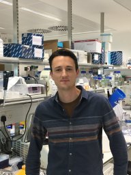 Clift Dean合作论文数Medical Research Council Laboratory of Molecular Biology (MRC LMB), Cambridge CB2 0QH, UK.6Flavia De Lima Alves合作论文数6Eris Duro合作论文数6Olga O. Nerusheva合作论文数5Juan Zou合作论文数Hunan Province Key Laboratory of Tumor Cellular & Molecular Pathology, Cancer Research Institute, University of South China, Hengyang, Hunan 421001, PR China5
 Josefin Fernius合作论文数5Dariusz Abramczyk合作论文数4
 Kevin Corbett合作论文数Department of Chemistry and Biochemistry, Division of Physical Sciences, University of California, San Diego4Menglu Wang合作论文数4
 Kerr Alastair Rw合作论文数Wellcome Trust Centre for Cell Biology;University of Edinburgh;Edinburgh;Wellcome Trust Centre for Cell Biology, University of Edinburgh4
 Charles L. Asbury合作论文数University of Washington4
 Krishna K. Sarangapani合作论文数University of Washington4Colette Fox合作论文数3Bonnie Alver合作论文数3Yi Deng合作论文数3
 Bettina P Mihalas合作论文数University of Edinburgh3Kiburz Brendan M合作论文数3Kwaku N. Opoku合作论文数3
 Weronika Borek合作论文数3
 Andrew McAinsh合作论文数Warwick Medical School, Faculty of Science, Engineering and Medicine, The University of Warwick3
 Jeffery Errington合作论文数School of Medical Sciences, University of Sydney;Institute of Infectious Diseases, Faculty of Medicine and Health, University of Sydney3
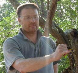 David T Kelly合作论文数University of Sheffield3Sarah M. Richardson合作论文数2Xue Bessie Su合作论文数2
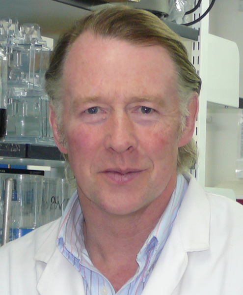 Ronald Hay合作论文数The Division of Molecular, Cell and Developmental Biology, School of Life Sciences, University of Dundee;College of Life Sciences, University of Dundee2Brian H. Lee合作论文数2Stephen McColm合作论文数2Lisa-Marie Kuhl合作论文数2Claudia Schaffner合作论文数2
 Fengji Tan合作论文数2Yisha Luo合作论文数2Adrien Paul-Dubois-Taine合作论文数2Andreas Wallek合作论文数2Yanqun Fan合作论文数2Lucia F. Massari合作论文数2
 Zhouqing Luo (罗周卿)合作论文数School of Life Sciences, Xiamen University2Nadine Vincenten合作论文数2
 Lori B Koch合作论文数Howard Hughes Med Inst2Maria Del Carmen Sanchez-Olmos合作论文数2
 Adan A. Ramirez Rojas合作论文数2Yehui Wu合作论文数2




