Inherited aneuploidy is one of the most common causes of human genetic disease affecting approximately 20% of all human embryos and is the major cause of non-conception in older women. Inherited aneuploidies typically arise from meiotic chromosome segregation errors and are strongly affected by maternal age. Cohesin, a protein complex that mediates cohesion between sister chromatids, becomes depleted from oocyte chromosomes with age in mice, potentially contributing to the high rate of aneuploidy in older mouse oocytes. However, it remains unclear at which stage of oogenesis ageing affects chromatid cohesion in mice and whether ageing similarly affects cohesin in human oocytes. Here we use fluorescence in situ hybridisation to assess at which stages of oogenesis ageing alters chromatid separation in mice and in humans. We show that chromatid separation increases with age while oocytes remain dormant in primordial follicles in both mice and humans and that this effect of age on chromatid separation is detectable in fertile women of child-bearing age. Furthermore, we show that this age-dependent increase in chromatid separation is accompanied by age-dependent depletion of the acetylated SMC3-marked cohesive subpopulation of cohesin from oocytes at the dormant primordial follicle stage in both mice and humans. These data suggest that ageing impacts on oocyte chromosomes while they remain dormant in primordial follicles, and that this aspect of oocyte chromosome ageing is shared in both mice and humans. Improving our understanding of these pathways may allow strategies to slow or prevent this aspect of oocyte ageing to be developed. ### Competing Interest Statement The authors have declared no competing interest. Medical Research Council, https://ror.org/03x94j517, MC\_UU\_00007/6, MC\_UU\_00035/3, MC\_UU\_00035/7, G1100357 Wellcome Trust, 215625 Royal Thai Government, Postgraduate studentship
Mammalian fertility depends on the production of an oocyte capable of fertilization and supporting early embryo development. This requires both cytoplasmic and nuclear, i.e. chromosomal, competence, processes that were initiated decades prior to ovulation. Current demographic changes with delayed motherhood are increasingly in conflict with these biological processes. This brief review highlights the key stages in oocyte development, as well as recent findings that continue to inform on how the oocyte is able to maintain function over such a prolonged period. These include minimizing oocyte damage caused by the production of reactive oxygen species, the importance of intercellular communication with the surrounding somatic cells, and the molecular mechanisms that underpin the fidelity of chromosome cohesion and then separation at the resumption of meiosis. Some of these are already approaching clinical testing and interventions, with new approaches in the coming years potentially being able to 'put back the clock' to improve oocyte quality.
In brief:Although sheep have been widely used as a large animal model for human ovarian biology, unlike women, they display a marked seasonality of breeding activity, the underlying mechanisms and extent of ovarian changes of which remain largely undefined. This study reveals the active remodeling of the ovarian extracellular matrix across the reproductive season, which could be an additional driver responsible for the observed variations in ovarian morphometry and follicle dynamics. Abstract:Ovarian function requires dynamic tissue remodeling provided by its extracellular matrix (ECM). In seasonal breeders, ovaries undergo an additional circannual cycle of recrudescence and regression. While increasing evidence suggests that the ECM impacts normal ovarian cyclicity and function, how its components are remodeled across reproductive seasonality has not been explored in large mammals. Using immunohistological and in vitro experiments, we investigated the influence of reproductive seasonality on ovarian morphometry, ECM properties and follicle developmental potential in vitro. Ovarian weight and volume were reduced during anestrus (P < 0.001). Neither follicular density nor the proportion of preantral follicles and earlier stages of development were impacted by the season, but the percentage of antral follicles increased during anestrus (P = 0.028), while corpora lutea were only present in ovaries collected during the breeding season. Concomitantly, ovarian ECM composition was significantly remodeled, with stromal collagen and fibronectin significantly increased (P < 0.01) and laminin decreased (P = 0.032) during anestrus compared to the breeding season. This correlated with thicker collagen fibers both in the stroma and in the tunica albuginea during anestrus. In vitro, preantral follicles isolated from their native environment exhibited a season-dependent pattern of follicular integrity, survival, antrum formation and growth. These results suggest the establishment of a stiffer ovarian microenvironment during anestrus, which, together with endocrine changes, regulates follicle growth, demise and the ovulatory response.
BACKGROUND:Women are increasingly choosing to delay childbirth, and those with low ovarian reserves indicative of primary ovarian insufficiency are at risk for sub- and infertility and also the early onset of menopause. Experimental strategies that promise to extend the duration of ovarian function in women are currently being developed. One strategy is to slow the rate of loss of existing primordial follicles (PFs), and a second is to increase, or 'boost', the number of autologous PFs in the human ovary. In both cases, the duration of ovarian function would be expected to be lengthened, and menopause would be delayed. This might be accompanied by an extended production of mature oocytes of sufficient quality to extend the fertile lifespan. OBJECTIVE AND RATIONALE:In this work, we consider how slowing physiological ovarian aging might improve the health and well-being of patients, and summarize the current state-of-the-art of approaches being developed. We then use mathematical modeling to determine how interventions are likely to influence the duration of ovarian function quantitatively. Finally, we consider efficacy benchmarks that should be achieved so that individuals will benefit, and propose criteria that could be used to monitor ongoing efficacy in different patients as these strategies are being validated. SEARCH METHODS:Current methods to estimate the size of the ovarian reserve and its relationship to the timing of the menopausal transition and menopause were compiled, and publications establishing methods designed to slow loss of the ovarian reserve or to deliver additional ovarian PFs to patients were identified. OUTCOMES:We review our current understanding of the consequences of reproductive aging in women, and compare different approaches that may extend ovarian function in women at risk for POI. We also provide modeling of primordial reserve decay in the presence of therapies that slow PF loss or boost PF numbers. An interactive online tool is provided that estimates how different interventions would impact the duration of ovarian function across the natural population. Modeling output shows that treatments that slow PF loss would need to be applied as early as possible and for many years to achieve significant delay of menopause. In contrast, treatments that add additional PFs should occur as late as possible relative to the onset of menopause. Combined approaches slowing ovarian reserve loss while also boosting numbers of (new) PFs would likely offer some additional benefits in delaying menopause. WIDER IMPLICATIONS:Extending ovarian function, and perhaps the fertile lifespan, is on the horizon for at least some patients. Modeling ovarian aging with and without such interventions complements and helps guide the clinical approaches that will achieve this goal. REGISTRATION NUMBER:Not applicable.
Aneuploid human eggs (oocytes) are a major cause of infertility, miscarriage, and chromosomal disorders. Such aneuploidies increase greatly as women age, with defective linkages between sister chromatids (cohesion) in meiosis as a common cause. We found that loss of a specific pool of the cohesin protector protein, shugoshin 2 (SGO2), may contribute to this phenomenon. Our data indicate that SGO2 preserves sister chromatid cohesion in meiosis by protecting a "cohesin bridge" between sister chromatids. In human oocytes, SGO2 localizes to both sub-centromere cups and the pericentromeric bridge, which spans the sister chromatid junction. SGO2 normally colocalizes with cohesin; however, in meiosis II oocytes from older women, SGO2 is frequently lost from the pericentromeric bridge and sister chromatid cohesion is weakened. MPS1 and BUB1 kinase activities maintain SGO2 at sub-centromeres and the pericentromeric bridge. Removal of SGO2 throughout meiosis I by MPS1 inhibition reduces cohesion protection, increasing the incidence of single chromatids at meiosis II. Therefore, SGO2 deficiency in human oocytes can exacerbate the effects of maternal age by rendering residual cohesin at pericentromeres vulnerable to loss in anaphase I. Our data show that impaired SGO2 localization weakens cohesion integrity and may contribute to the increased incidence of aneuploidy observed in human oocytes with advanced maternal age.
Turner syndrome (TS) is characterized by the absence of one X chromosome and affects approximately 1 in 2,500 newborn women. Most cases show a mosaicism pattern, i.e., cells with a normal pattern of 46XX mixed with cells of altered karyotype. This is associated with accelerated depletion of follicles: <30% of patients with TS have spontaneous puberty, and premature ovarian insufficiency occurs at a mean (±SD) age of 29 years. Some will conceive naturally, but this may occur in only approximately 5% of women with TS, and pregnancy carries an increased risk of miscarriage and potentially life-threatening complications, notably aortic dissection. This very truncated reproductive lifespan with limited opportunity for having a family is a major concern for girls and women with TS and their parents (1Oktay K. Bedoschi G. Berkowitz K. Bronson R. Kashani B. McGovern P. et al.Fertility preservation in women with Turner syndrome: A comprehensive review and practical guidelines.J Pediatr Adolesc Gynecol. 2016; 29: 409Abstract Full Text Full Text PDF PubMed Scopus (105) Google Scholar). Although oocyte donation is the only established medical therapy to enhance the chances of pregnancy in women with TS and premature ovarian insufficiency, the possibility of ovarian tissue cryopreservation (OTC) and subsequent reimplantation offers a fertility preservation option because this can be performed in childhood or adolescence and provides the possibility to store primordial follicles within the ovarian tissue before their premature disappearance. It is generally accepted that OTC is an option mostly suited to patients with mosaic TS rather than those with monosomic karyotypes because patients with mosaic TS are more likely to have a greater number of follicles present (1Oktay K. Bedoschi G. Berkowitz K. Bronson R. Kashani B. McGovern P. et al.Fertility preservation in women with Turner syndrome: A comprehensive review and practical guidelines.J Pediatr Adolesc Gynecol. 2016; 29: 409Abstract Full Text Full Text PDF PubMed Scopus (105) Google Scholar, 2Mamsen L.S. Charkiewicz K. Anderson R.A. Telfer E.E. McLaughlin M. Kelsey T.W. et al.Characterization of follicles in girls and young women with Turner syndrome who underwent ovarian tissue cryopreservation.Fertil Steril. 2019; 111: 1217Abstract Full Text Full Text PDF PubMed Scopus (48) Google Scholar). However, the patient's karyotype is generally determined from peripheral blood lymphocytes and may not accurately reflect all tissues or indeed all cell types within a tissue, such as the ovary. It is known that granulosa cells in TS ovarian tissue can be monosomic (45X), although surrounding oocytes have the normal complement of two X chromosomes. However, it is not known how common this might be or how this somatic cell composition affects oocyte and follicle development. This study by Peek et al. (3Peek R. Nadesapillai S. Thi Nguyen T.Y. Vassart S. Smeets D. van de Zande G. et al.Assessment of folliculogenesis in ovarian tissue from young patients with Turner syndrome using a murine xenograft model.Fertil Steril. 2023; 120: 371-381Abstract Full Text Full Text PDF Scopus (1) Google Scholar), published in Fertility and Sterility, examines the impact of aneuploid granulosa and stromal cells on the development of immature ovarian follicles containing normal oocytes using a mouse xenograft model with ovarian tissue from patients with mosaic TS. Tissue from 18 patients with mosaic TS aged 5–19 years and 13 similarly aged control patients was prepared and xenografted for five months (3Peek R. Nadesapillai S. Thi Nguyen T.Y. Vassart S. Smeets D. van de Zande G. et al.Assessment of folliculogenesis in ovarian tissue from young patients with Turner syndrome using a murine xenograft model.Fertil Steril. 2023; 120: 371-381Abstract Full Text Full Text PDF Scopus (1) Google Scholar). Using fluorescence in situ hybridization on histologic sections to detect X chromosomes and chromosome 18 as a somatic control, samples taken from nongrafted TS tissue revealed that 97% of oocytes examined were XX but surrounded by predominantly aneuploid (monosomy X) somatic cells. After grafting, it was demonstrated that the composition of the somatic cells did not significantly affect follicle development. Follicles at all stages of development, including antral stages, were present after grafting, but interestingly, the proportion of monosomy X granulosa cells within the growing follicles was significantly reduced, suggesting that as the granulosa cells proliferate, 46XX cells preferentially divide; this is consistent with evidence that 45X cells are more susceptible to apoptosis and have a longer cell cycle (3Peek R. Nadesapillai S. Thi Nguyen T.Y. Vassart S. Smeets D. van de Zande G. et al.Assessment of folliculogenesis in ovarian tissue from young patients with Turner syndrome using a murine xenograft model.Fertil Steril. 2023; 120: 371-381Abstract Full Text Full Text PDF Scopus (1) Google Scholar). This is novel and important information that will be valuable to factor into the analysis of tissue before transplantation. These observations will form the basis of further detailed investigations on how somatic cell composition affects cell communication, the regulation of proliferation and differentiation of these cells, and oocyte quality. The investigators also commented on reduced antimüllerian hormone (AMH) expression (by immunohistochemistry) in granulosa cells from these TS samples, although it increased toward control levels as follicles grew. This observation is at variance with increased AMH concentrations in follicular fluid detected in some TS samples (2Mamsen L.S. Charkiewicz K. Anderson R.A. Telfer E.E. McLaughlin M. Kelsey T.W. et al.Characterization of follicles in girls and young women with Turner syndrome who underwent ovarian tissue cryopreservation.Fertil Steril. 2019; 111: 1217Abstract Full Text Full Text PDF PubMed Scopus (48) Google Scholar) but may reflect analysis of different stages of follicular development. This may be important to consider in the assessment of the potential suitability of such patients for OTC (1Oktay K. Bedoschi G. Berkowitz K. Bronson R. Kashani B. McGovern P. et al.Fertility preservation in women with Turner syndrome: A comprehensive review and practical guidelines.J Pediatr Adolesc Gynecol. 2016; 29: 409Abstract Full Text Full Text PDF PubMed Scopus (105) Google Scholar) as it suggests that the relationship between serum AMH and follicle number may differ from that in normal ovaries. Ultimately, the key to the success of OTC and reimplantation is the number of follicles present at the time of tissue cryopreservation. This article highlights the variability in follicle numbers and the correlation with pubertal stage, and the investigators argue that OTC should be performed before puberty to optimize success. A previous study published in Fertility and Sterility by Mamsen et al. (2Mamsen L.S. Charkiewicz K. Anderson R.A. Telfer E.E. McLaughlin M. Kelsey T.W. et al.Characterization of follicles in girls and young women with Turner syndrome who underwent ovarian tissue cryopreservation.Fertil Steril. 2019; 111: 1217Abstract Full Text Full Text PDF PubMed Scopus (48) Google Scholar) analyzed 15 patients with TS aged 5–22 years who had undergone OTC and showed that 9 of 15 ovarian sample biopsies contained ovarian follicles. Furthermore, where follicles were present, many showed abnormal morphology, which is likely to limit their development. It would be interesting to know whether some of the unusual morphology observed in the Mamsen et al. (2Mamsen L.S. Charkiewicz K. Anderson R.A. Telfer E.E. McLaughlin M. Kelsey T.W. et al.Characterization of follicles in girls and young women with Turner syndrome who underwent ovarian tissue cryopreservation.Fertil Steril. 2019; 111: 1217Abstract Full Text Full Text PDF PubMed Scopus (48) Google Scholar) article was linked to the ploidy composition of the somatic cells; this article by Peek et al. (3Peek R. Nadesapillai S. Thi Nguyen T.Y. Vassart S. Smeets D. van de Zande G. et al.Assessment of folliculogenesis in ovarian tissue from young patients with Turner syndrome using a murine xenograft model.Fertil Steril. 2023; 120: 371-381Abstract Full Text Full Text PDF Scopus (1) Google Scholar) provides a good base for these types of analyses. Although OTC is increasingly performed on young girls with TS, its effectiveness and efficiency in this patient group remain unknown, with no successful pregnancies reported. In a series of 100 patients with TS considering OTC, the majority chose to have the procedure, but only two autotransplantations were recorded, and neither achieved restoration of endocrine ovarian function (4Rodriguez-Wallberg K.A. Sergouniotis F. Nilsson H.P. Lundberg F.E. Trends and outcomes of fertility preservation for girls, adolescents and young adults with Turner syndrome: A prospective cohort study.Front Endocrinol. 2023; 14113524Crossref PubMed Scopus (2) Google Scholar). This contrasts with the very high rate of restoration of ovarian function after OTC and replacement in women who have normal ovaries but are facing exogenous gonadotoxic treatment, generally for cancer. That situation differs markedly from conditions such as TS, where the pathological process is within the ovary and will therefore continue when the cryopreserved ovarian tissue is replaced, with the further insults of cryopreservation and engraftment (the timepoint when most follicles are lost from normal ovaries) superimposed. Current guidelines therefore regard as "research only" the clinical application of OTC for fertility preservation in conditions such as TS (5Anderson R.A. Amant F. Braat D. D'Angelo A. Chuva de Sousa Lopes S.M. et al.ESHRE Guideline Group on Female Fertility PreservationESHRE guideline: Female fertility preservation.Hum Reprod Open. 2020; 2020hoaa052Crossref Google Scholar). There are few studies focused on detailing the developmental potential of this tissue, and other options to use this tissue may also need to be explored, such as harvesting immature oocytes (2Mamsen L.S. Charkiewicz K. Anderson R.A. Telfer E.E. McLaughlin M. Kelsey T.W. et al.Characterization of follicles in girls and young women with Turner syndrome who underwent ovarian tissue cryopreservation.Fertil Steril. 2019; 111: 1217Abstract Full Text Full Text PDF PubMed Scopus (48) Google Scholar) and in vitro growth of immature follicles within this tissue. This study by Peek et al. (3Peek R. Nadesapillai S. Thi Nguyen T.Y. Vassart S. Smeets D. van de Zande G. et al.Assessment of folliculogenesis in ovarian tissue from young patients with Turner syndrome using a murine xenograft model.Fertil Steril. 2023; 120: 371-381Abstract Full Text Full Text PDF Scopus (1) Google Scholar) advances the field and provides important information that should be added to the decision-making process for fertility preservation in girls and young women with TS.
Turner syndrome (TS) leads to a characteristic phenotype, including premature ovarian insufficiency and infertility. Ovarian tissue cryopreservation (OTC) is becoming an established fertility preservation strategy for both pre- and post-pubertal females and may offer the chance of having a biological family to selected patients with TS. To date, women with TS have had ovarian tissue cryopreserved but there are few reports of autologous re-implantation and none of pregnancy. We herein report, to our knowledge, the first clinical pregnancy in a patient with TS, conceived naturally following re-implantation of cryopreserved ovarian tissue which had been removed soon after spontaneous puberty. This provides proof of concept for OTC as a means of fertility preservation in TS.
Deciphering novel functions of anti-Müllerian hormone (AMH).
Mammalian eggs (oocytes) are formed during fetal life and establish associations with somatic cells to form primordial follicles that create a store of germ cells (the primordial pool). The size of this pool is influenced by key events during the formation of germ cells and by factors that influence the subsequent activation of follicle growth. These regulatory pathways must ensure that the reserve of oocytes within primordial follicles in humans lasts for up to 50 years, yet only approximately 0.1% will ever be ovulated with the rest undergoing degeneration. This review outlines the mechanisms and regulatory pathways that govern the processes of oocyte and follicle formation and later growth, within the ovarian stroma, through to ovulation with particular reference to human oocytes/follicles. In addition, the effects of aging on female reproductive capacity through changes in oocyte number and quality are emphasized, with both the cellular mechanisms and clinical implications discussed. Finally, the details of current developments in culture systems that support all stages of follicle growth to generate mature oocytes in vitro and emerging prospects for making new oocytes from stem cells are outlined.
STUDY QUESTION How does in vitro culture alter the human ovarian cortical extracellular matrix (ECM) network structure? SUMMARY ANSWER The ECM composition and architecture vary in the different layers of the ovarian cortex and are remodelled during in vitro culture. WHAT IS KNOWN ALREADY The ovarian ECM is the scaffold within which follicles and stromal cells are organized. Its composition and structural properties constantly evolve to accommodate follicle development and expansion. Tissue preparation for culture of primordial follicles within the native ECM involves mechanical loosening; this induces undefined modifications in the ECM network and alters cell-cell contact, leading to spontaneous follicle activation. STUDY DESIGN, SIZE, DURATION Fresh ovarian cortical biopsies were obtained from six women aged 28-38 years (mean +/- SD: 32.7 +/- 4.1 years) at elective caesarean section. Biopsies were cut into fragments of similar to 4 x 1 x 1 mm and cultured for 0, 2, 4, or 6 days (D). PARTICIPANTS/MATERIALS, SETTING, METHODS Primordial follicle activation, stromal cell density, and ECM-related protein (collagen, elastin, fibronectin, laminin) positive area in the entire cortex were quantified at each time point using histological and immunohistological analysis. Collagen and elastin content, collagen fibre characteristics, and follicle distribution within the tissue were further quantified within each layer of the human ovarian cortex, namely the outer cortex, the mid-cortex, and the cortex-medulla junction regions. MAIN RESULTS AND THE ROLE OF CHANCE Primordial follicle activation occurred concomitantly with a loosening of the ovarian cortex during culture, characterized by an early decrease in stromal cell density from 3.6 +/- 0.2 x 10(6) at day 0 (D0) to 2.8 +/- 0.1 x 10(6) cells/mm(3) at D2 (P = 0.033) and a dynamic remodelling of the ECM. Notably, collagen content gradually fell from 55.5 +/- 1.7% positive area at D0 to 42.3 +/- 1.1% at D6 (P = 0.001), while elastin increased from 1.1 +/- 0.2% at D0 to 1.9 +/- 0.1% at D6 (P = 0.001). Fibronectin and laminin content remained stable. Moreover, collagen and elastin distribution were uneven throughout the cortex and during culture. Analysis at the sub-region level showed that collagen deposition was maximal in the outer cortex and the lowest in the mid-cortex (69.4 +/- 1.2% versus 53.8 +/- 0.8% positive area, respectively, P < 0.0001), and cortical collagen staining overall decreased from D0 to D2 (65.2 +/- 2.4% versus 60.6 +/- 1.8%, P = 0.033) then stabilized. Elastin showed the converse distribution, being most concentrated at the cortex-medulla junction (3.7 +/- 0.6% versus 0.9 +/- 0.2% in the outer cortex, P < 0.0001), and cortical elastin peaked at D6 compared to D0 (3.1 +/- 0.5% versus 1.3 +/- 0.2%, P < 0.0001). This was corroborated by a specific signature of the collagen fibre type across the cortex, indicating a distinct phenotype of the ovarian cortical ECM depending on region and culture period that might be responsible for the spatio-temporal and developmental pattern of follicular distribution observed within the cortex. LARGE SCALE DATA N/A. LIMITATIONS, REASONS FOR CAUTION Ovarian cortical biopsies were obtained from women undergoing caesarean sections. As such, the data obtained may not accurately reflect the ECM distribution and structure of non-pregnant women. WIDER IMPLICATIONS OF THE FINDINGS Clarifying the composition and architecture signature of the human ovarian cortical ECM provides a foundation for further exploration of ovarian microenvironments. It is also critical for understanding the ECM-follicle interactions regulating follicle quiescence and awakening, leading to improvements in both in vitro activation and in vitro growth techniques. STUDY FUNDING/COMPETING INTEREST(S) Medical Research Council grant MR/R003246/1 and Wellcome Trust Collaborative Award in Science: 215625/Z/19/Z. The authors have no conflicts to declare.
The amino acid metabolism of bovine follicles during in vitro growth (IVG) was evaluated to identify potential indicators of health during culture. The bovine ovarian cortex was sliced, prepared as strips, and cultured for 6 days. Tissue samples were examined histologically before and after 6 days of culture, and the degree of follicle activation was classified as either high or low based on the number of growing secondary follicles present (high: 7~11; low: 0~1). In a separate experiment, secondary follicles (diameter range: 100~200 μm) were manually isolated and cultured, and their growth was monitored for 6 days. Cultured follicles were classified as growth or degenerate based on diameter change during culture (growth: +60.5~74.1 μm; degenerate: −28~15.2 μm). Free amino acids and their metabolites were measured in the spent culture medium from each group. In cultured ovarian cortical strips, the concentration of α-aminoadipic acid was significantly higher in the low activation group than in the high group (p < 0.05), while those of methionine, lysine, and arginine were higher in the high activation group. In cultured isolated secondary follicles, concentrations of methionine, tyrosine, histidine, and hydroxyproline were higher in the degenerate group (p ≤ 0.05). In conclusion, amino acid metabolism has the potential to serve as an indicator of primordial follicle activation and subsequent growth rate during bovine IVG.
STUDY QUESTION: What are the effects of cyclophosphamide exposure on the human ovary and can anti-Mullerian hormone (AMH) and rapamycin protect against these?SUMMARY ANSWER: Exposure to cyclophosphamide compromises the health of primordial and transitional follicles in the human ovarian cortex and upregulates PI3K signalling, indicating both direct damage and increased follicular activation; AMH attenuates both of these chemotherapy-induced effects, while rapamycin attenuates only PI3K signalling upregulation.WHAT IS KNOWN ALREADY: Studies primarily in rodents demonstrate that cyclophosphamide causes direct damage to primordial follicles or that the primordial follicle pool is depleted primarily through excessive initiation of follicle growth. This increased follicular activation is mediated via upregulated PI3K signalling and/or reduced local levels of AMH production due to lost growing follicles. Furthermore, while rodent data show promise regarding the potential benefits of inhibitors/protectants alongside chemotherapy treatment to preserve female fertility, there is no information about the potential for this in humans.STUDY DESIGN, SIZE, DURATION: Fresh ovarian cortical biopsies were obtained from 17 healthy women aged 21-41 years (mean +/- SD: 31.8 +/- 4.9 years) at elective caesarean section. Biopsies were cut into small fragments and cultured for 24 h with either vehicle alone (DMSO), the active cyclophosphamide metabolite 4-hydroperoxycyclophosphamide (4-HC) alone, 4-HC + rapamycin or 4-HC+AMH. Two doses of 4-HC were investigated, 0.2 and 2 mu M in separate experiments, using biopsies from seven women (aged 27-41) and six women (aged 21-34), respectively. Biopsies from four women (aged 28-38) were used to investigate the effect of rapamycin or AMH only.PARTICIPANTS/MATERIALS, SETTING, METHODS: Histological analysis of ovarian tissue was undertaken for follicle staging and health assessment. Western blotting and immunostaining were used to assess activation of PI3K signalling by measuring phosphorylation of AKT and phosphorylated FOXO3A staining intensity, respectively.MAIN RESULTS AND THE ROLE OF CHANCE Exposure to either dose of 4-HC caused an increase in the proportion of unhealthy primordial (P < 0.0001, both doses) and transitional follicles (P < 0.01 for low dose and P < 0.01 for high dose) compared to vehicle. AMH significantly reduced follicle damage by approximately half in both of the investigated doses of 4-HC (P < 0.0001), while rapamycin had no protective effect on the health of the follicles. Culture with AMH or rapamycin alone had no effect on follicle health. Activation of PI3K signalling following 4-HC exposure was demonstrated by both Western blotting data showing that 4-HC increased in AKT phosphorylation and immunostaining showing increased phosphorylated FOXO3A staining of non-growing oocytes. Treatment with rapamycin reduced the activation of PI3K signalling in experiments with low doses of 4-HC while culture with AMH reduced PI3K activation (both AKT phosphorylation and phosphorylated FOXO3A staining intensity) across both doses investigated.LIMITATIONS, REASONS FOR CAUTION: These in vitro studies may not replicate in vivo exposures. Furthermore, longer experiment durations are needed to determine whether the effects observed translate into irreparable deficits of ovarian follicles.WIDER IMPLICATIONS OF THE FINDINGS: These data provide a solid foundation on which to explore the efficacy of AMH in protecting non-growing ovarian follicles from gonadotoxic chemotherapies. Future work will require consideration of the sustained effects of chemotherapy treatment and potential protectants to ensure these agents do not impair the developmental competence of oocytes or lead to the survival of oocytes with accumulated DNA damage, which could have adverse consequences for potential offspring.STUDY FUNDING/COMPETING INTEREST(S): This work was supported by grants from TENOVUS Scotland, the Academy of Medical Sciences (to R.R.), the Medical Research Council (G1100357 to R.A.A., MR/N022556/1 to the MRC Centre for Reproductive Health), and Merck Serono UK (to R.A.A.). R.R., H.L.S., N.S., and E.E.T. declare no conflicts of interest. R.A.A. reports grants and personal fees from Roche Diagnostics and Ferring Pharmaceuticals, and personal fees from IBSA and Merck outside the submitted work.
Androgens are essential in normal ovarian function and follicle health, but hyperandrogenism, as seen in polycystic ovary syndrome, is associated with disordered follicle development. There are few data on the effect of long-term exposure to high levels of testosterone as found in transgender men receiving gender-affirming endocrine therapy. In this study, we investigate the effect of testosterone on the development, morphological health and DNA damage and repair capacity of human ovarian follicles in vivo and their survival in vitro. Whole ovaries were obtained from transgender men (mean age: 27.6 ± 1.7 years; range: 20–34 years, n = 8) at oophorectomy taking pre-operative testosterone therapy. This was compared to cortical biopsies from age-matched healthy women obtained at caesarean section (mean age: 31.8 ± 1.5 years; range: 25–35 years, n = 8). Cortical tissues were dissected into fragments and either immediately fixed for histological analysis or cultured for 6 days and subsequently fixed. Follicle classification and morphological health were evaluated from histological sections stained with hematoxylin and eosin and expression of γH2AX as a marker of DNA damage by immunohistochemistry (IHC). In uncultured tissue, testosterone exposure was associated with reduced follicle growth activation, poor follicle health and increased DNA damage. After 6 days of culture, there was enhanced follicle activation compared to the control with further deterioration in morphological health and increased DNA damage. These data indicate that high circulating concentrations of testosterone have effects on the primordial and small-growing follicles of the ovary. These results may have implications for transgender men receiving gender-affirming therapy prior to considering pregnancy or fertility preservation measures. Lay summary As part of gender transitioning, transgender men take testosterone therapy. While androgens like testosterone are essential to maintain ovarian health, the effects of long-term testosterone treatment on the ovary are unclear. This study examines whether testosterone impacts ovarian follicle growth activation, follicle health and whether it causes DNA damage. It also looks at how well these follicles grow in tissue culture. The results showed there was a higher proportion of non-growing ovarian follicles in the ovaries of trans men, they appeared less healthy and there were higher levels of DNA damage. After 6 days of tissue culture, there were more growing follicles in transgender ovaries compared to control, but follicle health further deteriorated and there are increased levels of DNA damage. These results identify new effects of testosterone on the ovary and highlight the importance of discussing fertility preservation options prior to starting testosterone.
Abstract Study question Does gender-affirming testosterone therapy alter the composition of the extra-cellular matrix (ECM) within the ovarian stroma and subsequently affect follicle activation in vivo Summary answer Ovarian stroma of trans men is more collagenous and less elastic, indicating fibrotic change. This may affect in vivo follicle growth activation What is known already Changes in the ovarian stroma have been demonstrated in the ovaries of transgender men taking testosterone, including thickening of the tunica albuginea, stromal cell hyperplasia and stromal cell luteinisation. Ovaries of trans men also have increased cortical stiffness. These changes are similar to those seen in female patients with PCOS and in physiological ovarian aging, which has been attributed to accumulation of collagen in the ECM. Increasing stiffness of the supportive follicular microenvironment has been shown to reduce follicle growth activation in vitro Study design, size, duration Whole ovaries were obtained from transgender men (mean age 27.6 ± 1.7 years, n = 8) with informed consent at oophorectomy. All patients had received 1000mg testosterone undecanoate intramuscularly at 12-16 week intervals for a minimum of 18 months pre-operatively (range 18 months-10 years). Cortical tissue was dissected into small fragments (≈1x1x0.5mm) and fixed for histological and immunohistochemical analysis. Testosterone-treated ovaries were compared to cortical biopsies from age-matched healthy women obtained at caesarean section (mean age 31.8±1.5, n = 8). Participants/materials, setting, methods Follicle number, classification of developmental stage, non-growing follicle density (NGFD) and stromal cell density were evaluated by histological analysis of ovarian cortical tissue. Sections were stained with Picrosirius red (PSR) to analyse total collagen content using brightfield microscopy. Polarised light was also used to analyse the collagen birefringence, which allows quantification of collagen fibre thickness into thick, medium or thin. Total elastin content was evaluated using immunofluorescence. Main results and the role of chance 4526 follicles were analysed. Transgender ovary showed a higher proportion of non-growing follicles found compared to control (93.9±1.2% vs 84.6±1.5% p < 0.05): the proportions of primary (4.7±0.9% vs 10.6±1.5%, p = 0.2) and secondary (1.4±0.4% vs 4.6±0.7%, p = 0.1) follicles tended to be lower. Stromal cell density was significantly higher in transgender ovarian cortex than control (2.5±0.1 x10⁶cells/mm3 vs 1.7±0.1 x10⁶cells/mm3), indicating stromal cell hyperplasia. Combined data from control and transgender groups showed a positive correlation between NGFD and stromal density (r = 0.64, p = 0.01). Transgender ovary had a higher total collagen content (77.2±1.2%) compared to control (31.3±3.3%, p < 0.005). Analysis of collagen birefringence showed that transgender ovaries had similar quantities of thick collagen fibres (0.014±0.005 vs 0.010±0.009, p = 0.1), more medium thickness collagen fibres (45.1±6.6%vs 14.4±4.9%, p < 0.05) and fewer thinner fibres (41.5±9.6% vs 27.7±2.8%, p = 0.08) than control. The total elastin content in transgender ovaries was lower than control (1.3±0.1% vs 3.6±0.6%, p < 0.005) and subsequently, the collagen/elastin ratio was significantly higher (63.1±7.9 vs 10±1.3, p < 0.005). Limitations, reasons for caution The impact of these findings on in vivo follicle growth are unclear. The effect of duration of testosterone treatment has not investigated. Wider implications of the findings More collagenous, less elastic ovarian stroma in trans men indicates fibrotic change; these findings are similar to women with PCOS and with reproductive ageing. These stromal changes may alter follicle growth activation and may contribute value to our understanding of the regulation of follicle function in a range of conditions. Trial registration number nil
Despite centuries of lessons from history, war endures. Across Earth, during nearly every year from the beginning of the twentieth century to present day, over 30 wars have been fought resulting in 187 million casualties, excluding the most recent conflict, which is the impetus for this essay (Timeline of 20th and 21st century wars). We are, sadly, a war-mongering people. The word “war” word infiltrates our vernacular, e.g., the war on poverty, on drugs, on cancer, on COVID, and, apropos, on terror. How did rational approaches to disagreement and conflict evade the world’s progress? Reproductive physicians and scientists are dedicated to safeguard lives and build families. Violence is antithetical to our mission as professionals, and moral integrity as humans. We are deeply concerned for, and stand in unity with, our Ukrainian colleagues—the embryologists, scientists, OBGYN and REI physicians, infertility patients, and all people under siege. Reproductive health services for Ukrainians (as with many other war-torn regions) have collapsed. Deeply disturbing reports have emerged that cite civilian hospitals (including maternity centers) being targeted. Liquid nitrogen supplies are scarce. Pregnant mothers and gestational carriers are at emergent risk of delivering in extremely harsh conditions, cold underground bunkers and refugee queues.
 Richard Anderson合作论文数Centre for Reproductive Health, Institute for Regeneration and Repair, The University of Edinburgh58
 Marie McLaughlin合作论文数43
 W. H. B. Wallace合作论文数Department of Haematology/Oncology, Royal Hospital for Sick Children12R.G. Gosden合作论文数11Dunlop Cheryl E合作论文数9
 Norah Spears合作论文数Centre for Discovery Brain Sciences, Edinburgh Medical School,The University of Edinburgh9
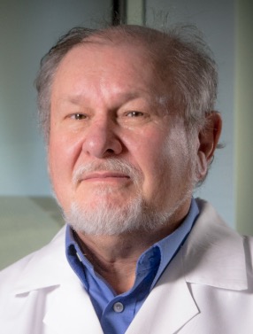 David Albertini合作论文数Hacettepe Üni̇versi̇tesi̇;Bedford Research Foundation;Marine Biological Laboratory Corporation8
 Claus Yding Andersen合作论文数Laboratory of Reproductive Biology, Juliane Marie Center for Children,;Women and Reproduction, University Hospital of Copenhagen;Faculty of Health Science, University of Copenhagen6Fiona H. Thomas合作论文数6Rosario Roseanne合作论文数5
 Jonathan L. Tilly合作论文数Northeastern University5
 Maidarti Mila合作论文数5Webb Robert合作论文数5B. K. Campbell合作论文数5
 Johanne Grosbois合作论文数Institute of Cell Biology, University of Edinburgh4Kirsty Walters合作论文数School of Medicine, Faculty of Medicine and Health, The University of Sydney4Woods Dori C合作论文数Dept Biol, Northeastern Univ4Clarkson Yvonne L合作论文数4Malcolm Faddy合作论文数Faculty of Science, School of Mathematical Sciences, Queensland University of Technology4
 David Baird合作论文数Centre for Reproductive Health, Institute for Regeneration and Repair, The University of Edinburgh4
 Roger Gosden合作论文数Biology Department, College of Arts & Sciences, William & Mary4S. Zahra合作论文数3C TORRANCE合作论文数3
 Joshua Johnson合作论文数3
 Thomas W Kelsey合作论文数3McNatty Kenneth P合作论文数3
 Linn Salto Mamsen合作论文数University Hospital of Copenhagen, University of Copenhagen3Martin Waterfall合作论文数3Grieve Kelsey合作论文数3Anne Grete Byskov合作论文数3
 David G. Armstrong合作论文数Keck School of Medicine, University of Southern California3J.P. Binnie合作论文数3
 Kok Joo Thong合作论文数3Helen M. Picton合作论文数3
 Outi Hovatta合作论文数Department of Clinical Science, Intervention and Technology, Karolinska Institutet3Tom Kelsey合作论文数School of Computer Science|University of St Andrews3E Bailie合作论文数2salina yahya saddick合作论文数2R Leask合作论文数2Ellen Drew合作论文数2Vanessa R Kay合作论文数2Janelle Luk合作论文数2Anne McLaren合作论文数2
 Aleksandar Jovanovic合作论文数University of Nicosia Medical School2
 Paul Skehel合作论文数Centre for Discovery Brain Sciences, College of Medicine and Veterinary Medicine,The University of Edinburgh2
 Hugh Clarke合作论文数Division of Experimental Medicine, Department of Medicine, Faculty of Medicine and Health Sciences, McGill University2
 Pasquale Patrizio合作论文数Department of Obstetrics, Gynecology & Reproductive Sciences, University of Miami Health Systems2Sukhodub Andriy合作论文数2
 Mary B Zelinski合作论文数2




