Functional Magnetic Resonance Imaging (fMRI) in rodents is pivotal for understanding the mechanisms underlying Blood Oxygen Level-Dependent (BOLD) signals and phenotyping animal models of disorders, among other applications. Despite its growing use, comparing rodent fMRI results across different research sites remains challenging due to variations in experimental protocols. Here, we aggregated and analyzed 22 sensory-evoked rat fMRI datasets from 12 imaging centers, totaling scans from 220 rats, to get a snapshot of the current acquisitions in the field. This retrospective analysis highlights common practices and parameters to inform future cross-laboratory standardization efforts. We applied a standardized preprocessing pipeline and evaluated the impact of different hemodynamic response function models on group- and individual-level activity patterns. Our analysis revealed inter-dataset variability attributed to differences in experimental design, anesthesia protocols, and imaging parameters. We identified robust activation clusters in all (22/22) datasets. The comparison between stock human models implemented in software and rat-specific models showed significant variations in the resulting statistical maps. Our findings emphasize the necessity for standardized protocols and collaborative efforts to improve the reproducibility and reliability of rodent fMRI studies. We provide open access to all datasets and analysis code to foster transparency and further research in the field.
Hemispheric brain asymmetry is a basic organizational principle of the human brain and has been implicated in various psychiatric conditions, including autism spectrum disorder. Brain asymmetry is not a uniquely human feature and is observed in other species such as the mouse. Yet, asymmetry patterns are generally nuanced, and substantial sample sizes are required to detect these patterns. In this pre-registered study, we use a mouse dataset from the Province of Ontario Neurodevelopmental Network, which comprises structural MRI data from over 2000 mice, including genetic models for autism spectrum disorder, to reveal the scope and magnitude of hemispheric asymmetry in the mouse. Our findings demonstrate the presence of robust hemispheric asymmetry in the mouse brain, such as larger right hemispheric volumes towards the anterior pole and larger left hemispheric volumes toward the posterior pole, opposite to what has been shown in humans. This suggests the existence of species-specific traits. Further clustering analysis identified distinct asymmetry patterns in autism spectrum disorder models, a phenomenon that is also seen in atypically developing participants. Our study shows potential for the use of mouse models to understand the biological bases of typical and atypical brain asymmetry but also warrants caution as asymmetry patterns seem to differ between humans and mice.
Magnetic resonance imaging (MRI) is a valuable tool for studying brain structure and function in animal and clinical studies. With the growth of public MRI repositories, access to data has finally become easier. However, filtering large datasets for potential poor-quality outliers can be a challenge. We present AIDAqc, a machine-learning-assisted automated Python-based command-line tool for small animal MRI quality assessment. Quality control features include signal-to-noise ratio (SNR), temporal SNR, and motion. All features are automatically calculated and no regions of interest are needed. Automated outlier detection for a given dataset combines the interquartile range and the machine-learning methods one-class support vector machine, isolation forest, local outlier factor, and elliptic envelope. To evaluate the reliability of individual quality control metrics, a simulation of noise (Gaussian, salt and pepper, speckle) and motion was performed. In outlier detection, single scans with induced artifacts were successfully identified by AIDAqc. AIDAqc was challenged in a large heterogeneous dataset collected from 19 international laboratories, including data from mice, rats, rabbits, hamsters, and gerbils, obtained with different hardware and at different field strengths. The results show that the manual inter-rater agreement (mean Fleiss Kappa score 0.17) is low when identifying poor-quality data. A direct comparison of AIDAqc results, therefore, showed only low-to-moderate concordance. In a manual post hoc validation of AIDAqc output, precision was high (>70%). The outlier data can have a significant impact on further postprocessing, as shown in representative functional and structural connectivity analysis. In summary, this pipeline optimized for small animal MRI provides researchers with a valuable tool to efficiently and effectively assess the quality of their MRI data, which is essential for improved reliability and reproducibility.
Task-free functional connectivity in animal models provides an experimental framework to examine connectivity phenomena under controlled conditions and allows for comparisons with data modalities collected under invasive or terminal procedures. Currently, animal acquisitions are performed with varying protocols and analyses that hamper result comparison and integration. Here we introduce StandardRat, a consensus rat functional magnetic resonance imaging acquisition protocol tested across 20 centers. To develop this protocol with optimized acquisition and processing parameters, we initially aggregated 65 functional imaging datasets acquired from rats across 46 centers. We developed a reproducible pipeline for analyzing rat data acquired with diverse protocols and determined experimental and processing parameters associated with the robust detection of functional connectivity across centers. We show that the standardized protocol enhances biologically plausible functional connectivity patterns relative to previous acquisitions. The protocol and processing pipeline described here is openly shared with the neuroimaging community to promote interoperability and cooperation toward tackling the most important challenges in neuroscience. The authors pooled resources to identify best practices and develop a new standardized protocol for estimating functional connectivity in rats with magnetic resonance imaging.
Multiple molecular pathways and cellular processes have been implicated in the neurobiology of autism and other neurodevelopmental conditions. There is a current focus on synaptic gene conditions, or synaptopathies, which refer to clinical conditions associated with rare genetic variants disrupting genes involved in synaptic biology. Synaptopathies are commonly associated with autism and developmental delay and may be associated with a range of other neuropsychiatric outcomes. Altered synaptic biology is suggested by both preclinical and clinical studies in autism based on evidence of differences in early brain structural development and altered glutamatergic and GABAergic neurotransmission potentially perturbing excitatory and inhibitory balance. This review focusses on the NRXN-NLGN-SHANK pathway, which is implicated in the synaptic assembly, trans-synaptic signalling, and synaptic functioning. We provide an overview of the insights from preclinical molecular studies of the pathway. Concentrating on NRXN1 deletion and SHANK3 mutations, we discuss emerging understanding of cellular processes and electrophysiology from induced pluripotent stem cells (iPSC) models derived from individuals with synaptopathies, neuroimaging and behavioural findings in animal models of Nrxn1 and Shank3 synaptic gene conditions, and key findings regarding autism features, brain and behavioural phenotypes from human clinical studies of synaptopathies. The identification of molecular-based biomarkers from preclinical models aims to advance the development of targeted therapeutic treatments. However, it remains challenging to translate preclinical animal models and iPSC studies to interpret human brain development and autism features. We discuss the existing challenges in preclinical and clinical synaptopathy research, and potential solutions to align methodologies across preclinical and clinical research. Bridging the translational gap between preclinical and clinical studies will be necessary to understand biological mechanisms, to identify targeted therapies, and ultimately to progress towards personalised approaches for complex neurodevelopmental conditions such as autism.
Task-free functional connectivity in animal models provides an experimental framework to examine connectivity phenomena under controlled conditions and allows comparison with invasive or terminal procedures. To date, animal acquisitions are performed with varying protocols and analyses that hamper result comparison and integration. We introduce StandardRat , a consensus rat functional MRI acquisition protocol tested across 20 centers. To develop this protocol with optimized acquisition and processing parameters, we initially aggregated 65 functional imaging datasets acquired in rats from 46 centers. We developed a reproducible pipeline for the analysis of rat data acquired with diverse protocols and determined experimental and processing parameters associated with a more robust functional connectivity detection. We show that the standardized protocol enhances biologically plausible functional connectivity patterns, relative to pre-existing acquisitions. The protocol and processing pipeline described here are openly shared with the neuroimaging community to promote interoperability and cooperation towards tackling the most important challenges in neuroscience.
 Joanes Grandjean合作论文数6
 J.R. Homberg (Judith)合作论文数Faculty of Medical Sciences, Radboud University Medical Center;Department of Cognitive Neuroscience, Group Behavioural Neurogenetics, Donders Institute for Brain, Cognition, and Behaviour, Radboud University Medical Centre5
 Ileana O Jelescu合作论文数Center for Biomedical Imaging, New York University Langone Medical Center4
 Guadalupe Soria合作论文数Institut d'Investigatigacions Biomèdiques Agust Pi i Sunyer, Institut d'Investigacions Biomèdiques de Barcelona4
 Eugene Kim合作论文数3
 Patricia Wenk合作论文数3Yujian Diao合作论文数3Henning M Reimann合作论文数3Sandra Strobelt合作论文数3Xin Yu合作论文数3Roel M Vrooman合作论文数3Ho Sung Lee合作论文数3Johannes Kaesser合作论文数3Danielle J Houwing合作论文数3
 Henriette Lambers合作论文数3Zhang Baogui (张宝贵)合作论文数3Gabriel Desrosiers-Gregoire合作论文数3Isabel Wank合作论文数3
 Eilidh MacNicol合作论文数Institute of Psychiatry, Psychology & Neuroscience, King’s College London3Erwan Selingue合作论文数3Yanyan Liu合作论文数3Nikoloz Sirmpilatze合作论文数3
 Diana Cash合作论文数Department of Neuroimaging, School of Neuroscience, Institute of Psychiatry, Psychology & Neuroscience, King’s College London3
 Gianluigi Forloni合作论文数Laboratory of Biology of Neurodegenerative Disorders, Mario Negri Institute;Department of Neuroscience, Mario Negri Institute3
 Ryuta Kawashima合作论文数Ryuta Kawashima Laboratory, Institute of Development, Aging and Cancer, Tohoku University3Tudela Raúl合作论文数3
 Yen-Yu Ian Shih合作论文数Department of Neurology, School of Medicine, University of North Carolina at Chapel Hill3
 Giovanna D. Ielacqua合作论文数3Broadwater Margaret A合作论文数3Wachsmuth Lydia合作论文数3
 Andreas Hess合作论文数3
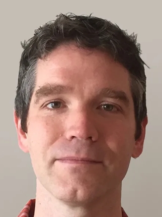 Jason Lerch合作论文数Preclinical Imaging Group, Nuffield Department of Clinical Neurosciences, Medical Sciences Division, University of Oxford;Wellcome Centre for Integrative Neuroimaging, University of Oxford3
 Susann Boretius合作论文数Biomedizinische NMR Forschungs GmbH, Max-Planck-Institut für biophysikalische Chemie3Thoralf Niendorf合作论文数3Yongzhi Zhang合作论文数3
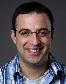 Noam Shemesh合作论文数Champalimaud Centre for the Unknown, Champalimaud Foundation3
 Edoardo Micotti合作论文数Biology of Neurodegenerative Disorders Laboratory, Mario Negri Institute for Pharmacological Research3
 Jurgen Baudewig合作论文数Abteilung für Klinische Neurophysiologie, Georg-August-Universität3
 Tom Scheenen合作论文数Department of Radiology, University Medical Center3
 Cornelius Faber合作论文数Department of Experimental Physics, University of Würzburg3
 Nick Todd合作论文数Audit Working Group3
 Rick Dijkhuizen合作论文数Division Imaging & Oncology, University Medical Center Utrecht3
 Gabriel a Devenyi合作论文数3
 Eike Budinger合作论文数3
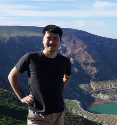 Akira Sumiyoshi合作论文数Graduate School of Pharmaceutical Sciences, Nagoya City University3Annette Van Der Toorn合作论文数3
 Emma Muñoz-Moreno合作论文数Laboratorio de Procesado de Imagen, Universidad de Valladolid3Xavier López-Gil合作论文数3
 Mallar Chakravarty合作论文数Brain Imaging Center, Computational Brain Anatomy Laboratory, Douglas Mental Health University Institute;Department of Psychiatry, McGill University;Department of Biomedical Engineering, McGill University;Cerebral Imaging Centre, Douglas Mental Health University Institute3




