Sciatic nerve injury associated with total hip arthroplasty (THA) confers chronic and progressive disability. Mechanisms of injury are heterogeneous and management nuances are often case-specific. We discuss a Sunderland Type 4 sciatic nerve transection by femoral cerclage wire from prior THA to highlight optimal clinical strategies when approaching complex cases. A 65-year-old woman presented to the neurosurgery clinic with worsening, medically refractory right sciatic sensorimotor neuropathy that began 1 year after ipsilateral hip arthroplasty. Neurological examination detected weakness in ankle dorsiflexion/plantarflexion and foot inversion/eversion (motor scale 2-3/5), toe extension/flexion (1/5), foot numbness, and hyperesthesia. Electromyogram confirmed sciatic neuropathy. Magnetic resonance neurogram (MRN) showed a thickened right sciatic nerve abutting a femoral cerclage wire, which appeared contiguous on reconstructed computed tomography imaging. Intraoperatively, the wire was discovered to have clearly transected and remained lodged within the sciatic nerve, requiring orthopedic surgery consultation and wire cutdown at the transection site. The surrounding neuroma was excised and the defect was reconstructed using nerve allograft interposition. Intraoperative neurophysiology monitoring (IONM) signals remained stable. Radiographs confirmed uncomplicated wire disconnection. The patient was discharged home the next day and reported significant symptomatic relief at 1-month follow-up. Delayed presentation of sciatic nerve transection by femoral cerclage wire with ongoing neural compression is rare. The anatomy of injury can be high risk, impelling thoughtful operative planning in THA as well as neuroplasty cases. Strategies include preoperative MRN to evaluate the pathoanatomy of nerve injury, neurosurgery and orthopedic surgery comanagement, and multimodal IONM to reduce risks of intraoperative neural injury and optimize outcomes.
OBJECTIVE Depression has been implicated with worse immediate postoperative outcomes in adult spinal deformity (ASD) correction, yet the specific impact of depression on those patients undergoing minimally invasive surgery (MIS) requires further clarity. This study aimed to evaluate the role of depression in the recovery of patients with ASD after undergoing MIS. METHODS Patients who underwent MIS for ASD with a minimum postoperative follow-up of 1 year were included from a prospectively collected, multicenter registry. Two cohorts of patients were identified that consisted of either those affirming or denying depression on preoperative assessment. The patient-reported outcome measures (PROMs) compared included scores on the Oswestry Disability Index (ODI), numeric rating scale (NRS) for back and leg pain, Scoliosis Research Society Outcomes Questionnaire (SRS-22), SF-36 physical component summary, SF-36 mental component summary (MCS), EQ-5D, and EQ-5D visual analog scale. RESULTS Twenty-seven of 147 (18.4%) patients screened positive for preoperative depression. The nondepressed cohort had an average of 4.83 levels fused, and the depressed cohort had 5.56 levels fused per patient (p = 0.267). At 1-year follow-up, 10 patients still reported depression, representing a 63% decrease. Postoperatively, both cohorts demonstrated improvement in their PROMs; however, at 1-year follow-up, those without depression had statistically better outcomes based on the EQ-5D, MCS, and SRS-22 scores (p < 0.05). Patients with depression continued to experience higher NRS leg scores at 1-year follow-up (3.63 vs 2.22, p = 0.018). After controlling for covariates, the authors found that depression significantly impacted only 1-year follow-up MCS scores (beta = 8.490, p < 0.05). CONCLUSIONS Depressed and nondepressed patients reported similar improvements after MIS surgery, except MCS scores were more likely to improve in nondepressed patients.
Abstract Social determinants of health are known to influence health outcomes, yet the interactive effects of patient-specific socioeconomic variables on overall survival remain poorly characterized in patients with glioblastoma (GBM). We aimed to assess socioeconomic factors associated with overall survival in a clinically homogenous GBM population. Patients with newly diagnosed isocitrate dehydrogenase wild-type glioblastoma, who underwent maximal safe resection of contrast-enhanced (CE) and non-contrast enhanced tissue (NC) tissue with adjuvant chemoradiation from 1997-2017 were selected for inclusion. Socioeconomic factors were collected through retrospective chart review. Variables associated with overall survival on univariate cox proportional hazards models (p<0.05) were selected for multivariate analysis. 272 patients (42.6% female, 14.3% minority) were included. On univariate logistic regression, older age (HR: 1.23, p<0.001), greater residual CE tissue (HR: 3.95, p<0.001), were associated with reduced survival. Greater extent of resection NCE tissue (HR 0.94, p=0.028), higher postoperative KPS (HR 0.89, p=0.034), and unemployment (HR: 0.67, p=0.022), were associated with longer overall survival. On multivariate analysis, age was associated with reduced survival (HR: 1.31, p<0.001), while unemployment (HR:0.69, p=0.049) was associated with longer overall survival. Subgroup multivariate analyses was performed dividing patients into those <65 (n=185, 45% female, 11.9% minority, 48% employed) and those ≥65 (n=87, 37% female, 14% minority, 20% employed). In the <65 cohort, protective factors included retirement (HR: 0.49 p=0.004), and unemployment (HR:.57, p=0.006). Older age (HR: 1.03, p=0.004), and greater residual CE tissue (HR: 1.16, p=0.03) continued to be associated with poorer survival. No variables correlated with outcomes in the ≥65 cohort. Active employment was associated with reduced survival in GBM patients treated with standard care, potentially serving as a proxy for access to subsequent treatment.
OBJECTIVE:Posterior fossa arteriovenous malformations (AVMs) represent 7% to 15% of all intracranial AVMs and are associated with an increased risk of hemorrhage, morbidity, and mortality compared with supratentorial AVMs, thus prompting urgent and definitive treatment. Cerebellopontine angle (CPA) AVMs are a unique group of posterior fossa AVMs incorporating characteristics of brainstem and cerebellar lesions, which are particularly amenable to microsurgical resection. This study reports the clinical, radiological, operative, and outcome features of patients with CPA AVMs in a large cohort. METHODS:The authors conducted a single-surgeon, 2-institution retrospective cohort study of all consecutive patients with CPA AVMs treated with microsurgical resection during a 25-year period. RESULTS:CPA AVMs represented 22% (38 of 176) of all infratentorial AVMs resected by the senior author. Overall, 38 patients (22 [58%] male and 16 [42%] female) met the study inclusion criteria and were analyzed. Most patients presented with hemorrhage (n = 29, 76%). The median age at surgery was 56 (range 6-82) years. Subtypes included 22 (58%) petrosal cerebellar AVMs, 11 (29%) lateral pontine AVMs, and 5 (13%) AVMs involving both the brainstem and cerebellum. Most AVM niduses were small (< 3 cm; n = 35, 92%) and compact (n = 31, 82%). Fourteen (37%) patients harbored flow-related aneurysms. Twenty (53%) patients underwent preoperative embolization. Complete angiographic obliteration was achieved with microsurgery in 35 (92%) patients. Five (13%) patients with poor neurological conditions at presentation died before hospital discharge. Of the 7 (18%) patients with new postoperative neurological deficits, 5 had transient deficits. The median (interquartile range) follow-up was 1.7 (0.5-3.2) years; 32 (84%) patients were alive at last follow-up, and 30 (79%) had achieved a favorable neurological outcome (modified Rankin Scale [mRS] score 0-2). The only independent predictor of unfavorable postoperative outcome (mRS score 3-6) was the preoperative mRS score (p = 0.002). CONCLUSIONS:CPA AVMs are unique posterior fossa lesions, including petrosal cerebellar and lateral pontine AVMs. The "backdoor resection" technique provides a safe and efficient strategy with high obliteration rates and a low risk of treatment-related morbidity. Microsurgical resection should be considered the frontline treatment for most CPA AVMs, except for those with a significant diffuse brainstem component.
Abstract Specialized neocortical circuits selectively organize neuronal signals and encode features of cognitive processing via local and long-range connections. Since glioma-infiltrated cortex is excitable and can participate in cognitive processing, the underlying laminar structure and functionality may still be preserved despite infiltration. Moreover, since glioma cells remodel existing neural circuits and microenvironmental factors drive invasion and proliferation, neuron-glioma interactions may demonstrate layer specificity. As glioma-infiltrated cortex remains poorly understood, we sought to characterize these regions using electrophysiological, structural, and genomic approaches. We assessed the power spectra of normal-appearing and glioma-infiltrated cortex using magnetoencephalography and subdural high-density electrode arrays. Immunohistochemistry of formalin-fixed paraffin-embedded (FFPE) samples of infiltrated cortex enabled protein-level neuronal and glioma identification to determine laminar preservation and spatial invasion patterns. Spatial transcriptomics of FFPE tissues and single-nucleus RNA-sequencing were used to identify cell populations, genomic alterations, and cell-cell communication within and across samples at testing and validation sites across the US and Europe. Increased delta range (1-4 Hz) power and decreased power in the beta range (12-20 Hz), activity thought to originate from deep layers, were identified as robust features of infiltrated cortex which were maintained across glioma subtypes (n = 139 patients) and preserved in a validation cohort (n = 8 patients). Immunohistochemistry revealed greater tumor burden within deep laminae regardless of glioma subtype (n = 20 samples). Tissue proteomics and spatial genomics analyses performed using glioma-infiltrated cortical samples (n = 10 samples; 29,011 spots) confirmed routine preservation of laminar organization, greater tumor burden in deep layers, as well as layer specific differences in glioma-related expression programs. Cell-cell communication analyses demonstrated increased interactions in glioma-infiltrated cortex across layers. These findings suggest that laminar structure may be preserved in glioma-infiltrated cortex while remodeling of neural circuits alters spatiotemporal activity in a predictable manner and support a deep to superficial invasion pattern. Thus, investigating glioma infiltration using layer-centric approaches may facilitate novel insights into neuron-glioma interactions.
INTRODUCTION: In patients with medulloblastomas, the combination of CSF cytology and MR imaging is believed to be superior to either tool alone in detecting leptomeningeal dissemination (LMD). However, MRI has drastically improved since the initial assessment of this approach. METHODS: A retrospective review of 117 unique patients with medulloblastomas from a single institution was conducted. CSF samples and MR images obtained within 36 days of each other were included. A 3-tier system (negative, atypical/suspicious, positive) was utilized to account for equivocal findings. A total of 253 CSF-MRI pairs (range: 1-12 pairs per patient) obtained between 2000-2021 were analyzed. RESULTS: The mean (SD) age of this cohort was 14.1 (13.5) years (62.4% were male). Gross total resection with surgery was achieved in 64.5%, 83.7% received radiotherapy, 81.5% received chemotherapy. 16.2% were deceased, with the mean (SD) time till death being 4.6 (4.7) years following the first operation. The molecular subtypes were: SHH (17.0%), WNT (3.6%), non-WNT/non-SHH (46.6%), unknown (32.8%). There was a 60.5% concordance between CSF cytology and MRI, with no significant differences by molecular subtype. In 57.3% of cases, CSF cytology and MRI were both negative. In 1.2% of cases, CSF cytology and MRI were both equivocal. 26.9% of cases were positive on CSF cytology and/or MRI. MRI had an 86.8% sensitivity and CSF cytology had a 20.6% sensitivity for detecting LMD that was positive in CSF, MRI, or both. In 10 cases (4.0%), MRI was negative when CSF was atypical or positive. In 80 cases (31.6%), CSF was negative when MRI was positive or suspicious. In 94.5% of cases, MRI demonstrated greater or equal ability to CSF cytology in detecting LMD. CONCLUSIONS: MRI is superior to CSF cytology in detecting LMD but misses a small percentage of cases.
INTRODUCTION: To date, there are no tools to intraoperatively predict postoperative visual function following endoscopic endonasal surgery (EES) for suprasellar lesions. The development of such a tool would impact postoperative care for patients at risk of new visual deficits. METHODS: Patients undergoing EES for resection of suprasellar lesions were administered 5 mg of ICG diluted in 10 mL of saline after tumor removal. Time between luminescence of the anterior cerebral artery and the superior hypophyseal artery branches enveloping the optic chiasm was noted, and the percentage of optic chiasm vessels that luminesced was recorded. Postoperative exams and imaging studies were then used to assess visual function. Patients with and without new deficits were compared to examine trends in ICG findings. RESULTS: A total of seven trials were performed on six patients. Mean time to chiasm peak luminescence was 3.8 seconds, and 81.8% of chiasm vessels luminesced. Patients with stable or improved vision after resection demonstrated over 90% chiasm luminescence in every case, and mean time to chiasm luminescence in these post-resection ICG administrations was 4.0 seconds. One patient experienced new postoperative visual deficits; on review of their ICG administration, 11.5% of chiasm vessels luminesced, and the chiasm itself failed to display robust luminescence after 30 seconds of direct observation. CONCLUSIONS: This pilot study showed the capability of intraoperative indocyanine green angiography to demonstrate perfusion of the optic chiasm during endoscopic endonasal surgery for resection of suprasellar lesions. Chiasm times under 5 seconds and over 90% chiasm vessel illumination may reflect adequate chiasm perfusion, while those with delayed or absent chiasm luminescence may have compromised chiasm perfusion.
BACKGROUND AND OBJECTIVES:Direct cortical stimulation (DCS) mapping enables the identification of functional language regions within and around gliomas before tumor resection. Intraoperative mapping is required because glioma-infiltrated cortex engages in synchronous activity during task performance in a manner similar to normal-appearing cortex but has decreased ability to encode information for complex tasks. It is unknown whether task complexity influenced DCS mapping results. We aim to understand correlations between audiovisual picture naming (PN) task complexity and DCS error rate. We also asked what functional and oncological factors might be associated with higher rates of erroneous responses. METHODS:We retrospectively reviewed intraoperative PN and word reading (WR) task performance during awake DCS language mapping for resection of dominant hemisphere World Health Organization grade 2 to 4 gliomas. The complexity of word tested in PN/WR tasks, patient characteristics, and tumor characteristics were compared between correct and incorrect trials. RESULTS:Between 2017 and 2021, 74 patients met inclusion criteria. At median 18.6 months of follow-up, 73.0% were alive and 52.7% remained recurrence-free. A total of 2643 PN and 978 WR trials were analyzed. A greater number of syllables in PN was associated with a higher DCS error rate ( P = .001). Multivariate logistic regression found that each additional syllable in PN tasks independently increased odds of error by 2.40 ( P < .001). Older age was also an independent correlate of higher error rate ( P < .043). World Health Organization grade did not correlate with error rate ( P = .866). More severe language impairment before surgery correlated with worse performance on more complex intraoperative tasks ( P < .001). A higher error rate on PN testing did not correlate with lower extent of glioma resection ( P = .949). CONCLUSION:Word complexity, quantified by the number of syllables, is associated with higher error rates for intraoperative PN tasks but does not affect extent of resection.
Abstract BACKGROUND For medulloblastoma, CSF cytology and MRI together are considered to be superior to either modality alone in identifying leptomeningeal dissemination (LMD). However, MRI has vastly improved since the initial assessment of this approach. METHODS A retrospective medical record review of 130 patients (36.2% female) with medulloblastoma was performed. CSF samples and MR images (6.3% utilized FIESTA) obtained within 1 month of each other from 2000-2023 were analyzed. There were 302 CSF-MRI pairs (range: 1-12 pairs per patient) in total. A 3-tier system (negative, atypical/suspicious, positive) was utilized to account for equivocal findings. RESULTS The mean (SD) age at surgery was 13.2 (13.0) years. Mean (SD) postoperative follow-up time was 7.0 (5.2) years. 71.5% underwent gross or near-total resection, 80.8% received radiotherapy, and 93.1% received chemotherapy. The molecular subtypes were: SHH (24.6%), WNT (4.6%), non-WNT/non-SHH (38.5%), unknown (32.3%). MRI had an 89.8% sensitivity and CSF cytology had a 22.7% sensitivity for detecting LMD that was positive on CSF and/or MRI. MRI was positive in 23.1% of CSF negative cases. CSF was positive in 2.2% of MRI negative cases. For all CSF positive-MRI negative pairs, the CSF samples and MR images were obtained within 10 days of surgery. MRI was positive in 38.9% of CSF atypical cases. CSF was positive in 11.9% of MRI suspicious cases. MRI demonstrated greater or equal ability to CSF in 94.7% of pairs. MRI was more likely to exceed CSF at 3T versus 1.5T (51.3% vs. 27.4%; p<0.01). The overall concordance between CSF cytology and MRI was 61.3%, and this differed by subtype (75.4% SHH vs. 80.0% WNT vs. 52.3% NWNS; p<0.01). CONCLUSIONS Understanding the ability of CSF cytology and MRI to detect LMD is important. While MRI is superior overall, LMD detection varies by MRI Tesla, medulloblastoma subtype, and time since surgery.
Abstract BACKGROUND We recently demonstrated that glioma-infiltrated cortex engages in task-specific computations but may encode less information while doing so. The mechanistic underpinnings of glioma burden and its behavioral impact remains unknown OBJECTIVE To investigate the relationship between glioma burden and degree of cognitive impairments. METHODS One-hundred-eight patients with newly diagnosed WHO 2-4 diffuse glioma were recruited from a UCSF prospective registry. MRI FLAIR and T1 post-gadolinium tumor volume masks were demarcated. A “Composite” mask was generated from the sum of T1 post-gadolinium and FLAIR volume masks. Participants completed language assessments using the Quick Aphasia Battery (QAB). Glioma burden was determined by tumor-specific marker expression (IDH-1 mutation and P53) and glioma proliferation (Ki-67 and MIB-1 proliferation index). For each mask type, support-vector regression voxel-lesion symptom mapping (SVR-LSM) and linear regression analyses were performed to identify anatomic regions of interest (ROI) and glioma-specific factors associated with lower QAB scores. RESULTS SVR-LSM analysis by mask type revealed significant clusters in the left superior temporal gyrus: composite lesions within a cluster of 41,762 voxels and T1 post-gadolinium lesions within a cluster of 42,570 voxels. Both were independently predictive of lower QAB scores. SVR-LSM analysis of FLAIR masks failed to produce any significant clusters. Multivariate linear regression analysis of composite lesions was utilized to control for composite lesion voxel volume, glioma grade, and tumor-specific marker expression. The presence of voxels overlapping with the composite lesion ROI scores (beta = -0.13; p < 0.001), and, relative to a low glioma proliferation group (< 2%), glioma proliferation between 31-50% (beta = -2.00, p = 0.044) were independently and significantly predictive of lower QAB scores. CONCLUSION Relative to a low glioma proliferation group (< 2%), glioma proliferation between 31-50% significantly predicts language impairments, with a linear relationship between voxel overlap with ROI and impairment severity.
INTRODUCTION: It has been shown recently that infiltration of gliomas into healthy neural tissue alters local neuronal spiking patterns, leading to circuit dysfunction. It is unknown, however, if glioma infiltration also impacts the coordination of local neuronal computations with other networks during behavioral tasks. Cross-frequency coupling of high gamma (HG) (80-150 Hz) amplitude with theta (Th) (4-7Hz) phase is a well-established mechanism for this type of neural integration. METHODS: Patients (n = 13) with dominant hemisphere perisylvian gliomas were intraoperatively implanted with an ECoG grid and performed a picture naming task. Electrodes overlying regions of tumor infiltration and those overlying normal parenchyma were identified using the T2-FLAIR signal. Following a widely accepted method, Kullback–Leibler (KL) divergence of HG amplitude distribution over Th phases from a uniform distribution was used to quantify Th-HG coupling of each electrode during speech production. Statistical significance was determined by comparing to a null distribution of KL divergence values corresponding to the original HG time series randomly cut in ten places and permuted, while preserving the theta time series. This destroys the cyclostationarity of theta-gamma coupling while preserving other non-stationarities. RESULTS: 43 out of 114 electrodes overlying normal parenchyma and 104 out of 300 electrodes overlying tumor-infiltrated regions exhibited significant Th-HG coupling after FDR correction. Electrodes overlying tumor regions and normal parenchyma had similar spatial coverage at the group level. Electrodes with Th-HG coupling at a preferred theta phase of π radians exhibited greater coupling. No significant difference in KL divergence was found between tumor-overlying electrodes and non-tumor overlying electrodes at the group level. CONCLUSIONS: Th-HG phase-amplitude coupling is preserved in glioma-infiltrated brain tissue during speech production. This suggests that glioma-infiltrated tissue participates in the coordination of spatially and temporally distributed processes during complex behaviors.
Abstract Glioblastoma, the most common primary brain tumor in adults, is a major cause of neurological morbidity and mortality with no effective therapies. Its proliferation and invasion are regulated by direct and paracrine-mediated neuronal activity. In this study, we uncover molecular targets and protein-protein signaling driving activity dependent proliferation. To identify candidate molecular pathways, we used high-density electrode arrays in vivo to record human local field potentials. Single-cell RNA sequencing identified Netrin-G1 (NTNG1) and thrombospondin-1 (TSP1) elevation in glioblastoma clonal population that proliferates in response to the presence of neurons. The amino acid sequence of NTNG1 and the TSR1 domain of TSP1 were utilized to predict the 3D interaction surface of NTNG1 and TSP1. Compared to structure predictions of NTNG1 and its known binding partner NGL1, NTNG1 and TSP1 complex predictions showed agreement between machine learning predicted models and lower predicted aligned error in interdomain regions of the complex, suggesting higher confidence in the relative position of NTNG1-TSP1 as compared to NTNG1-NGL1. We performed 45 protein structure predictions and two molecular dynamics simulations of mutant and WT TSP1 in complex with NTNG1. Each simulation box consisted of >263,000 atoms, each run for 50 million 2 femtosecond timesteps for a total simulation time of 200 ns. These calculations confirmed a stable complex in the WT condition and significant conformational change in the R46A condition, emphasizing the importance of this residue in mediating NTNG1-TSP1 binding. Computational affinity models between NTNG1 and TSP1 were validated by pull-down assays and immunofluorescence labelling using cerebral organoid and human glioma-neuronal coculture models. Finally, we design small molecule and fusion-protein inhibitors disrupting this interaction. Here we advance the development of targeted precision-medicine therapies to treat glioblastoma proliferation through activity-dependent mechanisms.
OBJECTIVE Prior studies have revealed that a body mass index (BMI) ≥ 30 is associated with worse outcomes following surgical intervention in grade 1 lumbar spondylolisthesis. Using a machine learning approach, this study aimed to leverage the prospective Quality Outcomes Database (QOD) to identify a BMI threshold for patients undergoing surgical intervention for grade 1 lumbar spondylolisthesis and thus reliably identify optimal surgical candidates among obese patients. METHODS Patients with grade 1 lumbar spondylolisthesis and preoperative BMI ≥ 30 from the prospectively collected QOD lumbar spondylolisthesis module were included in this study. A 12-month composite outcome was generated by performing principal components analysis and k-means clustering on four validated measures of surgical outcomes in patients with spondylolisthesis. Random forests were generated to determine the most important preoperative patient characteristics in predicting the composite outcome. Recursive partitioning was used to extract a BMI threshold associated with optimal outcomes. RESULTS The average BMI was 35.7, with 282 (46.4%) of the 608 patients from the QOD data set having a BMI ≥ 30. Principal components analysis revealed that the first principal component accounted for 99.2% of the variance in the four outcome measures. Two clusters were identified corresponding to patients with suboptimal outcomes (severe back pain, increased disability, impaired quality of life, and low satisfaction) and to those with optimal outcomes. Recursive partitioning established a BMI threshold of 37.5 after pruning via cross-validation. CONCLUSIONS In this multicenter study, the authors found that a BMI ≤ 37.5 was associated with improved patient outcomes following surgical intervention. These findings may help augment predictive analytics to deliver precision medicine and improve prehabilitation strategies.
BACKGROUND: Postoperative hemorrhage is a rare but potentially serious complication after pituitary surgery. The risk factors for this complication are mostly unknown, and further knowledge would help guide postoperative management. OBJECTIVE: To investigate the perioperative risks and clinical presentation of significant postoperative hemorrhage (SPH) after endonasal surgery for pituitary neuroendocrine tumors. METHODS: A population of 1066 patients undergoing endonasal (microscopic and endoscopic) surgery for pituitary neuroendocrine tumor resection at a high-volume academic center was reviewed. SPH cases were defined as postoperative hematoma evident on imaging requiring return to the operating room for evacuation. Patient and tumor characteristics were analyzed with uni- and multivariable logistic regression, and postoperative courses were descriptively examined. RESULTS: Ten patients were found to have SPH. On univariable analysis, these cases were significantly more likely to present with apoplexy ( P = .004), have larger tumors ( P < .001), and lower gross total resection rates ( P = .019). A multivariate regression analysis showed that tumor size (odds ratio 1.94, P = .008) and apoplexy at presentation (odds ratio 6.00, P = .018) were significantly associated with higher odds of SPH. The most common symptoms for patients with SPH were vision deficits and headache, and the median time for symptom onset was 1 day after surgery. CONCLUSION: Larger tumor size and presentation with apoplexy were associated with clinically significant postoperative hemorrhage. Patients presenting with pituitary apoplexy are more likely to experience a significant postoperative hemorrhage and should be carefully monitored for headache and vision changes in the days after surgery.
Gliomas synaptically integrate into neural circuits 1 , 2 . Previous research has demonstrated bidirectional interactions between neurons and glioma cells, with neuronal activity driving glioma growth 1 – 4 and gliomas increasing neuronal excitability 2 , 5 – 8 . Here we sought to determine how glioma-induced neuronal changes influence neural circuits underlying cognition and whether these interactions influence patient survival. Using intracranial brain recordings during lexical retrieval language tasks in awake humans together with site-specific tumour tissue biopsies and cell biology experiments, we find that gliomas remodel functional neural circuitry such that task-relevant neural responses activate tumour-infiltrated cortex well beyond the cortical regions that are normally recruited in the healthy brain. Site-directed biopsies from regions within the tumour that exhibit high functional connectivity between the tumour and the rest of the brain are enriched for a glioblastoma subpopulation that exhibits a distinct synaptogenic and neuronotrophic phenotype. Tumour cells from functionally connected regions secrete the synaptogenic factor thrombospondin-1, which contributes to the differential neuron–glioma interactions observed in functionally connected tumour regions compared with tumour regions with less functional connectivity. Pharmacological inhibition of thrombospondin-1 using the FDA-approved drug gabapentin decreases glioblastoma proliferation. The degree of functional connectivity between glioblastoma and the normal brain negatively affects both patient survival and performance in language tasks. These data demonstrate that high-grade gliomas functionally remodel neural circuits in the human brain, which both promotes tumour progression and impairs cognition.
Objective: Postoperative hemorrhage is a rare but potentially severe complication after pituitary surgery. The risk factors for this complication are mostly unknown, and further knowledge would help define blood pressure targets and placement after surgery. Here we queried a large database of patients undergoing endonasal removal of pituitary adenomas and describe preoperative risks for significant postoperative hemorrhage (SPH).
OBJECTIVE:Venous thromboembolism (VTE) following traumatic spinal cord injury (SCI) is a significant clinical concern. This study sought to determine the incidence of VTE and hemorrhagic complications among patients with SCI who received low-molecular-weight heparin (LMWH) within 24 hours of injury or surgery and identify variables that predict VTE using the prospective Transforming Research and Clinical Knowledge in SCI (TRACK-SCI) database.METHODS:The TRACK-SCI database was queried for individuals with traumatic SCI from 2015 to 2022. Primary outcomes of interest included rates of VTE (including deep vein thrombosis [DVT] and pulmonary embolism [PE]) and in-hospital hemorrhagic complications that occurred after LWMH administration. Secondary outcomes included intensive care unit and hospital length of stay, discharge location type, and in-hospital mortality.RESULTS:The study cohort consisted of 162 patients with SCI. Fifteen of the 162 patients withdrew from the study, leading to loss of data for certain variables for these patients. One hundred thirty patients (87.8%) underwent decompression and/or fusion surgery for SCI. DVT occurred in 11 (7.4%) of 148 patients, PE in 9 (6.1%) of 148, and any VTE in 18 (12.2%) of 148 patients. The analysis showed that admission lower-extremity motor score (p = 0.0408), injury at the thoracic level (p = 0.0086), admission American Spinal Injury Association grade (p = 0.0070), and younger age (p = 0.0372) were significantly associated with VTE. There were 3 instances of postoperative spine surgery-related bleeding (2.4%) in the 127 patients who had spine surgery with bleeding complication data available, with one requiring return to surgery (0.8%). Thirteen (8.8%) of 147 patients had a bleeding complication not related to spine surgery. There were 2 gastrointestinal bleeds associated with nasogastric tube placement, 3 cases of postoperative non-spine-related surgery bleeding, and 8 cases of other bleeding complications (5.4%) not related to any surgery.CONCLUSIONS:Initiation of LMWH within 24 hours was associated with a low rate of spine surgery-related bleeding. Bleeding complications unrelated to SCI surgery still occur with LMWH administration. Because neurosurgical intervention is typically the limiting factor in initializing chemical DVT prophylaxis, many of these bleeding complications would have likely occurred regardless of the protocol.
Importance Central nervous system (CNS)-penetrant systemic therapies have significantly advanced care for patients with melanoma brain metastases. However, improved understanding of the molecular landscape and microenvironment of these lesions is needed to both optimize patient selection and advance treatment approaches.Objective To evaluate how bulk and single-cell genomic features of melanoma brain metastases are associated with clinical outcome and treatment response.Design, Setting, and Participants This cohort study analyzed bulk DNA sequencing and single nuclear RNA-sequencing data from resected melanoma brain metastases and included 94 consecutive patients with a histopathologically confirmed diagnosis of melanoma brain metastasis who underwent surgical resection at a single National Comprehensive Cancer Network cancer center in San Francisco, California, from January 1, 2009, to December 31, 2022.Exposure A Clinical Laboratory Improvement Amendments-certified targeted sequencing assay was used to analyze tumor resection specimens, with a focus on BRAF V600E alteration. For frozen pathologic specimens from CNS treatment-naive patients undergoing surgical resection, commercial single nuclear RNA sequencing approaches were used.Main Outcomes and Measures The primary outcome was overall survival (OS). Secondary outcomes included CNS progression-free survival (PFS), microenvironmental composition with decreased T-cell and macrophage populations, and responses to immunotherapy.Results To correlate molecular status with clinical outcome, Kaplan-Meier survival analysis of 94 consecutive patients (median age, 64 years [range, 24-82 years]; 70 men [74%]) with targeted BRAF alteration testing showed worse median intracranial PFS (BRAF variant: 3.6 months [IQR, 0.1-30.6 months]; BRAF wildtype: 11.0 months [IQR, 0.8-81.5 months]; P < .001) and OS (BRAF variant: 9.8 months [IQR, 2.5-69.4 months]; BRAF wildtype: 23.2 months [IQR, 1.1-102.5 months]; P = .005; log-rank test) in BRAF V600E variant tumors. Multivariable Cox proportional hazards regression analysis revealed that BRAF V600E status was an independent variable significantly associated with both PFS (hazard ratio [HR], 2.65; 95% CI, 1.54-4.57; P < .001) and OS (HR, 1.96; 95% CI, 1.08-3.55; P = .03). For the 45 patients with resected melanoma brain metastases undergoing targeted DNA sequencing, molecular classification recapitulated The Cancer Genome Atlas groups (NRAS variant, BRAF variant, NF1 variant, and triple wildtype) with no subtype enrichment within the brain metastasis cohort. On a molecular level, BRAF V600E variant lesions were found to have a significantly decreased tumor mutation burden. Moreover, single nuclear RNA sequencing of treatment-naive BRAF V600E variant (n = 3) brain metastases compared with BRAF wildtype (n = 3) brain metastases revealed increased immune cell populations in BRAF wildtype tumors (mean [SD], 11% [4.1%] vs 3% [1.6%] CD45-positive cells; P = .04). Survival analysis of postoperative immunotherapy responses by BRAF status revealed that BRAF wildtype lesions were associated with a response to checkpoint inhibition (median OS: with immunotherapy, undefined; without immunotherapy, 13.0 months [range, 1.1-61.7 months]; P = .001; log-rank test) while BRAF variant lesions (median OS: with immunotherapy, 9.8 months [range, 2.9-39.8 months]; without immunotherapy, 9.5 months [range, 2.5-67.2 months]; P = .81; log-rank test) were not. Conclusions and Relevance This molecular analysis of patients with resected melanoma brain metastases found that BRAF V600E alteration is an important translational biomarker associated with worse clinical outcomes, differential microenvironmental composition, and benefit from immunotherapy. Patients with BRAF V600E variant melanoma brain metastases may thus benefit from alternative CNS-penetrant systemic regimens.
 Shawn Hervey-Jumper合作论文数School of Medicine, University of California, San Francisco;Weill Institute for Neurosciences, University of California, San Francisco;Medical School, University of Michigan30
 Jacob Young合作论文数17
 Jasleen Kaur合作论文数Dept Neurol Surg, Univ Calif San Francisco14
 David Brang合作论文数Department of Psychology, College of Literature, Science, and the Arts, University of Michigan-Ann Arbor14
 Mitchel Berger合作论文数Brain Tumor Center, University of California, San Francisco;School of Medicine, University of California, San Francisco11
 Krishna Saritha合作论文数Department of Physiology and Pharmacology, College of Veterinary Medicine.11
 Sofia Kakaizada合作论文数9
 Manish Aghi合作论文数Department of Neurological Surgery, School of Medicine, University of California, San Francisco8Praveen V. Mummaneni合作论文数Dept. of Neurosurgery, Emory University8Morshed Ramin A合作论文数The Brain Tumor Center, The University of Chicago Pritzker School of Medicine7
 Philip Theodosopoulos合作论文数Mayfield Clinic7
 Agarwal Nitin合作论文数6
 Daniel Andy G S合作论文数Brain and Mind Research Institute, Weill Cornell Medical College6
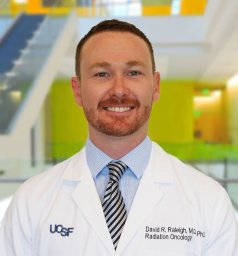 David R Raleigh合作论文数6
 Benjamin Lipkin合作论文数Massachusetts Institute of Technology5
 Srikantan Nagarajan合作论文数Department of Radiology and Biomedical Imaging, University of California, San Francisco;Biomagnetic Imaging Lab, University of California, San Francisco;Department of Bioengineering and Therapeutic Sciences, University of California, San Francisco5Anne M. Findlay合作论文数5Dean Chou合作论文数5Mulki Mehari合作论文数4
 Anthony M DiGiorgio合作论文数Institute for Health Policy Studies, University of California4Robert C. Osorio合作论文数4Joanna Phillips合作论文数Department of Neurological Surgery, Helen Diller Research Center, University of California San Francisco, San Francisco, CA, USA4
 Ryan R L Phelps合作论文数Dept Neurosurg, UCSF4
 Daniel Weissman合作论文数Department of Psychology, College of Literature, Science, and the Arts, University of Michigan-Ann Arbor4
 Ezequiel Goldschmidt合作论文数4
 John Yue合作论文数Weill Institute for Neurosciences, University of California, San Francisco;The Philip R. Lee Institute for Health Policy Studies, University of California, San Francisco4
 Chan Andrew K合作论文数From the Department of Neurological Surgery, Columbia University Medical Center;Weill Cornell Medical College, Cornell University;Department of Neurology, Columbia University Medical Center;Columbia University Medical Center, Columbia University Medical Center4
 Lee Anthony J合作论文数School of Psychology, University of Queensland4
 Saman Shabani合作论文数Department of Neurological Surgery, the Neurosciences Institute4
 Geoffrey Manley合作论文数Department of Neurological Surgery, School of Medicine, University of California, San Francisco;Brain And Spinal Injury Center;Zuckerberg San Francisco General Hospital4
 Annette Molinaro合作论文数Helen Diller Family Comprehensive Cancer Center, University of California, San Francisco;Department of Neurological Surgery, School of Medicine, University of California, San Francisco;Brain Tumor Research Center, University of California, San Francisco4
 Nishanth Krishnan合作论文数3Vardhaan Ambati合作论文数3Claudia Valdivia合作论文数3Cecilia Dalle Ore合作论文数3Abraham Dada合作论文数3Xuan Duong-Fernandez合作论文数3Debra D Hemmerle合作论文数3Poojan Shukla合作论文数Div HIV Infect Dis & Global Med, Univ Calif San Francisco3Gray Umbach合作论文数Univ Texas Southwestern Med Ctr Dallas, Dept Neurol Surg, Dallas, TX 75390 USA3
 Edward Chang (张复伦)合作论文数Weill Institute for Neurosciences, University of California, San Francisco;Department of Neurological Surgery, School of Medicine, University of California, San Francisco3Eunseon Ahn合作论文数Emory Univ, Emory Vaccine Ctr, Sch Med, Atlanta, GA 30322 USA3
 Vijay Letchuman合作论文数Department of Neurological Surgery, University of California3
 Vineeta Singh合作论文数Dept Neurol Surg, Univ Calif San Francisco3
 Christopher Ignatius Shaffrey合作论文数Department of Neurosurgery, School of Medicine, Duke University3
 Sanjay S Dhall合作论文数Dept Neurol Surg, Univ Calif San Francisco3
 Kai-Ming Fu合作论文数Department of Neurological Surgery, Weill Cornell Medical College3
 Adam R. Abate合作论文数Department of Bioengineering and Therapeutic Sciences, School of Pharmacy, University of California, San Francisco3Chou Austin合作论文数Brain & Spinal Injury Ctr, Univ Calif San Francisco3




