INTRODUCTION: In patients with medulloblastomas, the combination of CSF cytology and MR imaging is believed to be superior to either tool alone in detecting leptomeningeal dissemination (LMD). However, MRI has drastically improved since the initial assessment of this approach. METHODS: A retrospective review of 117 unique patients with medulloblastomas from a single institution was conducted. CSF samples and MR images obtained within 36 days of each other were included. A 3-tier system (negative, atypical/suspicious, positive) was utilized to account for equivocal findings. A total of 253 CSF-MRI pairs (range: 1-12 pairs per patient) obtained between 2000-2021 were analyzed. RESULTS: The mean (SD) age of this cohort was 14.1 (13.5) years (62.4% were male). Gross total resection with surgery was achieved in 64.5%, 83.7% received radiotherapy, 81.5% received chemotherapy. 16.2% were deceased, with the mean (SD) time till death being 4.6 (4.7) years following the first operation. The molecular subtypes were: SHH (17.0%), WNT (3.6%), non-WNT/non-SHH (46.6%), unknown (32.8%). There was a 60.5% concordance between CSF cytology and MRI, with no significant differences by molecular subtype. In 57.3% of cases, CSF cytology and MRI were both negative. In 1.2% of cases, CSF cytology and MRI were both equivocal. 26.9% of cases were positive on CSF cytology and/or MRI. MRI had an 86.8% sensitivity and CSF cytology had a 20.6% sensitivity for detecting LMD that was positive in CSF, MRI, or both. In 10 cases (4.0%), MRI was negative when CSF was atypical or positive. In 80 cases (31.6%), CSF was negative when MRI was positive or suspicious. In 94.5% of cases, MRI demonstrated greater or equal ability to CSF cytology in detecting LMD. CONCLUSIONS: MRI is superior to CSF cytology in detecting LMD but misses a small percentage of cases.
Abstract BACKGROUND For medulloblastoma, CSF cytology and MRI together are considered to be superior to either modality alone in identifying leptomeningeal dissemination (LMD). However, MRI has vastly improved since the initial assessment of this approach. METHODS A retrospective medical record review of 130 patients (36.2% female) with medulloblastoma was performed. CSF samples and MR images (6.3% utilized FIESTA) obtained within 1 month of each other from 2000-2023 were analyzed. There were 302 CSF-MRI pairs (range: 1-12 pairs per patient) in total. A 3-tier system (negative, atypical/suspicious, positive) was utilized to account for equivocal findings. RESULTS The mean (SD) age at surgery was 13.2 (13.0) years. Mean (SD) postoperative follow-up time was 7.0 (5.2) years. 71.5% underwent gross or near-total resection, 80.8% received radiotherapy, and 93.1% received chemotherapy. The molecular subtypes were: SHH (24.6%), WNT (4.6%), non-WNT/non-SHH (38.5%), unknown (32.3%). MRI had an 89.8% sensitivity and CSF cytology had a 22.7% sensitivity for detecting LMD that was positive on CSF and/or MRI. MRI was positive in 23.1% of CSF negative cases. CSF was positive in 2.2% of MRI negative cases. For all CSF positive-MRI negative pairs, the CSF samples and MR images were obtained within 10 days of surgery. MRI was positive in 38.9% of CSF atypical cases. CSF was positive in 11.9% of MRI suspicious cases. MRI demonstrated greater or equal ability to CSF in 94.7% of pairs. MRI was more likely to exceed CSF at 3T versus 1.5T (51.3% vs. 27.4%; p<0.01). The overall concordance between CSF cytology and MRI was 61.3%, and this differed by subtype (75.4% SHH vs. 80.0% WNT vs. 52.3% NWNS; p<0.01). CONCLUSIONS Understanding the ability of CSF cytology and MRI to detect LMD is important. While MRI is superior overall, LMD detection varies by MRI Tesla, medulloblastoma subtype, and time since surgery.
BACKGROUND AND OBJECTIVES: Anecdotal cases of rapidly progressing dementia in patients with dural arteriovenous fistulas (dAVFs) have been reported in small series. However, large series have not characterized these dAVFs. We conducted an analysis of the largest cohort of dAVFs presenting with cognitive impairment (dAVFs-CI), aiming to provide a detailed characterization of this subset of dAVFs. METHODS: Patients with dAVFs-CI were analyzed from the CONDOR Consortium, a multicenter repository comprising 1077 dAVFs. A propensity score matching analysis was conducted to compare dAVFs-CI with Borden type II and type III dAVFs without cognitive impairment (controls). Logistic regression was used to identify angiographic characteristics specific to dAVFs-CI. Furthermore, post-treatment outcomes were analyzed. RESULTS: A total of 60 patients with dAVFs-CI and 60 control dAVFs were included. Outflow obstruction leading to venous hypertension was observed in all dAVFs-CI. Sinus stenosis was significantly associated with dAVFs-CI (OR 2.85, 95% CI: 1.16-7.55, P = .027). dAVFs-CI were more likely to have a higher number of arterial feeders (OR 1.56, 95% CI 1.22-2.05, P < .001) and draining veins (OR 2.05, 95% CI 1.05-4.46, P = .004). Venous ectasia increased the risk of dAVFs-CI (OR 2.38, 95% CI 1.13-5.11, P = .024). A trend toward achieving asymptomatic status at follow-up was observed in patients with successful closure of dAVFs (OR 2.86, 95% CI 0.85-9.56, P = .09) CONCLUSION: Venous hypertension is a key angiographic feature of dAVFs-CI. Moreover, these fistulas present at a mean age of 58 years-old, and exhibit a complex angioarchitecture characterized by an increased number of arteriovenous connections and stenosed sinuses. The presence of venous ectasia further exacerbates the impaired drainage and contributes to the development of dAVFs-CI. Notably, in certain cases, closure of the dAVF has the potential to reverse symptoms.
The relationship between systemic inflammation and secondary injury in traumatic brain injury (TBI) is complex. We investigated associations between inflammatory markers and clinical confirmation of TBI diagnosis and prognosis. The prospective TRACK-TBI Pilot (Transforming Research and Clinical Knowledge in Traumatic Brain Injury Pilot) study enrolled TBI patients triaged to head computed tomography (CT) and received blood draw within 24 h of injury. Healthy controls (HCs) and orthopedic controls (OCs) were included. Thirty-one inflammatory markers were analyzed from plasma. Area under the receiver operating characteristic curve (AUC) was used to evaluate discriminatory ability. AUC >0.7 was considered acceptable. Criteria included: TBI diagnosis (vs. OC/HC); moderate/severe vs. mild TBI (Glasgow Coma Scale; GCS); radiographic TBI (CT positive vs. CT negative); 3- and 6-month Glasgow Outcome Scale-Extended (GOSE) dichotomized to death/greater relative disability versus less relative disability (GOSE 1-4/5-8); and incomplete versus full recovery (GOSE <8/ = 8). One-hundred sixty TBI subjects, 28 OCs, and 18 HCs were included. Markers discriminating TBI/OC: HMGB-1 (AUC = 0.835), IL-1b (0.795), IL-16 (0.784), IL-7 (0.742), and TARC (0.731). Markers discriminating GCS 3-12/13-15: IL-6 (AUC = 0.747), CRP (0.726), IL-15 (0.720), and SAA (0.716). Markers discriminating CT positive/CT negative: SAA (AUC = 0.767), IL-6 (0.757), CRP (0.733), and IL-15 (0.724). At 3 months, IL-15 (AUC = 0.738) and IL-2 (0.705) discriminated GOSE 5-8/1-4. At 6 months, IL-15 discriminated GOSE 1-4/5-8 (AUC = 0.704) and GOSE <8/ = 8 (0.711); SAA discriminated GOSE 1-4/5-8 (0.704). We identified a profile of acute circulating inflammatory proteins with potential relevance for TBI diagnosis, severity differentiation, and prognosis. IL-15 and serum amyloid A are priority markers with acceptable discrimination across multiple diagnostic and outcome categories. Validation in larger prospective cohorts is needed. ClinicalTrials.gov Registration: NCT01565551
BACKGROUND: Virtual reality (VR) is increasingly used for trajectory planning in neurosurgery. OBJECTIVE: To describe a case series showing the application of immersive VR involving both "top-down" from skull to lesion and "bottom-up" from lesion to skull approaches for trajectory planning in pediatric neurosurgical patients. METHODS: We detail the preoperative and intraoperative application of VR and clinical courses of 5 children (aged 7-14 years) with anatomically challenging intraparenchymal lesions that posed operative risks to nearby vascular anatomy and fiber tracts. Preoperative planning consisted of standard presurgical evaluation with computed tomography and magnetic resonance imaging used to render 3-dimensional models that could be viewed and manipulated using desktop software and immersive VR headsets and hand controllers by the surgeon and family. Patient satisfaction was evaluated by survey. Surgical outcomes were degree of seizure control or extent of resection. RESULTS: Three patients underwent lesion resection and 2 laser ablation. Modifications to 2-dimensional and "top-down" VR trajectory plans were made after "bottom-up" navigation in all cases. All families reported that the VR enhanced their understanding of the procedure. There were no complications, and no patients suffered permanent neurological deficits postoperatively. Gross total resection was achieved in all lesional cases, and patients with epilepsy achieved seizure freedom at 2 years postoperatively. CONCLUSION: Immersive VR allows operative corridors to be virtually traveled and viewed from a "top-down" and "bottom-up" perspective, as if looking up from under a forest canopy of overlying anatomy, for optimal trajectory planning and improvement of family understanding in pediatric neurosurgery.
BackgroundAnterior cranial fossa dural arteriovenous fistulas (ACF-dAVFs) are aggressive vascular lesions. The pattern of venous drainage is the most important determinant of symptoms. Due to the absence of a venous sinus in the anterior cranial fossa, most ACF-dAVFs have some degree of drainage through small cortical veins. We describe the natural history, angiographic presentation and outcomes of the largest cohort of ACF-dAVFs.MethodsThe CONDOR consortium includes data from 12 international centers. Patients included in the study were diagnosed with an arteriovenous fistula between 1990–2017. ACF-dAVFs were selected from a cohort of 1077 arteriovenous fistulas. The presentation, angioarchitecture and treatment outcomes of ACF-dAVF were extracted and analyzed.Results60 ACF-dAVFs were included in the analysis. Most ACF-dAVFs were symptomatic (38/60, 63%). The most common symptomatic presentation was intracranial hemorrhage (22/38, 57%). Most ACF-dAVFs drained through cortical veins (85%, 51/60), which in most instances drained into the superior sagittal sinus (63%, 32/51). The presence of cortical venous drainage predicted symptomatic presentation (OR 9.4, CI 1.98 to 69.1, p=0.01). Microsurgery was the most effective modality of treatment. 56% (19/34) of symptomatic patients who were treated had complete resolution of symptoms. Improvement of symptoms was not observed in untreated symptomatic ACF-dAVFs.ConclusionMost ACF-dAVFs have a symptomatic presentation. Drainage through cortical veins is a key angiographic feature of ACF-dAVFs that accounts for their malignant course. Microsurgery is the most effective treatment. Due to the high risk of bleeding, closure of ACF-dAVFs is indicated regardless of presentation.
AbstractDeregulation of neuroblastoma-derived myc (N-myc) is a leading cause of malignant brain tumors in children. To target N-myc-driven medulloblastoma, most research has focused on identifying genomic alterations or on the analysis of the medulloblastoma transcriptome. Here, we have broadly characterized the translatome of medulloblastoma and shown that N-myc unexpectedly drives selective translation of transcripts that promote protein homeostasis. Cancer cells are constantly exposed to proteotoxic stress associated with alterations in protein production or folding. It remains poorly understood how cancers cope with proteotoxic stress to promote their growth. Here, our data revealed that N-myc regulates the expression of specific components (∼5%) of the protein folding machinery at the translational level through the major cap binding protein, eukaryotic initiation factor eIF4E. Reducing eIF4E levels in mouse models of medulloblastoma blocked tumorigenesis. Importantly, targeting Hsp70, a protein folding chaperone translationally regulated by N-myc, suppressed tumor growth in mouse and human medulloblastoma xenograft models. These findings reveal a previously hidden molecular program that promotes medulloblastoma formation and identify new therapies that may have impact in the clinic.Significance:Translatome analysis in medulloblastoma shows that N-myc drives selective translation of transcripts that promote protein homeostasis and that represent new therapeutic vulnerabilities.
Background: Cranial dural arteriovenous fistulas with cortical venous drainage are rare lesions that can present with hemorrhage. A high rate of rebleeding in the early period following hemorrhage has been reported, but published long-term rates are much lower. No study has examined how risk of rebleeding changes over time. Our objective was to quantify the relative incidence of rebleeding in the early and later periods following hemorrhage. Methods: Patients with dural arteriovenous fistula and cortical venous drainage presenting with hemorrhage were identified from the multinational CONDOR (Consortium for Dural Fistula Outcomes Research) database. Natural history follow-up was defined as time from hemorrhage to first treatment, rebleed, or last follow-up. Rebleeding in the first 2 weeks and first year were compared using incidence rate ratio and difference. Results: Of 1077 patients, 250 met the inclusion criteria and had 95 cumulative person-years natural history follow-up. The overall annualized rebleed rate was 7.3% (95% CI, 3.2–14.5). The incidence rate of rebleeding in the first 2 weeks was 0.0011 per person-day; an early rebleed risk of 1.6% in the first 14 days (95% CI, 0.3–5.1). For the remainder of the first year, the incidence rate was 0.00015 per person-day; a rebleed rate of 5.3% (CI, 1.7–12.4) over 1 year. The incidence rate ratio was 7.3 (95% CI, 1.4–37.7; P , 0.026). Conclusions: The risk of rebleeding of a dural arteriovenous fistula with cortical venous drainage presenting with hemorrhage is increased in the first 2 weeks justifying early treatment. However, the magnitude of this increase may be considerably lower than previously thought. Treatment within 5 days was associated with a low rate of rebleeding and appears an appropriate timeframe.
Background Although the liquid embolic agent, Onyx, is often the preferred embolic treatment for cerebral dural arteriovenous fistulas (DAVFs), there have only been a limited number of single-center studies to evaluate its performance. Objective To carry out a multicenter study to determine the predictors of complications, obliteration, and functional outcomes associated with primary Onyx embolization of DAVFs. Methods From the Consortium for Dural Arteriovenous Fistula Outcomes Research (CONDOR) database, we identified patients who were treated for DAVF with Onyx-only embolization as the primary treatment between 2000 and 2013. Obliteration rate after initial embolization was determined based on the final angiographic run. Factors predictive of complete obliteration, complications, and functional independence were evaluated with multivariate logistic regression models. Results A total 146 patients with DAVFs were primarily embolized with Onyx. Mean follow-up was 29 months (range 0–129 months). Complete obliteration was achieved in 80 (55%) patients after initial embolization. Major cerebral complications occurred in six patients (4.1%). At last follow-up, 84% patients were functionally independent. Presence of flow symptoms, age over 65, presence of an occipital artery feeder, and preprocedural home anticoagulation use were predictive of non-obliteration. The transverse-sigmoid sinus junction location was associated with fewer complications, whereas the tentorial location was predictive of poor functional outcomes. Conclusions In this multicenter study, we report satisfactory performance of Onyx as a primary DAVF embolic agent. The tentorium remains a more challenging location for DAVF embolization, whereas DAVFs located at the transverse-sigmoid sinus junction are associated with fewer complications.
OBJECTIVE:Healthcare disparities are widely described in adults, but barriers affecting access to care for pediatric patients with moyamoya disease (MMD) are unknown. Understanding socioeconomic factors impacting hospital access and outcomes is necessary to address pediatric healthcare disparities. METHODS:In this cross-sectional observational study, the Kids' Inpatient Database was used to identify patients admitted with a primary diagnosis of MMD from 2003 to 2016. Patients ≤ 18 years with a primary diagnosis of MMD based on International Classification of Diseases (ICD) codes were included. Hospital admissions were queried for use of cerebral revascularization based on ICD procedure codes. RESULTS:Query of the KID yielded 1449 MMD hospitalizations. After multivariable regression, Hispanic ethnicity (OR 0.52 [95% CI 0.33-0.81], p = 0.004) was associated with lack of surgical revascularization. Private insurance (OR 1.56 [95% CI 1.15-2.13], p = 0.004), admissions at medium- and high-volume centers (OR 2.01 [95% CI 1.42-2.83], p < 0.001 and OR 2.84 [95% CI 1.95-4.14], p < 0.001, respectively), and elective hospitalization (OR 3.37 [95% CI 2.46-4.64], p < 0.001) were positively associated with revascularization. Compared with Caucasian race, Hispanic ethnicity was associated with increased mean (± SEM) length of stay by 2.01 ± 0.70 days (p = 0.004) and increased hospital charges by $24,333.61 ± $7918.20 (p = 0.002), despite the decreased utilization of surgical revascularization. Private insurance was associated with elective admission (OR 1.50 [95% CI 1.10-2.05], p = 0.01) and admission to high-volume centers (OR 1.90 [95% CI 1.26-2.88], p = 0.002). African American race was associated with the development of in-hospital complications (OR 2.52 [95% CI 1.38-4.59], p = 0.003). CONCLUSIONS:Among pediatric MMD hospitalizations, multiple socioeconomic factors were associated with access to care, whether surgical treatment is provided, and whether in-hospital complications occur. These results suggest that socioeconomic factors are important drivers of healthcare disparities in children with MMD and warrant further study.
Pituitary somatotroph adenomas are the primary cause of acromegaly and can have devastating long-term sequelae if unrecognized or untreated. Neurosurgical intervention is typically the first-line treatment. Important preoperative considerations include accuracy of diagnosis, imaging interpretation, anesthetic evaluation, and possible premedication. Several aspects of the operative procedure include the surgical approach, the effect of acromegaly on the surgical approach, the maximal extent of resection that can be safely achieved, and appropriate use of advanced technology to assist with aspects of the surgery that are anticipated to be the most complex warrant thorough consideration. A high level of vigilance should be maintained for several intraoperative or postoperative complications, including vascular injury, cranial nerve injury, and hormonal disturbances. With optimal diagnosis, preoperative planning, surgical technique, and postoperative care (sometimes including adjunctive medication or radiation therapy), these patients can recover well and attain relatively normal lifespan and quality of life.
Introduction The management of recurrent craniopharyngioma is complex with limited data to guide decision-making. Some reports suggest reoperation should be avoided due to an increased complication profile, while others have demonstrated that safe reoperation can be performed. For other types of skull base lesions, maximal safe resection followed by adjuvant therapy has replaced radical gross total resection due to the favorable morbidity profiles. Methods Seventy-one patients underwent resection over a 9-year period for craniopharyngioma and were retrospectively reviewed. Patients were separated into primary resection and reoperation cohorts and stratified by surgical approach (endonasal vs. cranial) and survival analyses were performed based on cohort and surgical approach. Results Fifty patients underwent primary resection, while 21 underwent reoperation for recurrence. Fifty endonasal transsphenoidal surgeries and 21 craniotomies were performed. Surgical approaches were similarly distributed across cohorts. Subtotal resection was achieved in 83% of all cases. There were no differences in extent of resection, visual outcomes, subsequent neuroendocrine function, and complications across cohorts and surgical approaches. The median time to recurrence was 87 months overall, and there were no differences by cohort and approach. The 5-year survival rate was 81.1% after reoperation versus 93.2% after primary resection. Conclusion Compared with primary resection, reoperation for craniopharyngioma recurrence is associated with similar functional and survival outcomes in light of individualized surgical approaches. Maximal safe resection followed by adjuvant radiotherapy for residual tumor likely preserves vision and endocrine function without sacrificing overall patient survival.
OBJECTIVE Cranial dural arteriovenous fistulas (dAVFs) are rare lesions, hampering efforts to understand them and improve their care. To address this challenge, investigators with an established record of dAVF investigation formed an international, multicenter consortium aimed at better elucidating dAVF pathophysiology, imaging characteristics, natural history, and patient outcomes. This report describes the design of the Consortium for Dural Arteriovenous Fistula Outcomes Research (CONDOR) and includes characterization of the 1077-patient cohort. METHODS Potential collaborators with established interest in the field were identified via systematic review of the literature. To ensure uniformity of data collection, a quality control process was instituted. Data were retrospectively obtained. RESULTS CONDOR comprises 14 centers in the United States, the United Kingdom, the Netherlands, and Japan that have pooled their data from 1077 dAVF patients seen between 1990 and 2017. The cohort includes 359 patients (33%) with Borden type I dAVFs, 175 (16%) with Borden type II fistulas, and 529 (49%) with Borden type III fistulas. Overall, 852 patients (79%) presented with fistula-related symptoms: 427 (40%) presented with nonaggressive symptoms such as tinnitus or orbital phenomena, 258 (24%) presented with intracranial hemorrhage, and 167 (16%) presented with nonhemorrhagic neurological deficits. A smaller proportion (224 patients, 21%), whose dAVFs were discovered incidentally, were asymptomatic. Many patients (85%, 911/1077) underwent treatment via endovascular embolization (55%, 587/1077), surgery (10%, 103/1077), radiosurgery (3%, 36/1077), or multimodal therapy (17%, 184/1077). The overall angiographic cure rate was 83% (758/911 treated), and treatment-related permanent neurological morbidity was 2% (27/1467 total procedures). The median time from diagnosis to follow-up was 380 days (IQR 120-1038.5 days). CONCLUSIONS With more than 1000 patients, the CONDOR registry represents the largest registry of cranial dAVF patient data in the world. These unique, well-annotated data will enable multiple future analyses to be performed to better understand dAVFs and their management.
Introduction: Pial arteriovenous fistulas are characterized by an abnormal connection between an intracranial artery and vein without an intervening nidus. Their predominant symptoms largely arise from mass effect, shunting, or hemorrhage. Most conservatively managed cases progress to death, but endovascular and/or surgical intervention is often successful. Case Presentation: We present the unique case of a 15-year-old girl with spontaneous intracranial hemorrhage from a single-vessel arteriovenous fistula. Although preoperative imaging failed to show a distinct nidus, intraoperative indocyanine green angiography performed after successful clipping of the primary fistulous site revealed residual shunting from a peri-lesional arteriovenous malformation. Discussion/Conclusion: This case demonstrates the importance of intraoperative imaging and meticulous circumferential inspection of these lesions to detect residual vascular shunting.
INTRODUCTION: Return to work (RTW) is an important milestone of mild traumatic brain injury (mTBI) recovery. METHODS: Adult subjects from the prospective multicenter TRACK-TBI Pilot study with mTBI (GCS 13-15) who were employed at baseline, with completed three-and six-month RTW status, and three-month Acute Concussion Evaluation (ACE), were extracted. Univariate and multivariable analyses were performed for six-month RTW, focusing on baseline employment, three-month RTW, and three-month ACE domains (physical, cognitive, sleep, and/or emotional postconcussional symptoms (PCS)). Odds ratios (OR) and 95% confidence intervals [CI] were reported. RESULTS: In 152 patients aged 40.7 ± 15.0 years, 72% were employed full-time at baseline. Three- and six-month RTW were 77.6% and 78.9%, respectively. At three months, 59.2%, 47.4%, 46.1% and 31.6% scored positive for ACE physical, cognitive, sleep, and emotional PCS domains, respectively. Three-month RTW predicted six-month RTW (OR = 19.80, 95% CI [7.61-51.52]). On univariate analysis, scoring positive in any three-month ACE domain predicted inability for six-month RTW (OR = 0.10-0.11). On multivariable analysis, emotional symptoms predicted inability to six-month RTW (OR = 0.19 [0.04-0.85]). Subjects who scored positive in all four ACE domains were more likely to be unable to RTW at six-months (4 domains: RTW = 58.3%, vs. 0-to-3 domains: RTW = 9.5%; multivariable OR = 0.09 [0.02-0.33]). CONCLUSION: Three-month post-injury is an important time point at which RTW status and PCS should be assessed, as both are prognostic markers for six-month RTW. Clinicians should be particularly vigilant of patients presenting with emotional symptoms and/or with symptoms across multiple PCS categories, as these patients are at further risk of inability to RTW and may benefit from targeted evaluation and support.
BACKGROUND: Low-grade intracranial dural arteriovenous fistulas (dAVF) have a benign natural history in the majority of cases. The benefit from treatment of these lesions is controversial. OBJECTIVE: To compare the outcomes of observation versus intervention for low-grade dAVFs. METHODS: We retrospectively reviewed dAVF patients from institutions participating in the CONsortium for Dural arteriovenous fistula Outcomes Research (CONDOR). Patients with low-grade (Borden type I) dAVFs were included and categorized into intervention or observation cohorts. The intervention and observation cohorts were matched in a 1:1 ratio using propensity scores. Primary outcome was modified Rankin Scale (mRS) at final follow-up. Secondary outcomes were excellent (mRS 0-1) and good (mRS 0-2) outcomes, symptomatic improvement, mortality, and obliteration at final follow-up. RESULTS: The intervention and observation cohorts comprised 230 and 125 patients, respectively. We found no differences in primary or secondary outcomes between the 2 unmatched cohorts at last follow-up (mean duration 36 mo), except obliteration rate was higher in the intervention cohort (78.5% vs 24.1%, P < .001). The matched intervention and observation cohorts each comprised 78 patients. We also found no differences in primary or secondary outcomes between the matched cohorts except obliteration was also more likely in the matched intervention cohort (P < .001). Procedural complication rates in the unmatched and matched intervention cohorts were 15.4% and 19.2%, respectively. CONCLUSION: Intervention for low-grade intracranial dAVFs achieves superior obliteration rates compared to conservative management, but it fails to improve neurological or functional outcomes. Our findings do not support the routine treatment of low-grade dAVFs.
 Ethan Winkler合作论文数Weill Institute for Neurosciences, University of California, San Francisco;School of Medicine, University of California, San Francisco16
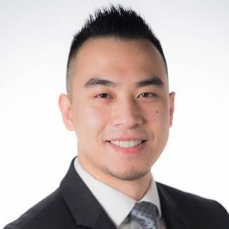 John Yue合作论文数Weill Institute for Neurosciences, University of California, San Francisco;The Philip R. Lee Institute for Health Policy Studies, University of California, San Francisco11
 Adib A. Abla合作论文数Neurological Surgery, UCSF Weill Institute for Neurosciences10
 Hansen Deng合作论文数Dept Neurol Surg, Univ Pittsburgh9
 Mukherjee Pratik合作论文数7
 Geoffrey Manley合作论文数Department of Neurological Surgery, School of Medicine, University of California, San Francisco;Brain And Spinal Injury Center;Zuckerberg San Francisco General Hospital7
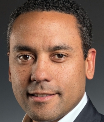 David O. Okonkwo合作论文数Neurotrauma Clinical Trials Center, Department of Neurological Surgery, School of Medicine, University of Pittsburgh7
 Minako Hayakawa合作论文数6Amanda Kwasnicki合作论文数6
 Hester F Lingsma合作论文数6Pui Man Rosalind Lai合作论文数6Ridhima Guniganti合作论文数Dept Neurol Surg, Washington Univ6
 David M Schnyer合作论文数Department of Psychology, College of Liberal Arts, University of Texas at Austin6Kunal P Raygor合作论文数6
 Stephanie Chen合作论文数Department of Neurosurgery, University of Miami Miller School of Medicine6
 Jason Sheehan合作论文数Department of Biomedical Engineering, School of Engineering and Applied Science, University of Virginia;Department of Radiation Oncology, School of Medicine, University of Virginia;UVA Health6
 Andrew Durnford合作论文数Department of Neurosurgery, University Hospital Southampton NHS Foundation Trust6
 Adriaan R E Potgieser合作论文数6
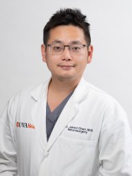 Chen Ching-Jen合作论文数Departments of 1 Neurological Surgery and.6
 Kansagra Akash P合作论文数School of Medicine, University of California6
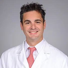 Isaac J. Abecassis合作论文数University of Louisville;Brain & Spine Institute, UofL Health;Abraham Flexner Outpatient Center, UofL Health6
 W. Christopher Fox合作论文数6Alex B. Valadka合作论文数6
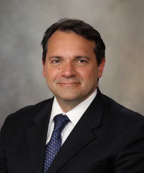 Giuseppe Lanzino合作论文数Department of Neurologic Surgery, Mayo Clinic;Department of Radiology, Mayo Clinic;Department of Neurosurgery, Mayo Clinic6
 Colin P. Derdeyn合作论文数Department of Radiology, University of Iowa6Robert M. Starke合作论文数6
 Rose Du合作论文数6
 Ali Alaraj合作论文数Department of Neurosurgery, College of Medicine, University of Illinois6
 Mary Vassar合作论文数Brain and Spinal Injury Center, University of California, San Francisco6
 Sepideh Amin-Hanjani合作论文数Department of Neurosurgery, University of Illinois at Chicago6
 Edgar A Samaniego合作论文数Stanford University, Stanford University6
 Diederik O. Bulters合作论文数Department of Neurosurgery, Addenbrooke’s Hospital6
 Esther Yuh合作论文数Department of Radiology & Biomedical Imaging, University of California, San Francisco;School of Medicine, University of California, San Francisco6Yoshiteru Tada合作论文数6
 J. Marc C. Van Dijk合作论文数University Medical Center Groningen, University of Groningen6
 Bradley A. Gross合作论文数The Feinberg School of Medicine and McGaw Medical Center, Northwestern University6
 Michael Levitt合作论文数University of Washington6
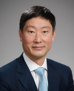 Louis J. Kim合作论文数University of Washington6Junichiro Satomi合作论文数6
 Gregory J. Zipfel合作论文数Division of Pediatric Neurosurgery, Department of Neurosurgery, Washington University School of Medicine in St. Louis;Roy and Diana Vagelos Division of Biology & Biomedical Sciences, Washington University School of Medicine in St. Louis;Barnes-Jewish Hospital;St. Louis Children’s Hospital;Missouri Baptist Medical Center6Megan Holt合作论文数5
 Adrian Contreras合作论文数5Nicole Nasholm合作论文数5
 Adam Polifka合作论文数5Shannon Wong-Michalak合作论文数5Robin Lea合作论文数5
 Jon D. Larson合作论文数5Joanna Phillips合作论文数Department of Neurological Surgery, Helen Diller Research Center, University of California San Francisco, San Francisco, CA, USA5Miller Huang合作论文数Div Hematol Oncol & Blood & Marrow Transplantat, Childrens Hosp Los Angeles5




