Lineage tracing based on modern live imaging approaches enables to visualize, reconstruct, and analyze the developmental history, fate, and dynamic behaviors of cells in vivo in a direct, comprehensive, and quantitative manner. Light-sheet fluorescence microscopy (LSFM) has greatly boosted lineage tracing efforts, because fluorescently labeled specimens can be imaged in their entirety, over long periods of time, with high spatiotemporal resolution and minimal photodamage. In addition, an increasing arsenal of commercial and open-source software solutions for cell and nuclei segmentation and tracking can be employed to convert data from pixel-based to object-based representations, and to reconstruct the lineages of cells in their native context as they organize in tissues, organs, and whole organisms. This chapter describes the preparation of LSFM image datasets and the use of three freely available platforms, namely, the Fiji/ImageJ plugins Massive Multiview Tracker (MaMuT), Mastodon and TrackMate, for small-scale and large-scale lineage tracing purposes using manual, semi-automated, and fully automated pipelines for nuclei or cell tracking. Lineage tracing with these tools is described on LSFM image datasets of fluorescently labeled embryos from the crustacean model Parhyale hawaiensis that lends itself to multi-scale investigations of development and regeneration.
Visual computing is an indispensable tool in biology, revolutionizing the way researchers interact with complex biological data. The integration of high-resolution Virtual Reality offers rich visualization experience, crucial for advancing our knowledge in areas such as genomics, proteomics, and cellular biology. Recent VR-based biological visualization often deploys lower-resolution Head Mounted Displays (HMDs), limiting the observation of fine structural detail. Moreover, the simultaneous exploration of multiple datasets in an interactive manner is still a research challenge. This work presents an interactive 3D reconstruction and visualization system for biological data in ultra-high-definition VR. The system supports volume rendering, flexible rendering, and the simultaneous visualization of multiple datasets, all within an 8K VR environment. Intricate cellular and subcellular structures as well as detailed protein folding patterns and subtle variations in tissue density are visualized, critical for accurate medical diagnosis and research. Our system’s performance is evaluated based on loading time, frames per second, and device latency, while handling large biological datasets. A think aloud study provided expert feedback.
How does growth encode form in developing organisms? Many different spatiotemporal growth profiles may sculpt tissues into the same target 3D shapes, but only specific growth patterns are observed in animal and plant development. In particular, growth profiles may differ in their degree of spatial variation and growth anisotropy; however, the criteria that distinguish observed patterns of growth from other possible alternatives are not understood. Here we exploit the mathematical formalism of quasiconformal transformations to formulate the problem of "growth pattern selection" quantitatively in the context of 3D shape formation by growing 2D epithelial sheets. We propose that nature settles on growth patterns that are the "simplest" in a certain way. Specifically, we demonstrate that growth pattern selection can be formulated as an optimization problem and solved for the trajectories that minimize spatiotemporal variation in areal growth rates and deformation anisotropy. The result is a complete prediction for the growth of the surface, including not only a set of intermediate shapes, but also a prediction for cell displacement along those surfaces in the process of growth. Optimization of growth trajectories for both idealized surfaces and those observed in nature show that relative growth rates can be uniformized at the cost of introducing anisotropy. Minimizing the variation of programmed growth rates can therefore be viewed as a generic mechanism for growth pattern selection and may help us to understand the prevalence of anisotropy in developmental programs.
Hybrid fluorescence and optoacoustic microscopy systems have recently emerged as powerful imaging modalities concurrently capturing both radiative and non-radiative molecular relaxations in biological tissues. Nevertheless, such approaches provide limited information as specimens are imaged exclusively from one side, not permitting the acquisition of their full anatomical, structural, or functional features in multiple views of interest. Herein we present a bimodal optical and optoacoustic multiview (BOOM) cost-efficient microscope operating in the frequency-domain for the comprehensive label-free imaging of established and emerging model organisms. Thus, the capabilities of BOOM microscopy have been proven suitable for highly demanding observations in developmental biology and embryology.
Frequency-domain photoacoustic microscopy (FD-PAM) constitutes a powerful cost-efficient imaging method integrating intensity-modulated laser beams for the excitation of single-frequency photoacoustic waves. Nevertheless, FD-PAM provides an extremely small signal-to-noise ratio (SNR), which can be up to two orders of magnitude lower than the conventional time-domain (TD) systems. To overcome this inherent SNR limitation of FD-PAM, we utilize a U-Net neural network aiming at image augmentation without the need for excessive averaging or the application of high optical power. In this context, we improve the accessibility of PAM as the system's cost is dramatically reduced, and we expand its applicability to demanding observations while retaining sufficiently high image quality standards.
Parhyale hawaiensis is a marine crustacean which has emerged as a powerful model organism to study molecular and cellular mechanisms linked to embryonic development and regenerative processes. Recently, several fluorescence-based optical microscopy techniques have been employed for the study of Parhyale to obtain anatomical descriptions, analyze gene expression patterns and reconstruct cell lineages. Aiming at the expansion of the imaging repertoire for this emerging model organism, we introduce a low-cost hybrid diagnostic system which integrates confocal fluorescence and frequency domain photoacoustic (FDPA) microscopy modalities, concurrently capturing both the radiative and radiationless relaxations of molecules following their excitation by an intensity-modulated laser source. We initially characterize the hybrid microscope in terms of PA signal generation efficiency and lateral spatial resolution, and subsequently evaluate its capabilities for the in vivo imaging of unlabelled and fluorescently labelled Parhyale embryos found at different stages of development. The recorded hybrid images have revealed a remarkable contrast complementarity between the integrated imaging modes, providing valuable information regarding cells’ structure, nuclei location, cellular membranes and yolk distribution. Our findings may pave the way for the broader adoption of inexpensive hybrid optical and PA microscopy methods in developmental biology, significantly upgrading the capabilities of the currently used technologies.
Morphogenesis, the process through which genes generate form, establishes tissue-scale order as a template for constructing the complex shapes of the body plan. The extensive growth required to build these ordered substrates is fuelled by cell proliferation, which, naively, should destroy order. Understanding how active morphogenetic mechanisms couple cellular and mechanical processes to generate order—rather than annihilate it—remains an outstanding question in animal development. We show that cell divisions are the primary drivers of tissue flow, leading to a fourfold orientationally ordered phase. Waves of anisotropic cell proliferation propagate across the embryo with precise patterning. Defects introduced into the nascent lattice by cell divisions are moved out of the tissue bulk towards the boundary by subsequent divisions. Specific cell proliferation rates and orientations enable cell divisions to organize rather than fluidize the tissue. We observe this using live imaging and tissue cartography to analyse the dynamics of fourfold tissue ordering in the trunk segmental ectoderm of the crustacean Parhyale hawaiensis beginning 72 h after egg lay. The result is a robust, active mechanism for generating global orientational order in a non-equilibrium system that sets the stage for the subsequent development of shape and form.
How the size and shape of developing tissues is encoded in the genome has been a longstanding riddle for biologists. Constituent cells integrate several genetic and mechanical signals to decide whether to divide, die, change shape or position. We review here how morphogenetic cell behaviors contribute to leg formation from imaginal disc epithelia in the insect Drosophila melanogaster, as well as to direct embryonic limb outgrowths in the non-insect pancrustacean Parhyale hawaiensis. Considering the deep conservation of developmental programs for limb patterning among arthropods and other bilaterians, moving forward, it will be exciting to see how these genetic similarities reflect at the cellular and tissue mechanics level.
We present the application of a low‐cost frequency domain photoacoustic (FDPA) microscope for the label‐free imaging of live developing embryos of the crustacean model organism Parhyale hawaiensis . By modulating the intensity of a continuous wave laser source at 9.5 MHz, we achieve the excitation of monochromatic PA waves, which are detected to provide amplitude and phase recordings. The data are subsequently processed to generate accurate maximum amplitude projection and surface reconstructions, delineating the morphological features of the embryos with high resolution and contrast. The findings of this study pave the way for the broader adoption of inexpensive PA diagnostic techniques in developmental biology, shedding light on various fundamental processes in established and emerging model organisms.
This chapter looks back over the last 20 years that witnessed the emergence of the marine crustacean Parhyale hawaiensis as an attractive experimental model organism for comparative developmental, genetic and genomic research. It covers the unique ethological, anatomical, embryological and developmental characteristics of Parhyale that enabled the establishment of diverse functional manipulations and standardized resources in this species. Multidisciplinary studies in Parhyale have helped resolve longstanding issues in biology, such as the molecular and cellular mechanisms underlying body patterning and appendage diversification during arthropod development and evolution. The chapter looks also forward and identifies key fields of biological and biomedical research, including regenerative biology, quantitative developmental biology, ecotoxicology and biotechnology, where Parhyale research could make major contributions in the future.
Many animal embryos pull and close an epithelial sheet around the ellipsoidal egg surface during a gastrulation process known as epiboly. The ovoidal geometry dictates that the epithelial sheet first expands and subsequently compacts. Moreover, the spreading epithelium is mechanically stressed and this stress needs to be released. Here we show that during extraembryonic tissue (serosa) epiboly in the insect Tribolium castaneum, the non-proliferative serosa becomes regionalized into a solid-like dorsal region with larger non-rearranging cells, and a more fluid-like ventral region surrounding the leading edge with smaller cells undergoing intercalations. Our results suggest that a heterogeneous actomyosin cable contributes to the fluidization of the leading edge by driving sequential eviction and intercalation of individual cells away from the serosa margin. Since this developmental solution utilized during epiboly resembles the mechanism of wound healing, we propose actomyosin cable-driven local tissue fluidization as a conserved morphogenetic module for closure of epithelial gaps.
Crustaceans have been favored in developmental biology for the study of the diversification of body plans and their associated appendages, which exhibit remarkable diversity within and between species. Until recently, because of technical limitations, crustacean studies were restricted in scope to the comparison of appendage morphologies and expression patterns of candidate limb patterning genes already known from classic developmental animal models. To remedy this limitation and explore their full potential, a few select crustacean experimental models have been reinforced with powerful genomic and transcriptomic resources, new methods for forward and reverse genetic investigations, and for live imaging of entire embryos, or cell and tissue-specific markers, with exceptional spatial and temporal resolution. These models include the malacostracan amphipod Parhyale hawaiensis and the branchiopod cladocerans Daphnia magna and Daphnia pulex, which display collectively all the different uniramous, biramous, and phyllopodous crustacean limb types. Within the past couple years, important discoveries have been made on the molecular and cellular basis of embryonic limb development and postembryonic limb regeneration. In Parhyale alone, gain and loss-of-function studies of Hox genes have revealed the combinatorial logic used by these genes for appendage specialization, whereas the reconstruction of single-cell-resolution fate maps of developing and regenerating appendages have identified the lineage restrictions and cellular behaviors driving both morphogenetic processes. Century-old questions regarding the conservation and divergence of appendage patterning mechanisms across arthropods and bilaterians, or how these mechanisms can be used and reused throughout the lifetime of an organism, can now be addressed productively with crustaceans.
Many animal embryos pull and close an epithelial sheet around the spherical or ellipsoidal egg surface during a gastrulation process known as epiboly. The ovoidal geometry dictates that the epithelial sheet first expands and subsequently compacts. Moreover, the epithelial sheet spreading over the sphere is mechanically stressed and this stress needs to be released. Here we show that during extraembryonic tissue (serosa) epiboly in the red flour beetle Tribolium castaneum , the non-proliferative serosa becomes regionalized into two distinct territories: a dorsal region under higher tension away from the leading edge with larger non-rearranging cells, and a more fluid ventral region under lower tension surrounding the leading edge with smaller cells undergoing cell intercalation. Our results suggest that fluidization of the leading edge is caused by a heterogeneous actomyosin cable that drives sequential eviction and intercalation of individual cells away from the serosa margin. Since this developmental solution utilized during epiboly resembles the mechanism of wound healing in other systems, we propose actomyosin cable-driven local tissue fluidization as a conserved morphogenetic module for closure of epithelial gaps.
During gastrulation, physical forces reshape the simple embryonic tissue to form the complex body plans of multicellular organisms 1 . These forces often cause large-scale asymmetric movements of the embryonic tissue 2 , 3 . In many embryos, the gastrulating tissue is surrounded by a rigid protective shell 4 . Although it is well-recognized that gastrulation movements depend on forces that are generated by tissue-intrinsic contractility 5 , 6 , it is not known whether interactions between the tissue and the protective shell provide additional forces that affect gastrulation. Here we show that a particular part of the blastoderm tissue of the red flour beetle ( Tribolium castaneum ) tightly adheres in a temporally coordinated manner to the vitelline envelope that surrounds the embryo. This attachment generates an additional force that counteracts tissue-intrinsic contractile forces to create asymmetric tissue movements. This localized attachment depends on an αPS2 integrin (inflated), and the knockdown of this integrin leads to a gastrulation phenotype that is consistent with complete loss of attachment. Furthermore, analysis of another integrin (the αPS3 integrin, scab) in the fruit fly ( Drosophila melanogaster ) suggests that gastrulation in this organism also relies on adhesion between the blastoderm and the vitelline envelope. Our findings reveal a conserved mechanism through which the spatiotemporal pattern of tissue adhesion to the vitelline envelope provides controllable, counteracting forces that shape gastrulation movements in insects.
During development, coordinated cell behaviors orchestrate tissue and organ morphogenesis. Detailed descriptions of cell lineages and behaviors provide a powerful framework to elucidate the mechanisms of morphogenesis. To study the cellular basis of limb development, we imaged transgenic fluorescently-labeled embryos from the crustacean Parhyale hawaiensis with multi-view light-sheet microscopy at high spatiotemporal resolution over several days of embryogenesis. The cell lineage of outgrowing thoracic limbs was reconstructed at single-cell resolution with new software called Massive Multi-view Tracker (MaMuT). In silico clonal analyses suggested that the early limb primordium becomes subdivided into anterior-posterior and dorsal-ventral compartments whose boundaries intersect at the distal tip of the growing limb. Limb-bud formation is associated with spatial modulation of cell proliferation, while limb elongation is also driven by preferential orientation of cell divisions along the proximal-distal growth axis. Cellular reconstructions were predictive of the expression patterns of limb development genes including the BMP morphogen Decapentaplegic.
SUMMARYDuring development coordinated cell behaviors orchestrate tissue and organ morphogenesis to suit the lifestyle of the organism. We have used here the crustaceanParhyale hawaiensisto study the cellular basis of limb development. TransgenicParhyaleembryos with fluorescently labeled nuclei were imaged at high spatiotemporal resolution with multi-view light-sheet fluorescence microscopy over several days of embryogenesis spanning appendage morphogenesis from early specification up to late differentiation stages. Cell tracking with a new tool called Massive Multi-view Tracker (MaMuT) enabled the reconstruction of the complete cell lineage of an outgrowing thoracic limb with single-cell resolution.In silicoclonal analyses suggested that the limb primordium becomes subdivided from an early stage first into anterior-posterior and then into dorsal-ventral compartments whose boundaries intersect at the distal tip of the growing limb. Limb bud formation is associated with the spatial modulation of cell proliferation, while limb elongation is also driven by the preferential orientation of division of epidermal cells along the proximal-distal axis of growth. Cellular reconstructions were predictive of the expression patterns of limb development genes including the Decapentaplegic (Dpp) morphogen.HIGHLIGHTSMulti-view light-sheet microscopy of crustacean embryos from speciesParhyale hawaiensisare ideal for cellular-level analysis of organ morphogenesis.Lineages of 3-dimensional organs were reconstructed at single-cell resolution with the Fiji/ImageJ plugin Massive Multi-view Tracker.TheParhyalelimb primordium undergoes early lineage restrictions associated with particular cell behaviors and patterns of gene expression.Differential rates of cell proliferation and oriented cell divisions guide appendage proximal-distal outgrowth.
The amphipod crustacean Parhyale hawaiensis is a blossoming model system for studies of developmental mechanisms and more recently regeneration. We have sequenced the genome allowing annotation of all key signaling pathways, transcription factors, and non-coding RNAs that will enhance ongoing functional studies. Parhyale is a member of the Malacostraca clade, which includes crustacean food crop species. We analysed the immunity related genes of Parhyale as an important comparative system for these species, where immunity related aquaculture problems have increased as farming has intensified. We also find that Parhyale and other species within Multicrustacea contain the enzyme sets necessary to perform lignocellulose digestion ('wood eating'), suggesting this ability may predate the diversification of this lineage. Our data provide an essential resource for further development of Parhyale as an experimental model. The first malacostracan genome will underpin ongoing comparative work in food crop species and research investigating lignocellulose as an energy source.
Full text Figures and data Side by side Abstract eLife digest Introduction Results and discussion Materials and methods Data availability References Decision letter Author response Article and author information Metrics Abstract The amphipod crustacean Parhyale hawaiensis is a blossoming model system for studies of developmental mechanisms and more recently regeneration. We have sequenced the genome allowing annotation of all key signaling pathways, transcription factors, and non-coding RNAs that will enhance ongoing functional studies. Parhyale is a member of the Malacostraca clade, which includes crustacean food crop species. We analysed the immunity related genes of Parhyale as an important comparative system for these species, where immunity related aquaculture problems have increased as farming has intensified. We also find that Parhyale and other species within Multicrustacea contain the enzyme sets necessary to perform lignocellulose digestion ('wood eating'), suggesting this ability may predate the diversification of this lineage. Our data provide an essential resource for further development of Parhyale as an experimental model. The first malacostracan genome will underpin ongoing comparative work in food crop species and research investigating lignocellulose as an energy source. https://doi.org/10.7554/eLife.20062.001 eLife digest The marine crustacean known as Parhyale hawaiensis is related to prawns, shrimps and crabs and is found at tropical coastlines around the world. This species has recently attracted scientific interest as a possible new model to study how animal embryos develop before birth and, because Parhyale can rapidly regrow lost limbs, how tissues and organs regenerate. Indeed, Parhyale has many characteristics that make it a good model organism, being small, fast-growing and easy to keep and care for in the laboratory. Several research tools have already been developed to make it easier to study Parhyale. This includes the creation of a system for using the popular gene editing technology, CRISPR, in this animal. However, one critical resource that is available for most model organisms was missing; the complete sequence of all the genetic information of this crustacean, also known as its genome, was not available. Kao, Lai, Stamataki et al. have now compiled the Parhyale genome – which is slightly larger than the human genome – and studied its genetics. Analysis revealed that Parhyale has genes that allow it to fully digest plant material. This is unusual because most animals that do this rely upon the help of bacteria. Kao, Lai, Stamataki et al. also identified genes that provide some of the first insights into the immune system of crustaceans, which protects these creatures from diseases. Kao, Lai, Stamataki et al. have provided a resource and findings that could help to establish Parhyale as a popular model organism for studying several ideas in biology, including organ regeneration and embryonic development. Understanding how Parhyale digests plant matter, for example, could progress the biofuel industry towards efficient production of greener energy. Insights from its immune system could also be adapted to make farmed shrimp and prawns more resistant to infections, boosting seafood production. https://doi.org/10.7554/eLife.20062.002 Introduction Very few members of the Animal Kingdom hold the esteemed position of major model system for understanding living systems. Inventions in molecular and cellular biology increasingly facilitate the emergence of new experimental systems for developmental genetic studies. The morphological and ecological diversity of the phylum Arthropoda makes them an ideal group of animals for comparative studies encompassing embryology, adaptation of adult body plans and life history evolution (Akam, 2000; Budd and Telford, 2009; Peel et al., 2005; Scholtz and Wolff, 2013). While the most widely studied group are Hexapods, reflected by over a hundred sequencing projects available in the NCBI genome database, genomic data in the other three sub-phyla in Arthropoda are still relatively sparse. Recent molecular and morphological studies have placed crustaceans along with hexapods into a pancrustacean clade (Figure 1A), revealing that crustaceans are paraphyletic (Mallatt et al., 2004; Cook et al., 2005; Regier et al., 2005; Ertas et al., 2009; Richter, 2002). Previously, the only available fully sequenced crustacean genome was that of the water flea Daphnia which is a member of the Branchiopoda (Colbourne et al., 2011). A growing number of transcriptomes for larger phylogenetic analyses have led to differing hypotheses of the relationships of the major pancrustacean groups (Figure 1B) (Meusemann et al., 2010; Regier et al., 2010; Oakley et al., 2013; von Reumont et al., 2012). The genome of the amphipod crustacean Parhyale hawaiensis addresses the paucity of high quality non-hexapod genomes among the pancrustacean group, and will help to resolve relationships within this group as more genomes and complete proteomes become available (Rivarola-Duarte et al., 2014; Kenny et al., 2014). Crucially, genome sequence data is also necessary to further advance research in Parhyale, currently the most tractable crustacean model system. This is particularly true for the application of powerful functional genomic approaches, such as genome editing (Cong et al., 2013; Serano et al., 2015; Martin et al., 2015; Mali et al., 2013; Jinek et al., 2012; Gilles and Averof, 2014). Figure 1 Download asset Open asset Introduction. (A) Phylogenetic relationship of Arthropods showing the Chelicerata as an outgroup to Mandibulata and the Pancrustacea clade which includes crustaceans and insects. Species listed for each clade have ongoing or complete genomes. Species include Crustacea: Parhyale hawaiensis, D. pulex; Hexapoda: Drosophila melanogaster, Apis mellifera, Bombyx mori, Aedis aegypti, Tribolium castaneum; Myriapoda: Strigamia maritima, Trigoniulus corallines; Chelicerata: Ixodes scapularis, Tetranychus urticae, Mesobuthus martensii, Stegodyphus mimosarum. (B) One of the unresolved issues concerns the placement of the Branchiopoda either together with the Cephalocarida, Remipedia and Hexapoda (Allotriocarida hypothesis A) or with the Copepoda, Thecostraca and Malacostraca (Vericrustacea hypothesis B). (C) Life cycle of Parhyale that takes about two months at 26C. Parhyale is a direct developer and a sexually dimorphic species. The fertilized egg undergoes stereotyped total cleavages and each blastomere becomes committed to a particular germ layer already at the 8-cell stage depicted in (D). The three macromeres Er, El, and Ep give rise to the anterior right, anterior left, and posterior ectoderm, respectively, while the fourth macromere Mav gives rise to the visceral mesoderm and anterior head somatic mesoderm. Among the 4 micromeres, the mr and ml micromeres give rise to the right and left somatic trunk mesoderm, en gives rise to the endoderm, and g gives rise to the germline. https://doi.org/10.7554/eLife.20062.003 Parhyale is a member of the diverse Malacostraca clade with thousands of extant species including economically and nutritionally important groups such as shrimps, crabs, crayfish and lobsters, as well as common garden animals like woodlice. They are found in all marine, fresh water, and higher humidity terrestrial environments. Apart from attracting research interest as an economically important food crop, this group of animals has been used to study developmental biology and the evolution of morphological diversity (for example with respect to Hox genes) (Martin et al., 2015; Averof and Patel, 1997; Liubicich et al., 2009; Pavlopoulos et al., 2009), stem cell biology (Konstantinides and Averof, 2014; Benton et al., 2014), innate immunity processes (Vazquez et al., 2009; Hauton, 2012) and recently the cellular mechanisms of regeneration (Konstantinides and Averof, 2014; Benton et al., 2014; Alwes et al., 2016). In addition, members of the Malacostraca, specifically both Amphipods and Isopods, are thought to be capable of 'wood eating' or lignocellulose digestion and to have microbiota-free digestive systems (King et al., 2010; Kern et al., 2013; Boyle and Mitchell, 1978; Zimmer et al., 2002). The life history of Parhyale makes it a versatile model organism amenable to experimental manipulations (Figure 1C) (Wolff and Gerberding, 2015). Gravid females lay eggs every 2 weeks upon reaching sexual maturity and hundreds of eggs can be easily collected at all stages of embryogenesis. Embryogenesis takes about 10 days at 26°C and has been described in detail with an accurate staging system (Browne et al., 2005). Early embryos display an invariant cell lineage with each blastomere at the 8-cell stage contributing to a specific germ layer (Figure 1D) (Browne et al., 2005; Gerberding et al., 2002). Embryonic and post-embryonic stages are amenable to experimental manipulations and direct observation in vivo (Gerberding et al., 2002; Extavour, 2005; Rehm et al., 2009a, 2009b, 2009c, 2009d; Price et al., 2010; Alwes et al., 2011; Hannibal et al., 2012; Kontarakis and Pavlopoulos, 2014; Nast and Extavour, 2014; Chaw and Patel, 2012; Pavlopoulos and Averof, 2005). These can be combined with transgenic approaches (Pavlopoulos and Averof, 2005; Kontarakis et al., 2011; Kontarakis and Pavlopoulos, 2014; Pavlopoulos et al., 2009), RNA interference (RNAi) (Liubicich et al., 2009) and morpholino-mediated gene knockdown (Ozhan-Kizil et al., 2009), and transgene-based lineage tracing (Konstantinides and Averof, 2014). Most recently the utility of the clustered regularly interspaced short palindromic repeats (CRISPR)/CRISPR-associated (Cas) system for targeted genome editing has been elegantly demonstrated during the systematic study of Parhyale Hox genes (Martin et al., 2015; Serano et al., 2015). This arsenal of experimental tools (Table 1) has already established Parhyale as an attractive model system for biological research. Table 1 Experimental resources. Available experimental resources in Parhyale and corresponding references. https://doi.org/10.7554/eLife.20062.004 Experimental ResourcesReferencesEmbryological manipulations Cell microinjection, isolation, ablation(Gerberding et al., 2002; Extavour, 2005; Price et al., 2010; Alwes et al., 2011; Hannibal et al., 2012; Rehm et al., 2009; Rehm et al., 2009; Kontarakis and Pavlopoulos, 2014; Nast and Extavour, 2014)Gene expression studies In situ hybridization, antibody staining(Rehm et al., 2009; Rehm et al., 2009)Gene knock-down RNA interference, morpholinos(Liubicich et al., 2009; Ozhan-Kizil et al., 2009)Transgenesis Transposon-based, integrase-based(Pavlopoulos and Averof, 2005; Kontarakis et al., 2011; Kontarakis and Pavlopoulos, 2014)Gene trapping Exon/enhancer trapping, iTRAC (trap conversion)(Kontarakis et al., 2011)Gene misexpressionHeat-inducible(Pavlopoulos et al., 2009)Gene knock-outCRISPR/Cas(Martin et al., 2015)Gene knock-in CRISPR/Cas homology-dependent or homology-independent(Serano et al., 2015)Live imaging Bright-field, confocal, light-sheet microscopy(Alwes et al., 2011; Hannibal et al., 2012; Chaw and Patel, 2012; Alwes et al., 2016) So far, work in Parhyale has been constrained by the lack of a reference genome and other standardized genome-wide resources. To address this limitation, we have sequenced, assembled and annotated the genome. At an estimated size of 3.6 Gb, this genome represents one of the largest animal genomes tackled to date. The large size has not been the only challenge of the Parhyale genome, that also exhibits some of the highest levels of sequence repetitiveness and polymorphism reported among published genomes. We provide information in our assembly regarding polymorphism to facilitate functional genomic approaches sensitive to levels of sequence similarity, particularly homology-dependent genome editing approaches. We analysed a number of key features of the genome as foundations for new areas of research in Parhyale, including innate immunity in crustaceans, lignocellulose digestion, non-coding RNA biology, and epigenetic control of the genome. Our data bring Parhyale to the forefront of developing model systems for a broad swathe of important bioscience research questions. Results and discussion Genome assembly, annotation, and validation The Parhyale genome contains 23 pairs (2n=46) of chromosomes (Figure 2) and with an estimated size of 3.6 Gb, it is currently the second largest reported arthropod genome after the locust genome (Parchem et al., 2010; Wang et al., 2014). Sequencing was performed on genomic DNA isolated from a single adult male taken from a line derived from a single female and expanded after two rounds of sib-mating. We performed k-mer analyses of the trimmed reads to assess the impact of repeats and polymorphism on the assembly process. We analyzed k-mer frequencies (Figure 3A) and compared k-mer representation between our different sequencing libraries. We observed a 93% intersection of unique k-mers among sequencing libraries, indicating that the informational content was consistent between libraries (Source code 1). The k-mer analysis revealed a bimodal distribution of error-free k-mers (Figure 3A). The higher-frequency peak corresponded to k-mers present on both haplotypes (i.e. homozygous regions), while the lower-frequency peak had half the coverage and corresponded to k-mers present on one haplotype (i.e. heterozygous regions) (Simpson and Durbin, 2012). We concluded that the single sequenced adult Parhyale exhibits very high levels of heterozygosity, similar to the highly heterozygous oyster genome (see below). Figure 2 Download asset Open asset Parhyale karyotype. (A) Frequency of the number of chromosomes observed in 42 mitotic spreads. Forty-six chromosomes were observed in more than half of all preparations. (B) Representative image of Hoechst-stained chromosomes. https://doi.org/10.7554/eLife.20062.005 Figure 3 Download asset Open asset Parhyale genome assembly metrics. (A) K-mer frequency spectra of all reads for k-lengths ranging from 20 to 50. (B) K-mer branching analysis showing the frequency of k-mer branches classified as variants compared to Homo sapiens (human), Crassostrea gigas (oyster), and Saccharomyces cerevisiae (yeast). (C) K-mer branching analysis showing the frequency of k-mer branches classified as repetitive compared to H. sapiens, C. gigas and S. cerevisiae. (D) Histogram of read coverages of assembled contigs. (E) The number of contigs with an identity ranging from 70–95% to another contig in the set of assembled contigs. (F) Collapsed contigs (green) are contigs with at least 95% identity with a longer primary contig (red). These contigs were removed prior to scaffolding and added back as potential heterozygous contigs after scaffolding. https://doi.org/10.7554/eLife.20062.006 In order to quantify global heterozygosity and repeat content of the genome we assessed the de-Bruijn graphs generated from the trimmed reads to observe the frequency of both variant and repeat branches (Simpson, 2014) (Figure 3B and C). We found that the frequency of the variant branches was 10x higher than that observed in the human genome and very similar to levels in the highly polymorphic genome of the oyster Crassostrea gigas (Zhang et al., 2012). We also observed a frequency of repeat branches approximately 4x higher than those observed in both the human and oyster genomes (Figure 3C), suggesting that the big size of the Parhyale genome can be in large part attributed to the expansion of repetitive sequences. These metrics suggested that both contig assembly and scaffolding with mate-pair reads were likely to be challenging due to high heterozygosity and repeat content. After an initial contig assembly we remapped reads to assess coverage of each contig. We observed a major peak centered around 75x coverage and a smaller peak at 150x coverage. Contigs with lower 75x coverage represent regions of the genome that assembled into separate haplotypes and had half the frequency of mapped sequencing reads, reflecting high levels of heterozygosity. This resulted in independent assembly of haplotypes for much of the genome (Figure 3D). One of the prime goals in sequencing the Parhyale genome was to achieve an assembly that could assist functional genetic and genomic approaches in this species. Different strategies have been employed to sequence highly heterozygous diploid genomes of non-model and wild-type samples (Kajitani et al., 2014). We aimed for an assembly representative of different haplotypes, allowing manipulations to be targeted to different allelic variants in the assembly. This could be particularly important for homology dependent strategies that are likely to be sensitive to polymorphism. However, the presence of alternative haplotypes could lead to poor scaffolding between contigs as many mate-pair reads may not map uniquely to one contig and distinguish between haplotypes in the assembly. To alleviate this problem we used a strategy to conservatively identify pairs of allelic contigs and proceeded to use only one in the scaffolding process. First, we estimated levels of similarity (identity and alignment length) between all assembled contigs to identify independently assembled allelic regions (Figure 3E). We then kept the longer contig of each pair for scaffolding using our mate-pair libraries (Figure 3F), after which we added back the shorter allelic contigs to produce the final genome assembly (Figure 4A). Figure 4 with 1 supplement see all Download asset Open asset Workflows of assembly, annotation, and proteome generation. (A) Flowchart of the genome assembly. Two shotgun libraries and four mate-pair libraries with the indicated average sizes were prepared from a single male animal and sequenced to a predicted depth of 115x coverage after read filtering, based on a predicted size of 3.6 Gbp. Contigs were assembled at two different k-lengths with Abyss and the two assemblies were merged with GAM-NGS. Filtered contigs were scaffolded with SSPACE. (B) The final scaffolded assembly was annotated with a combination of Evidence Modeler to generate 847 high quality gene models and Augustus for the final set of 28,155 predictions. These protein-coding gene models were generated based on a Parhyale transcriptome consolidated from multiple developmental stages and conditions, their homology to the species indicated, and ab initio predictions with GeneMark and SNAP. (C) The Parhyale proteome contains 28,666 entries based on the consolidated transcriptome and gene predictions. The transcriptome contains 292,924 coding and non-coding RNAs, 96% of which could be mapped to the assembled genome. https://doi.org/10.7554/eLife.20062.007 Figure 4—source data 1 Catalog of repeat elements in Parhyale genome assembly. Description of repeat content in the Parhyale genome. https://doi.org/10.7554/eLife.20062.008 Download elife-20062-fig4-data1-v3.xlsx Figure 4—source data 2 Software and Data. List of programs and bioinformatic tools and publicly available sequence data used in this study. https://doi.org/10.7554/eLife.20062.009 Download elife-20062-fig4-data2-v3.xlsx RepeatModeler and RepeatMasker were used on the final assembly to find repetitive regions, which were subsequently classified into families of transposable elements or short tandem repeats (Source code 2). We found 1473 different repeat element sequences representing 57% of the assembly (Figure 4—source data 1). The Parhyale assembly comprises of 133,035 scaffolds (90% of assembly), 259,343 unplaced contigs (4% of assembly), and 584,392 shorter, potentially allelic contigs (6% of assembly), with a total length of 4.02 Gb (Table 2). The N50 length of the scaffolds is 81,190 bp. The final genome assembly was annotated with Augustus trained with high confidence gene models derived from assembled transcriptomes, gene homology, and ab initio predictions. This resulted in 28,155 final gene models (Figure 4B; Source code 3) across 14,805 genic scaffolds and 357 unplaced contigs with an N50 of 161,819, bp and an N90 of 52,952 bp. Table 2 Assembly statistics. Length metrics of assembled scaffolds and contigs. https://doi.org/10.7554/eLife.20062.011 # sequencesN90N50N10Sum lengthMax length# Nsscaffolds133,03514,79981,190289,7053.63 GB1,285,3851.10 GBunplaced contigs259,3433046271779146 MB40,22223,431hetero. contigs584,3922654021038240 MB24,461627genic scaffolds15,16052952161,8194338361.49 GB1,285,385323 MB Parhyale has a mean coding gene size (introns and ORFs) of 20 kb (median of 7.2 kb), which is longer than D. pulex (mean: 2 kb, median: 1.2 kb), while shorter than genes in Homo sapiens (mean: 52.9 kb, median: 18.5 kb). This difference in gene length was consistent across reciprocal blast pairs where ratios of gene lengths revealed Parhyale genes were longer than Caenorhabditis elegans, D. pulex, and Drosophila melanogaster and similar to H. sapiens. (Figure 5A). The mean intron size in Parhyale is 5.4 kb, similar to intron size in H. sapiens (5.9 kb) but dramatically longer than introns in D. pulex (0.3 kb), D. melanogaster (0.3 kb) and C. elegans (1 kb) (Figure 5B). Figure 5 with 1 supplement see all Download asset Open asset Parhyale genome comparisons. (A) Box plots comparing gene sizes between Parhyale and humans (H. sapiens), water fleas (D. pulex), flies (D. melanogaster) and nematodes (C. elegans). Ratios were calculated by dividing the size of the top blast hit in each species with the corresponding Parhyale gene size. (B) Box plots showing the distribution of intron sizes in the same species used in A. (C) Comparison between Parhyale and representative proteomes from the indicated animal taxa. Colored bars indicate the number of blast hits recovered across various thresholds of E-values. The top hit value represents the number of proteins with a top hit corresponding to the respective species. (D) Cladogram showing the number of shared orthologous protein groups at various taxonomic levels, as well as the number of clade-specific groups. A total of 123,341 orthogroups were identified with Orthofinder across the 16 genomes used in this analysis. Within Pancrustacea, 37 orthogroups were shared between Branchiopoda and Hexapoda (supporting the Allotriocarida hypothesis) and 49 orthogroups were shared between Branchiopoda and Amphipoda (supporting the Vericrustacea hypothesis). https://doi.org/10.7554/eLife.20062.012 Figure 5—source data 1 List of proteins currently unique to Parhyale. List of proteins in Parhyale without identity to other species. https://doi.org/10.7554/eLife.20062.013 Download elife-20062-fig5-data1-v3.txt Figure 5—source data 2 List of genes likely to be specific to the Malacostraca List of genes likely to be specific to the Malacostraca. https://doi.org/10.7554/eLife.20062.014 Download elife-20062-fig5-data2-v3.txt Figure 5—source data 3 Orthofinder analysis. Orthofinder analysis using the Parhyale predicted proteome. https://doi.org/10.7554/eLife.20062.015 Download elife-20062-fig5-data3-v3.txt For downstream analyses of Parhyale protein coding content, a final proteome consisting of 28,666 proteins was generated by combining candidate coding sequences identified with TransDecoder (Haas et al., 2013) from mixed stage transcriptomes. Almost certainly the high number of predicted gene models and proteins is an overestimation due to fragmented genes, very different isoforms or unresolved alleles, that will be consolidated as annotation of the Parhyale genome improves. We also included additional high confidence gene predictions that were not found in the transcriptome (Figure 4C). The canonical proteome dataset was annotated with both Pfam, KEGG, and BLAST against Uniprot. Assembly quality was further evaluated by alignment to core eukaryotic genes defined by the Core Eukaryotic Genes Mapping Approach (CEGMA) database (Parra et al., 2007). We identified 244/248 CEGMA orthology groups from the assembled genome alone and 247/248 with a combination of genome and mapped transcriptome data (Figure 4—figure supplement 1). Additionally, 96% of over 280,000 identified transcripts, most of which are fragments that do not contain a large ORF, also mapped to the assembled genome. Together these data suggest that our assembly is close to complete with respect to protein coding genes and transcribed regions that are captured by deep RNA sequencing. High levels of heterozygosity and polymorphism in the Parhyale genome To estimate the level of heterozygosity in genes we first identified transcribed regions of the genome by mapping back transcripts to the assembly. Where these regions appeared in a single contig in the assembly, heterozygosity was calculated using information from mapped reads. Where these regions appeared in more than one contig, because haplotypes had assembled independently, heterozygosity was calculated using an alignment of the genomic sequences corresponding to mapped transcripts and information from mapped reads. This allowed us to calculate heterozygosity for each gene within the sequenced individual (Source code 4). We then calculated the genomic coverage of all transcribed regions in the genome and found, as expected, they fell broadly into two categories with higher and lower read coverage (Figure 6A; Source code 4). Genes that fell within the higher read coverage group had a lower mean heterozygosity (1.09% of bases displaying polymorphism), which is expected as more reads were successfully mapped. Genes that fell within the lower read coverage group had a higher heterozygosity (2.68%), as reads mapped independently to each haplotype (Figure 6B) (Simpson, 2014). Thus, we conclude that heterozygosity that influences read mapping and assembly of transcribed regions, and not just non-coding parts of the assembly. Figure 6 with 1 supplement see all Download asset Open asset Variation analyses of predicted genes. (A) A read coverage histogram of predicted genes. Reads were first mapped to the genome, then coverage was calculated for transcribed regions of each defined locus. (B) A coverage distribution plot showing that genes in the lower coverage region (<105x coverage, peak at 75x ) have a higher level of heterozygosity than genes in the higher coverage region (>105 coverage and <250, peak at approximately 150x coverage). (C) Distribution plot indicating that mean level of population variance is similar for genes in the higher and lower coverage regions. https://doi.org/10.7554/eLife.20062.017 Figure 6—source data 1 Polymorphism in Parhyale devlopmental genes. Description of polymorphism in previously identfied Parhyale developmental genes. https://doi.org/10.7554/eLife.20062.018 Download elife-20062-fig6-data1-v3.xlsx The assembled Parhyale transcriptome was derived from various laboratory populations, hence we expected to see additional polymorphism beyond that detected in the two haplotypes of the individual male we sequenced. Analysing all genes using the transcriptome we found additional variations in transcribed regions not found in the genome of the sequenced individual. In addition to polymorphisms that agreed with heterozygosity in the genome sequence we observed that the rate of additional variations is not substantially different between genes from the higher (0.88%) versus lower coverage group genes (0.73%; Figure 6C). This analysis suggests that within captive laboratory populations of Parhyale there is considerable additional polymorphism distributed across genes, irrespective of whether or not they have relatively low or high heterozygosity in the individual male we sequenced. In addition the single male we have sequenced provides an accurate reflection of polymorphism of the wider laboratory population and the established Chicago-F strain does not by chance contain unusually divergent haplotypes. We also performed an assessment of polymorphism on previously cloned Parhyale developmental genes, and found some examples of startling levels of variation. (Figure 6—figure supplement 1 and source data 1). For example, we found that the cDNAs of the germ line determinants, nanos (78 SNPS, 34 non-synonymous substitutions and one 6 bp indel) and vasa (37 SNPs, 7 non-synonymous substitutions and a one 6 bp indel) can have more variability within laboratory Parhyale populations than might be observed for orthologs between closely related species (Figure 6—source data 1). To further evaluate the extent of polymorphism across the genome, we mapped the genomic reads to a set of previously Sanger-sequenced BAC clones of the Parhyale Hox cluster from the same Chicago-F line from which we sequenced the genome of an adult male. (Serano et al., 2015). We detected SNPs at a rate of 1.3 to 2.5% among the BACs (Table 3) and also additional sequence differences between the BACs and genomic reads, confirming that additional polymorhism exists in the C
One of the key morphogenetic processes used during development is the controlled intercalation of cells between their neighbors. This process has been co-opted into a range of developmental events, and it also underlies an event that occurs in each major group of bilaterians: elongation of the embryo along the anterior-posterior axis [1]. In Drosophila, a novel component of this process was recently discovered by Pare et al., who showed that three Toll genes function together to drive cell intercalation during germband extension [2]. This finding raises the question of whether this role of Toll genes is an evolutionary novelty of flies or a general mechanism of embryonic morphogenesis. Here we show that the Toll gene function in axis elongation is, in fact, widely conserved among arthropods. First, we functionally demonstrate that two Toll genes are required for cell intercalation in the beetle Tribolium castaneum. We then show that these genes belong to a previously undescribed Toll subfamily and that members of this subfamily exhibit striped expression (as seen in Tribolium and previously reported in Drosophila [3-5]) in embryos of six other arthropod species spanning the entire phylum. Last, we show that two of these Toll genes are required for normal morphogenesis during anterior-posterior embryo elongation in the spider Parasteatoda tepidariorum, a member of the most basally branching arthropod lineage. From our findings, we hypothesize that Toll genes had a morphogenetic function in embryo elongation in the last common ancestor of all arthropods, which existed over 550 million years ago.
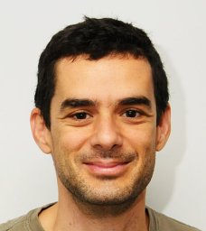 Michalis Averof合作论文数;Institute of Molecular Biology and Biotechnology (IMBB),, Foundation for Research and Technology Hellas (FoRTH);Institute of Molecular Biology and Biotechnology (IMBB),, Foundation for Research and Technology Hellas (FoRTH)10
 Kontarakis Zacharias合作论文数6Stamataki Evangelia合作论文数6
 Pavel Tomancak合作论文数Max Planck Institute of Molecular Cell Biology and Genetics;Central European Institute of Technology6
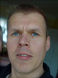 Carsten Wolff合作论文数Institut für Biologie/Vergleichende Zoologie, Humboldt-Universität zu Berlin5
 Akanksha Jain合作论文数Quantitative Developmental Biology Lab, Eidgenössische Technische Hochschule Zürich4
 Tserevelakis George J合作论文数Institute of Electronic Structure and Laser, Foundation of Research and Technology-Hellas4Konstantinides Nikolaos合作论文数Department of Biology, New York University4
 Benton Matthew A合作论文数Laboratory for Development and Evolution, University of Cambridge4
 Nipam Patel合作论文数The Marine Biological Laboratory, The University of Chicago;Department of Organismal Biology and Anatomy, The University of Chicago;Department of Molecular Genetics and Cell Biology, The University of Chicago4
 Giannis Zacharakis合作论文数Institute of Electronic Structure and Laser, Foundation for Research and, Technology—Hellas4
 Michael Akam合作论文数Darwin College, University of Cambridge;Department of Zoology, University of Cambridge4Ioannis Liaskas合作论文数3
 John Rallis合作论文数3
 Stephan W. Grill合作论文数<institution>Max-Planck-Institute of Molecular Cell Biology and Genetics</institution>, Dresden, <country>Germany</country>3
 Charalambos Savakis合作论文数Hellas Institute of Molecular Biology and Biotechnology, Foundation for Research and Technology3
 Benjamin Harich合作论文数2Andy Le合作论文数2Maria Kalogeridi合作论文数2Alessia Di Donfrancesco合作论文数2Stefan Oehler合作论文数2Dillon Cislo合作论文数2
 Andrew L Lemire合作论文数Howard Hughes Med Inst, Janelia Res Campus2
 Lokesh G Pimpale合作论文数Max Planck Inst Mol Cell Biol & Genet, Dresden, Germany2
 Michael Bruce Eisen合作论文数College of Letters & Science, University of California, Berkeley;Department of Cell Biology, Howard Hughes Medical Institute2
 Alvina Lai合作论文数Institute of Health Informatics, Faculty of Population Health Sciences, University College London2
 Heather Bruce合作论文数Department of Zoology, Faculty of Science, The University of British Columbia2
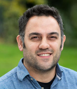 Aziz Aboobaker合作论文数Department of Zoology, University of Oxford;Department of Biology, University of Oxford2
 Peter Sarkies合作论文数2
 Siwanowicz Igor合作论文数2Kao Damian合作论文数2
 Marco Grillo合作论文数Institut de Biologia Molecular, Institut de Recerca Biomèdica de Barcelona2Rošić Silvana合作论文数2
 Jarvis Erin E合作论文数2
 Julia Serano合作论文数2
 Stefan Münster合作论文数Harvard2Marie Sémon合作论文数Smurfit Institute of Genetics, Trinity College Dublin2Danielle Liubicich合作论文数2
 Spencer L. Shorte合作论文数Laboratory of Molecular Dynamics;Department of Cell Biology;Medical University of South Carolina;Laboratory of Molecular Dynamics, Medical University of South Carolina2
 Philipp J. Keller合作论文数The Keller Lab, Howard Hughes Medical Institute2
 Florian Jug合作论文数Fondatione Human Technopole;Max Planck Institute of Molecular Cell Biology and Genetics2
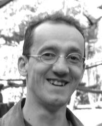 Jean-Yves Tinevez合作论文数Laboratoire Physico-Chimie Curie, Institut Curie Recherche2
 William E Browne合作论文数University of Chicago2Maria G. Kapetanaki合作论文数2
 Vladimir Ulman合作论文数Centre for Biomedical Image Analysis, Masaryk University2
 Robert Haase合作论文数Medical Faculty Carl Gustav Carus, TU Dresden2
 Stephan Preibisch合作论文数Max-Planck-Institute for Molecular Cell Biology and Genetics, D-01307 Dresden, Germany2
 Cassandra G. Extavour合作论文数Department of Organismic and Evolutionary Biology, Faculty of Arts and Sciences, Harvard University;Howard Hughes Medical Institute2Tobias Pietzsch合作论文数Technische Universitat Dresden2




