Efficient priming of antigen‑specific CD8⁺ T cells remains a major challenge for peptide‑ and protein‑based therapeutic vaccines targeting cancer and intracellular pathogens, despite the use of potent innate immune adjuvants such as synthetic double‑stranded RNA (dsRNA). In this study, we evaluated photochemical internalization (PCI), an endosomal escape technology, as a strategy to enhance vaccine‑induced immunity in combination with the dsRNA adjuvants poly(I:C) and poly‑ICLC. In mouse models, PCI combined with the SIINFEKL peptide and poly(I:C) increased circulating antigen‑specific CD8⁺ T cells by approximately 100‑fold compared with vaccination without PCI. Using a synthetic long HPV16 E7 peptide, PCI with poly(I:C) induced robust effector- and memory-phenotype CD8⁺ T‑cell responses and strong anti‑tumour activity in the TC‑1 HPV cancer model. PCI also enhanced CD8⁺ T‑cell priming induced by the clinically relevant telomerase vaccine UV1 when combined with poly‑ICLC. For larger protein antigens, including PPD, HBsAg, and KLH, PCI augmented CD4⁺ T‑cell responses and IgG production. Together, these findings establish PCI as a broadly applicable platform for enhancing peptide‑ and protein‑based vaccine immunity in combination with synthetic dsRNA adjuvants.
Breast cancer, a prevalent malignancy worldwide, includes the triple‐negative subtype (TNBC) characterized by poor treatment outcomes. TNBC has been shown to be sensitive to ferroptotic cell death, an iron‐dependent cell death mechanism involving reactive oxygen species (ROS) and lipid peroxidation. Herein, biodegradable tetraphenylchlorin‐conjugated chitosan nanoparticles (TPC‐CS NPs) in combination with the free ferroptosis inducer RSL3 is used in MCF7 (hormone receptor‐positive, epithelial) and MDA‐MB‐231 (hormone receptor‐negative, mesenchymal‐like) breast cancer cell lines. The results show that RSL3 treatment has no cytotoxic effect in MCF7 and there is no enhanced sensitivity when combined with TPC‐CS NPs, while the combination sensitizes MDA‐MB‐231 cells. Western blot analysis reveals that the combined treatment decreases and differently affects GPX4 levels and the ratio between the two GPX isoforms in the two cell lines. In MDA‐MB‐231 cells, the combined treatment shows enhanced effects on lipid peroxidation, mitochondrial potential, and basal and maximal respiration, as compared to single treatments. Finally, ferroptosis expression signatures distinguish breast cancer cell lines with an increasing score in mesenchymal‐like cells. Moreover, the signatures correlate with breast cancer subtypes, exhibiting the highest scores in subtypes rich in mesenchymal‐like cells, particularly basal‐like and claudin‐low tumors, suggesting their susceptibility to ferroptosis induction.
Triple-negative breast cancer (TNBC) is a subtype of breast cancer associated with poor prognosis and limited treatment options. While chemotherapy has traditionally been the main treatment, resistance frequently emerges, reducing its effectiveness. Recently, a range of immunotherapy strategies, including combinations of chemotherapy and immunotherapy, have expanded treatment possibilities. To further enhance the therapeutic efficacy, there is a desire to combine chemotherapy with other currently used modalities such as photodynamic therapy (PDT) or to encapsulate drugs into nanoparticles (NPs). Ferroptosis has been described as a highly immunogenic type of cell death, characterized by increased ROS and lipid peroxidation. Mesenchymal cancer cells have been reported to be more sensitive to ferroptosis than epithelial cells. We have previously shown that tetraphenylchlorin-conjugated chitosan nanoparticles (TPC-CS NPs) loaded with mertansine and cabazitaxel induce ferroptosis in TNBC.1 In this project, we investigated the effect of TPC-CS NPs together with free RSL3, a ferroptosis inducer. We show that such treatment induces cell death in a mesenchymal breast cancer cell line but not in an epithelial one. The combined treatment also significantly affects lipid peroxidation and mitochondrial function. Based on these results, we encapsulated RSL3 into TPC-CS NPs and used them in two different breast cancer cell lines. We explored the photodynamic effect of empty and RSL3 loaded TPC-CS NPs upon illumination on cell viability by using an MTS assay. Since we have not observed any beneficial effect of TPC-CS NPs-RSL3, we explored the uptake of only empty TPC-CS NPs by using fluorescence microscopy and flow cytometry in these two cell lines. Currently, a biodistribution study in immunocompetent animals bearing 4T1 tumors is being performed. We will perform the maximum tolerable dose experiments for illumination to determine the light dose for further experiments. Moreover, we will perform efficacy studies of TPC-CS NPs and investigate the immune cell composition of tumors and tumor microenvironment using a flow cytometry panel of 25 markers. 1.Pandya AD, Øverbye A, Sahariah P, et al. Drug-Loaded Photosensitizer-Chitosan Nanoparticles for Combinatorial Chemo- and Photodynamic-Therapy of Cancer. Biomacromolecules. 2020/04/13 2020;21(4):1489-1498. This project has received funding from the Europe Union’s Horizon 2020 research and innovation program under the Marie Sklodowska-Curie grant agreement 956544. Marek Feith, Abhilash D. Pandya, Astrid Hyldbakk, Anders Høgset, Kirsten Sandvig, Tore Skotland, Tore-Geir Iversen, Gunhild Mari Mælandsmo. Photodynamic therapy in breast cancer by photosensitizer-chitosan particles inducing ferroptosis [abstract]. In: Proceedings of the American Association for Cancer Research Annual Meeting 2025; Part 1 (Regular Abstracts); 2025 Apr 25-30; Chicago, IL. Philadelphia (PA): AACR; Cancer Res 2025;85(8_Suppl_1):Abstract nr 4483.
Vaccination can prevent infections and modulate immune-related diseases. Although conventional vaccines primarily stimulate CD4+ T cells and antibody production, effective antitumor immunity requires activation of CD8+ T cells. Photochemical internalization (PCI) is a promising technology that can facilitate cytosolic antigen delivery, promoting CD8+ T-cell responses. In this study, we studied early immune and cutaneous reactions after PCI-based vaccination with the photosensitizer disulphonated tetraphenyl chlorine. Mice were vaccinated intradermally with ovalbumin antigen and disulphonated tetraphenyl chlorine, followed by light treatment. We assessed cutaneous inflammatory reactions through histology, immunohistochemistry, fluorescence microscopy, and real-time PCR. Systemic inflammatory and immune reactions were analyzed in blood, lymph nodes, and spleen by flow cytometry, clinical chemistry, and ELISA. Disulphonated tetraphenyl chlorine was retained in cutaneous structures and accumulated in draining lymph nodes, and light activation triggered dose- and time-dependent cutaneous inflammatory reactions, including infiltration of innate myeloid CD11b+ and GR1+ macrophages, cross-presenting dendritic cells, and neutrophils, alongside enhanced local and systemic production of proinflammatory cytokines. High disulphonated tetraphenyl chlorine doses triggered severe cutaneous and systemic reactions, but PCI-treated skin showed a high degree of plasticity and healing. These mechanistic insights into local and systemic effects of PCI-based vaccination may contribute to translating PCI into clinical practice and wider application.
Glioblastoma multiforme (GBM) is one of the most aggressive cancers in humans, with low survival rates for many cancers after surgery, ionizing radiation, or chemotherapy. Because of that, the interest in developing new treatment technologies and using alternative substances is continuously increasing, and rodent brain tumor models are useful for developing effective therapies for GBM. Herin, the photosensitizing potential of the photosensitizer meso-tetraphenyl chlorin disulphonate (fimaporfin/TPCS[Formula: see text], AmphinexⓇ) has been evaluated by a rat orthotopic glioma model, 10 days after intracranial instillation of F98 cells in hippocampus of Fisher 344 rats. The injection of fimaporfin (3 mg/200 g rat) was performed in one of the tumor-bearing animals’ tail veins, followed by in vivo fluorescence imaging, whole heads, bodies, and ex vivoanalyses of glioma tumors and blood samples. The tumor localization and size in the hippocampus were documented by MRI 9 days after F98 cell instillation and the results indicate no fluorescence in the blood samples post 6 days after intravenous injection. Interestingly, the present model documents a clear fluorescence in the glioma ex vivo analyses 18 days after F98 cell instillation in the hippocampus (10 days post-PDT), which shows a fimaporfin accumulation in gliomas much longer than in the circulating blood.
Photodynamic therapy (PDT) is an effective treatment for both malignant and non-malignant diseases, and new photosensitizers / chromophores are studied by confocal imaging and biological techniques determining cell survival with/without light. During PDT the activated PS transfers energy to nearby oxygen molecules, generating singlet oxygen (1O2) resulting in oxidative stress (ROS), which further elicit cell death by necrosis and apoptosis. Protoporphyrin IX (PpIX), is an efficient and widely used PS for bladder superficial bladder cancer treatment; either endogenously produced in the cancer cells by e. g. aminolaevulinic acid (ALA) or exogenously added as e. g. hexyl-ALA. The effects in vitro and in vivo are present by using an orthotopic rat cancer model; also included pphotochemical internalization (PCI). This is a new strategy for local enhancement of various types of drug molecules by employing a photosensitising compound and illumination of a diseased area in the body. The possibility of using PCI to enhance effects of the cytotoxic drug bleomycin is investigated, together with photophysical determinations and outlines of a treatment for intravesical therapy of bladder cancer. In vitro experiments indicate that employment of PCI technology using the novel photosensitizer TPCS2a® enhance cytotoxic effects of bleomycin in bladder cancer cells. Furthermore, experiments in an orthotopic in vivo bladder cancer model show effective reduction in both necrotic area and bladder weight after TPCS2a based photodynamic therapy (PDT). The tumor selectivity and PDT effects may be sufficient to destroy tumors without damaging detrusor muscle layers. Our results present a possible new treatment strategy for non-muscle invasive bladder cancer, with intravesical instillation of photosensitizer and bleomycin followed by illumination through an optic fiber by using a catheter.
Photochemical internalization (PCI) is a promising new technology for site-specific drug delivery, developed from photodynamic therapy (PDT). In PCI, light-induced activation of a photosensitizer trapped inside endosomes together with e.g. chemotherapeutics, nucleic acids or immunotoxins, allows cytosolic delivery and enhanced local therapeutic effect. Here we have evaluated the photosensitizer meso-tetraphenyl chlorine disulphonate (TPCS2a/fimaporfin) in a proteome analysis of AY-27 rat bladder cancer cells in combination with the chemotherapeutic drug bleomycin (BML). We find that BLMPCI attenuates oxidative stress responses induced by BLM alone, while concomitantly increasing transcriptional repression and DNA damage responses. BLMPCI also mediates downregulation of bleomycin hydrolase (Blmh), which is responsible for cellular degradation of BLM, as well as several factors known to be involved in fibrotic responses. PCI-mediated delivery might thus allow reduced dosage of BLM and alleviate unwanted side effects from treatment, including pulmonary fibrosis.
Conventional vaccines are very efficient in the prevention of bacterial infections caused by extracellular pathogens due to effective stimulation of pathogen-specific antibodies. In contrast, considering that intracellular surveillance by antibodies is not possible, they are typically less effective in preventing or treating infections caused by intracellular pathogens such as Mycobacterium tuberculosis. The objective of the current study was to use so-called photochemical internalization (PCI) to deliver a live bacterial vaccine to the cytosol of antigen-presenting cells (APCs) for the purpose of stimulating major histocompatibility complex (MHC) I-restricted CD8 T-cell responses. For this purpose, Mycobacterium bovis BCG (BCG) was combined with the photosensitiser tetraphenyl chlorine disulfonate (TPCS2a) and injected intradermally into mice. TPCS2a was then activated by illumination of the injection site with light of defined energy. Antigen-specific CD4 and CD8 T-cell responses were monitored in blood, spleen, and lymph nodes at different time points thereafter using flow cytometry, ELISA and ELISPOT. Finally, APCs were infected and PCI-treated in vitro for analysis of their activation of T cells in vitro or in vivo after autologous vaccination of mice. Combination of BCG with PCI induced stronger BCG-specific CD4 and CD8 T-cell responses than treatment with BCG only or with BCG and TPCS2a without light. The overall T-cell responses were multifunctional as characterized by the production of IFN-γ, TNF-α, IL-2 and IL-17. Importantly, PCI induced cross-presentation of BCG proteins for stimulation of antigen-specific CD8 T-cells that were particularly producing IFN-γ and TNF-α. PCI further facilitated antigen presentation by causing up-regulation of MHC and co-stimulatory proteins on the surface of APCs as well as their production of TNF-α and IL-1β in vivo. Furthermore, PCI-based vaccination also caused local inflammation at the site of vaccination, showing strong infiltration of immune cells, which could contribute to the stimulation of antigen-specific immune responses. This study is the first to demonstrate that a live microbial vaccine can be combined with a photochemical compound and light for cross presentation of antigens to CD8 T cells. Moreover, the results revealed that PCI treatment strongly improved the immunogenicity of M. bovis BCG.
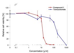
Glioblastoma multiforme is one of the most aggressive cancer forms in humans, and has low recovery rates after surgery, ionizing radiation and chemotherapy. Therefore, there is a high interest in the development of new treatment methods, as for instance photodynamic therapy (PDT). It is here presented results of the cytotoxic properties of the novel compound N1-(4-(4-(benzyl(methyl)amino)thieno[2,3-d]pyrimidin-6-yl)benzyl)-N2,N2-dimethylethane-1,2-diamine (1), which was found ten-fold more active on the rat glioma cell model F98 than the reference drug Temozolomide (TMZ). Further cell survival studies showed a profound increase in F98 cell death on UVA-radiation (330 nm, 0.5 mW/cm2). Photochemical internalization induced delivery of compound 1, but in contrast to the cytostatic drug Bleomycin, a higher cytotoxicity was not observed. Localization studies using fluorescence microscopy revealed that compound 1 readily internalized into the cytosol but did not enter the cell nucleus. The compound was shown to be a relatively weak epidermal growth factor receptor inhibitor, which is not likely to explain its cytotoxicity. However, the quantum efficiency for generation of singlet oxygen was 23%, suggesting generation of reactive oxygen species as one possible mechanism. Although more studies are needed to reveal detailed mode of action, compound 1 is a promising photosensitizer candidate for further development in tests of animal models.
Abstract Background Photochemical internalization (PCI) is a novel technology for light-induced enhancement of the local therapeutic effect of cancer drugs, utilizing a specially designed photosensitizing molecule (fimaporfin). The photosensitizing molecules are trapped in endosomes along with macromolecules or drugs. Photoactivation of fimaporfin disrupts the endosomal membranes so that drug molecules are released from endosomes inside cells and can reach their therapeutic target in the cell cytosol or nucleus. Compared with photodynamic therapy, the main cytotoxic effect with PCI is disruption of the endosomal membrane resulting in delivery of chemotherapy drug, and not to the photochemical reactions per se. In this study we investigated the effect of PCI with gemcitabine in patients with inoperable perihilar cholangiocarcinoma (CCA). Methods The in vitro cytotoxic effect of PCI with gemcitabine was studied on two CCA-derived cell lines. In a fimaporfin dose-escalation phase I clinical study, we administered PCI with gemcitabine in patients with perihilar CCA (n = 16) to establish a safe and tolerable fimaporfin dose and to get early signals of efficacy. The patients enrolled in the study had tumors in which the whole length of the tumor could be illuminated from the inside of the bile duct, using an optical fiber inserted via an endoscope (Fig. 1). Fimaporfin was administered intravenously at day 0; gemcitabine (i.v.) and intraluminal biliary endoscopic laser light application on day 4; followed by standard gemcitabine/cisplatin chemotherapy. Results Preclinical experiments showed that PCI enhanced the effect of gemcitabine. In patients with CCA, PCI with gemcitabine was well tolerated with no dose-limiting toxicities, and no unexpected safety signals. Disease control was achieved in 10 of 11 evaluable patients, with a clearly superior effect in the two highest dose groups. The objective response rate (ORR) was 42%, including two complete responses, while ORR at the highest dose was 60%. Progression-free survival at 6 months was 75%, and median overall survival (mOS) was 15.4 months, with 22.8 months at the highest fimaporfin dose. Conclusion Photochemical internalization with gemcitabine was found to be safe and resulted in encouraging response and survival rates in patients with unresectable perihilar CCA.
Background and AimsPhotochemical internalization (PCI) is a technology for inducing release of endocytosed antigens into the cell cytosolviaa light-induced process. Preclinical experiments have shown that PCI improves MHC class I antigen presentation, resulting in strongly enhanced CD8+ T-cell responses to polypeptide antigens. In PCI vaccination a mixture of the photosensitizing compound fimaporfin, vaccine antigens, and an adjuvant is administered intradermally followed by illumination of the vaccination site. This work describes an open label, phase I study in healthy volunteers, to assess the safety, tolerability, and immune response to PCI vaccination in combination with the adjuvant poly-ICLC (Hiltonol) (ClinicalTrials.govIdentifier: NCT02947854).MethodsThe primary objective of the study was to assess the safety and local tolerance of PCI mediated vaccination, and to identify a safe fimaporfin dose for later clinical studies. A secondary objective was to analyze the immunological responses to the vaccination. Each subject received 3 doses of HPV16 E7 peptide antigens and two doses of Keyhole Limpet Hemocyanin (KLH) protein. A control group received Hiltonol and vaccine antigens only, whereas the PCI groups in addition received fimaporfin + light. Local and systemic adverse effects were assessed by standard criteria, and cellular and humoral immune responses were analyzed by ELISpot, flow cytometry, and ELISA assays.Results96 healthy volunteers were vaccinated with fimaporfin doses of 0.75–50 µg. Doses below 17.5 µg were safe and tolerable, higher doses exhibited local tolerability issues in some study subjects, mainly erythema, and pain during illumination. There were few, and only mild and expected systemic adverse events. The employment of PCI increased the number of subjects exhibiting a T-cell response to the HPV peptide vaccine about 10-fold over what was achieved with the antigen/Hiltonol combination without PCI. Moreover, the use of PCI seemed to result in a more consistent and multifunctional CD8+ T-cell response. An enhancement of the humoral immune response to KLH vaccination was also observed.ConclusionsUsing PCI in combination with Hiltonol for intradermal vaccination is safe at fimaporfin doses below 17.5 µg, and gives encouraging immune responses to peptide and protein based vaccination.
Antigen cross-presentation to cytotoxic CD8+ T cells is crucial for the induction of anti-tumor and anti-viral immune responses. Recently, co-encapsulation of photosensitizers and antigens into microspheres and subsequent photochemical internalization (PCI) of antigens in antigen presenting cells has emerged as a promising new strategy for inducing antigen-specific CD8+ T cell responses in vitro and in vivo. However, the exact cellular mechanisms have hardly been investigated in vivo, i.e., which cell types take up antigen-loaded microspheres at the site of injection, or in which secondary lymphoid organ does T cell priming occur? We used spray-dried poly(lactic-co-glycolic acid) (PLGA) microspheres loaded with ovalbumin and the photosensitizer tetraphenyl chlorine disulfonate (TPCS2a) to investigate these processes in vivo. Intravital microscopy and flow cytometric analysis of the murine ear skin revealed that dendritic cells (DCs) take up PLGA microspheres in peripheral tissues. Illumination then caused photoactivation of TPCS2a and induced local tissue inflammation that enhanced CCR7-dependent migration of microsphere-containing DCs to tissue-draining lymph nodes (LNs), i.e., the site of CD8+ T cell priming. The results contribute to a better understanding of the functional mechanism of PCI-mediated vaccination and highlight the importance of an active transport of vaccine microspheres by antigen presenting cells to draining LNs.
A 77-year-old Caucasian male was diagnosed with squamous cell cancer of the right ear. The patient elected to take part in the first-in-man phase I TPCS2a based bleomycin photochemical internalization (PCI). On Day 0, The patient received the photosensitiser [Amphinex (TPCS2a)], by slow intravenous injection. Four days later, surface illumination based (PCI) was implemented 3 h after the slow infusion of Bleomycin. Four weeks following the infusion of the photosensitiser, the cancerous area turned into black rigid mass with clear demarcation from the macroscopically normal skin. The size of the treated area has been substantially reduced. Histopathologic assessment of the excised necrotic mass revealed no viable tumour and the excised margins (PCI-treated margins) were tumour-free. This case was a clear indication that PCI is a clinically relevant technique that has potential in the treatment of such cancers to avoid radical intervention.
Activation of sonosensitizers via focused ultrasound, i.e., sonodynamic therapy, has been proposed as an alternative to light-activated photodynamic therapy for the treatment of a number of conditions from cancer to bacterial infections. The use of focused ultrasound allows treatment to sites buried deep within tissues, overcoming one of the main limitations of light-based modalities. Photochemical internalization is a technique that utilizes the photochemical properties of photodynamic therapy for the release of trapped endo-lysosomal macromolecules into the cell cytoplasm, greatly enhancing their efficacy. We have examined ultrasonic activation of disulfonated tetraphenyl chlorin (fimaporfin) together with the anti-cancer agent bleomycin, termed sonochemical internalization, as an alternative to light-activated photochemical internalization. Our results indicate that, compared to drug or focused ultrasound treatment alone, focused ultrasound activation of fimaporfin together with BLM significantly inhibits the viability of glioma monolayers and the treated cells’ ability to form clonogenic colonies.
Photochemical internalization (PCI) is a further development of photodynamic therapy (PDT). In this report, we describe PCI as a potential tool for cellular internalization of chemotherapeutic agents or antigens and systematically review the ongoing research. Eighteen published papers described the pre-clinical and clinical developments of PCI-mediated delivery of chemotherapeutic agents or antigens. The studies were screened against pre-defined eligibility criteria. Pre-clinical studies suggest that PCI can be effectively used to deliver chemotherapeutic agents to the cytosol of tumor cells and, thereby, improve treatment efficacy. One Phase-I clinical trial has been conducted, and it demonstrated that PCI-mediated bleomycin treatment was safe and identified tolerable doses of the photosensitizer disulfonated tetraphenyl chlorin (TPCS2a). Likewise, PCI was pre-clinically shown to mediate major histocompatibility complex (MHC) class I antigen presentation and generation of tumor-specific cytotoxic CD8+ T-lymphocytes (CTL) and cancer remission. A first clinical Phase I trial with the photosensitizer TPCS2a combined with human papilloma virus antigen (HPV) was recently completed and results are expected in 2020. Hence, photosensitizers and light can be used to mediate cytosolic delivery of endocytosed chemotherapeutics or antigens. While the therapeutic potential in cancer has been clearly demonstrated pre-clinically, further clinical trials are needed to reveal the true translational potential of PCI in humans.
The possibility of using photochemical internalization (PCI) to enhance the effects of the cytotoxic drug bleomycin is investigated, together with photophysical determination and outlines of a possible treatment for intravesical therapy of bladder cancer. In vitro experiments indicated that the employment of PCI technology using the novel photosensitizer TPCS2a® can enhance the cytotoxic effect of bleomycin in bladder cancer cells. Furthermore, experiments in an orthotopic in vivo bladder cancer model show an effective reduction in both the necrotic area and the bladder weight after TPCS2a based photodynamic therapy (PDT). The tumor selectivity and PDT effects may be sufficient to destroy tumors without damaging the detrusor muscle layer. Our results present a possible new treatment strategy for non-muscle invasive bladder cancer, with the intravesical instillation of the photosensitizer and bleomycin followed by illumination through an optic fiber by using a catheter.
Photochemical internalization (PCI) depends on the delivery of sublethal photodynamic reaction to facilitate the work of a chemotherapeutic agent. We discuss our experience in managing a patient with extensive squamous cell carcinoma of the right face and scalp under the TPCS2a-based bleomycin PCI treatment protocol. In this case, an 84-year-old Caucasian received 0.25 mg kg(-1)of TPCS2a(Amphinex (R) PCI Biotech AS, Oslo, Norway). Surface illumination photochemical internalization was carried out after 4 days, which was preceded by the chemotherapeutic agent infusion (Bleomycin). After one week from the illumination time, tissue necrosis was evident and tumor shrinkage was most noticeable at day 14 postillumination. Follow-up at 6 weeks continued to show tissue healing and regeneration with no clinical evidence of recurrence. Multiple surgical biopsies were taken at 1 and 3 months postillumination and found to be tumor free. PCI's depth of effect has been very significant with negligible damage to the collateral tissues. This technology has a role in interventional oncology especially when managing challenging cases.
Photochemical internalisation (PCI) is a novel technology for release of endocytosed macromolecules into the cytosol. The technology is based on the use of photosensitizers that locate in endocytic vesicles, and that upon activation by light induce a release of macromolecules from the endocytic vesicles. PCI has been shown to stimulate delivery of a large variety of macromolecules and other molecules that do not readily penetrate the plasma membrane. The preclinical evaluation of PCI has been performed with aluminum phthalocyanine disulfonate (AlPcS2a) as photosensitizer. AlPcS2a, due to its large number of isomers potentially with batch-to-batch ratio variations, is not an optimal photosenstizer for clinical use. Disulfonated tetraphenyl chlorin (TPCS2a) has therefore been developed by di-imide reduction of disulfonated tetraphenyl porphine (TPPS2a). The synthesized TPCS2a contains 3 isomers as shown by HPLC with low (<4%) inter-batch variation with respect to isomer formation, less than 0.5% (w/w) of the starting material TPPS2a and absorbs light at 652 nm. As prerequisites for a PCI photosensitizer TPCS2a was found to localize in intracellular granules assumed to be endocytic vesicles. In cells in culture TPCS2a-PCI induced activation of gelonin as seen by enhanced cytotoxicity, increased transfection efficacy by an enhanced green fluorescence protein (EGFP)-encoding plasmid, induced gene silencing by siRNA towards EGFP and induced in a synergistic manner tumor growth delay by TPCS2a-mediated PCI of bleomycin in CT26.CL25 carcinomas growing subcutaneously in athymic mice. TPCS2a -PCI of bleomycin was found superior to meso-tetraphenyl chlorin-based photodynamic therapy (mTHPC-PDT) with respect to inhibition of tumor growth. The tumor growth delay by PCI of bleomycin was independent of the time of bleomycin administration between 3 h prior to light to immediately after light, while bleomycin administered 24 h prior to or 24 h after the light exposure induced suboptimal or only additive effects on tumor growth delay respectively. TPCS2a-PDT and -PCI induced indistinguishably strong edema the first 3–4 days after TPCS2a-administration and only weak erythema the first day after TPCS2a administration. In contrast, mTHPC-PDT induced moderate edema the first 7 days after mTHPC administration, but strong erythema resulting in open wounds and escar formation the first 2–3 days after mTHPC administration. The pharmacokinetic properties of TPCS2a were evaluated in athymic mice. The plasma pharmacokinetics was best fit to a 2-compartment model with half-lives of 0.78 and 36 hrs. TPCS2a was found to be a clinically suitable PCI photosensitizer for photochemical activation of molecules that do not readily penetrate the cellular plasma membrane.
In this study we have developed biodegradable polymeric nanoparticles (NPs) containing the cytostatic drugs mertansine (MRT) or cabazitaxel (CBZ). The NPs are based on chitosan (CS) conjugate polymers synthesized with different amounts of the photosensitizer tetraphenylchlorin (TPC). These TPC–CS NPs have high loading capacity and strong drug retention due to π–π stacking interactions between the drugs and the aromatic photosensitizer groups of the polymers. CS polymers with 10% of the side chains containing TPC were found to be optimal in terms of drug loading capacity and NP stability. The TPC–CS NPs loaded with MRT or CBZ displayed higher cytotoxicity than the free form of these drugs in the breast cancer cell lines MDA-MB-231 and MDA-MB-468. Furthermore, light-induced photochemical activation of the NPs elicited a strong photodynamic therapy effect on these breast cancer cells. Biodistribution studies in mice showed that most of the TPC–CS NPs accumulated in liver and lungs, but they were also found to be localized in tumors derived from HCT-116 cells. These data suggest that the drug-loaded TPC–CS NPs have a potential in combinatory anticancer therapy and as contrast agents.
Triple-negative breast cancer (TNBC) and malignant melanoma are highly aggressive cancers that widely express the cell surface chondroitin sulfate proteoglycan 4 (CSPG4/NG2). CSPG4 plays an important role in tumor cell growth and survival and promotes chemo- and radiotherapy resistance, suggesting that CSPG4 is an attractive target in cancer therapy. In the present work, we applied the drug delivery technology photochemical internalization (PCI) in combination with the novel CSPG4-targeting immunotoxin 225.28-saporin as an efficient and specific strategy to kill aggressive TNBC and amelanotic melanoma cells. Light-activation of the clinically relevant photosensitizer TPCS2a (fimaporfin) and 225.28-saporin was found to act in a synergistic manner, and was superior to both PCI of saporin and PCI-no-drug (TPCS2a + light only) in three TNBC cell lines (MDA-MB-231, MDA-MB-435 and SUM149) and two BRAFV600E mutated malignant melanoma cell lines (Melmet 1 and Melmet 5). The cytotoxic effect was highly dependent on the light dose and expression of CSPG4 since no enhanced cytotoxicity of PCI of 225.28-saporin compared to PCI of saporin was observed in the CSPG4-negative MCF-7 cells. The PCI of a smaller, and clinically relevant CSPG4-targeting toxin (scFvMEL-rGel) validated the CSPG4-targeting concept in vitro and induced a strong inhibition of tumor growth in the amelanotic melanoma xenograft A-375 model. In conclusion, the combination of the drug delivery technology PCI and CSPG4-targeting immunotoxins is an efficient, specific and light-controlled strategy for the elimination of aggressive cells of TNBC and malignant melanoma origin. This study lays the foundation for further preclinical evaluation of PCI in combination with CSPG4-targeting.
 Kristian Berg合作论文数68
 Pal K. Selbo合作论文数Norwegian Radium Hosp, Oslo Univ Hosp43Lina Prasmickaite合作论文数37Anette Bonsted合作论文数17Birgit Engesæter合作论文数Department of Cervical Cancer Screening, Cancer Registry of Norway16
 Anette Weyergang合作论文数The Norwegian Radium Hospital, Oslo University Hospital15
 Gunhild Mari Mælandsmo合作论文数Department of Tumor Biology, Institute for Cancer Research, Oslo University Hospital13
 Odrun A Gederaas合作论文数Faculty of Medicine, Norwegian University of Science and Technology12
 Colin Hopper合作论文数Eastman Dental Institute, Faculty of Medical Sciences, University College London12
 Monika Hakerud合作论文数11Kaare M. Gautvik合作论文数10
 Zaid Hamdoon合作论文数University of Mosul-College of Dentistry, Mosul, Iraq;
College of Dental Medicine, Oral and Craniofacial Health Sciences, University of Sharjah, Sharjah, United Arab
Emirates10Marit Hellum合作论文数10
Zaid Hamdoon合作论文数University of Mosul-College of Dentistry, Mosul, Iraq;
College of Dental Medicine, Oral and Craniofacial Health Sciences, University of Sharjah, Sharjah, United Arab
Emirates10Marit Hellum合作论文数10 Pal Johansen合作论文数Department of Dermatology, Universitäts Spital Zürich;University of Zurich10Waseem Jerjes合作论文数9
 Monica Bostad合作论文数7Ying Waeckerle-Men合作论文数7C.A. Mosse合作论文数6BN KAREEM合作论文数6Lilletvedt M合作论文数6Edwards Victoria Tudor合作论文数6Thomas M. Kundig合作论文数6Andreas Dietze合作论文数6Erik Rokkones合作论文数6
 Peter Alestrom合作论文数6Ahmed A Sultan合作论文数5Anne Grete Nedberg合作论文数5S. Kristensen合作论文数5
 Stephen Bown合作论文数Department of Surgical Biotechnology, Divsion of Surgery & Interventional Science, Faculty of Medical Sciences, University College London5
 Ernst Wagner合作论文数Center for System-based Drug Research, Department of Pharmacy, Ludwig-Maximilians-Universitat5
 Cathrine Olsen合作论文数4Tone Otterhaug合作论文数4Dawn M Carnell合作论文数4
 Erik Holmgren合作论文数4
 Cornelia Halin Winter合作论文数Department of Chemistry and Applied Biosciences, Eidgenössische Technische Hochschule Zürich4Blingsmo O R合作论文数4
 Kirsten Sandvig合作论文数Dept. of Molecular Cell Biology, Institute for Cancer Research, Oslo University Hospital;Institute for molecular Biosciences, University of Oslo;Institute for Cancer Research, The Norwegian Radium Hospital, Oslo4
 Mikael Lindgren合作论文数Department of Physics, Faculty of Natural Sciences, Norwegian University of Sciende and Technology4Richard Sturgess合作论文数4
 Már Másson合作论文数Faculty of Pharmacy, University of Iceland4
 Jan O. Gordeladze合作论文数Institute of Medical Biochemistry, University of Oslo4Øystein Fodstad合作论文数4Vigdis T. Gautvik合作论文数4
 Ellen Skarpen合作论文数4
 Luca Nardo合作论文数Department of Physics and Mathematics, University of Insubria and C.N.I.S.M.-C.N.R.4
 Vivek S. Gaware合作论文数School of Health Sciences, University of Iceland4T. Otterhaug合作论文数3L. Finnesand合作论文数3Zuzanna K Kotkowska合作论文数3




