Plasmacytoid dendritic cells (pDCs), a unique type of innate immune cells, are known to play an important role in fighting viral infections, especially due to their capacity to sense DNA/RNA nucleic acids by TLR7/-9, and production of type-I interferons. The role of pDCs in driving immunological effects in cancer development and mounting antitumoral responses remains to be elucidated, though few studies describe opposing outcomes in patients based on tumor infiltrating pDCs. These observations come with little evidence explaining the immunosuppressive or tumorigenic state of pDCs. To study fundamental anti-tumoral functions of pDCs we utilized a robust and validated in vitro stem-cell derived human pDC model. Importantly, this model permits genetic manipulation prior to pDC differentiation and enables large-scale co-culture studies with various cancer types. Using this model, we recently employed a CRISPR library screening approach to identify factors that aid the cytotoxic mechanisms of pDCs following co-culture with tumor cell lines. Initially, using co-culture assays consisting of hematological, lung, or breast cancer cell lines in combination with in vitro generated pDCs, we demonstrated a time-dependent cytotoxic effect using Incucyte live imaging. Importantly, cytotoxic effects were elevated when pDCs had been stimulated with TLR9 agonists. We demonstrated that a direct cell-to-cell interaction was essential for pDC cytotoxicity, and that promoting stronger cell-to-cell interaction, by genetically modifying pDCs to express scFv targeting a tumor antigen, increased the killing effects. CRISPR knock-out of Granzyme B in pDCs did not remove the cytotoxic capabilities of pDCs. We are currently conducting a genetic screen to identify universal factors vital for pDC interactions and cytotoxicity effects in cancer settings. This preliminary data will be presented at the AACR meeting. Using in vitro-generated pDCs we explore an important anti-tumoral function of pDCs. If successful, our work can open for potential new therapeutics and prognostic possibilities in cancer treatment, harnessing the cytotoxic effects of pDCs. Tobias Wang Bjerg, Ian Helstrup Nielsen, Sabina Sanchez Hernandez, Anders Laustsen, Isabella Barbutti, Shuli Svetitsky, James Lee, Rasmus Bak, Martin Jakobsen. Characterizing an immune synapse between plasmacytoid dendritic cells and cancer cells that drives cancer cytotoxicity [abstract]. In: Proceedings of the American Association for Cancer Research Annual Meeting 2025; Part 1 (Regular Abstracts); 2025 Apr 25-30; Chicago, IL. Philadelphia (PA): AACR; Cancer Res 2025;85(8_Suppl_1):Abstract nr 5861.
Plasmacytoid dendritic cells (pDCs) play a pivotal role in immune responses, particularly against viral infections. pDCs exhibit diverse functions, including interferon production, cytokine secretion, and antigen presentation. Here, we investigate the antigen cross-presentation capacity of pDCs and their role in CD8 + T cell activation. Utilizing a culturing system with CD8 + T cells and autologous pDCs derived from circulating CD34 + hematopoietic stem and progenitor cells, we demonstrate that pDCs efficiently activate CD8 + T cells via cross-presentation, promoting T cell expansion and cytotoxic activity. The antigen presentation capacity of pDCs is comparable to that of monocyte-derived dendritic cells (moDCs) and myeloid dendritic cells, which are known for their efficient antigen-presentation capacity. Transcriptomic analysis reveals genetic signatures in CD8 + T cells activated by pDCs distinct from moDCs, suggesting different activation mechanisms. These findings underscore the importance of pDCs in antigen presentation and their contribution to CD8 + T cell activation.
Background Plasmacytoid Dendritic Cells (pDCs) - sometimes referred to as type I interferon producing innate lymphocyte cells - are known as a master regulator of the immune system. The pDCs are responsible for the body’s Interferon (IFN) α production, and contain multiple direct effector functions that can mount strong anti-tumor responses. The low number of pDCs in tissues and their frailness have, however, impeded their use in the clinic as adoptive cell therapy. Here we present a first-in-class pDC cell therapy engineered for antigen-dependent activation that results in release of IFNs, direct killing of tumor cells, and recruitment of other innate and adaptive immune cells. Methods Hematopoietic stem and progenitor cells (HSPCs) were used as source material in a GMP-compliant manufacturing process of human pDCs. During early differentiation, HSPCs were genetically engineered with a Synthetic Notch (SynNotch) receptor by lentiviral transduction. In this system, tumor specificity was provided by a single-chain fragment variant (scFv) targeting tumor antigen (here used CD19) and antigen-dependent activation of using a genetic response element, based on gain-of-function STING gene (STINGGoF). In vitro co-culture assays including different CD19+cancer cell lines (REH, NALM6, RAJI) with genetic modified pDCs were used to assess functionality. An in vivo xenograft model of NOG (NOD.Cg-PrkdcscidIl2rgtm1Sug/JicTac) mice subcutaneously engrafted with NALM6 cells to mimic a solid tumor, was used to study the potency of the cell product. Results From bulk RNA-seq we demonstrate that in vitro co-cultures of pDC-STINGGoF with cancer cell lines elicit a strong and broad gene signature including Type I IFNs and a spectrum of inflammatory cytokines. In cytotoxicity assays, pDC-STINGGoF elicit antigen-dependent killing of all tested cancer cell lines to levels beyond that of NK cells (up to 80% in a 1:2 (T:E) ratio). In NOG mice xenografted with NALM6 tumors and receiving a single intravenous infusion of pDC-STINGGoF, we observed pDC tumor infiltration 48hrs post-injection which was supported by strong tumor regression. Mice treated with non-targeted pDCs exhibit tumor growth similar to control mice. Conclusions Our genetic engineered pDC is a novel cell therapy that may change the paradigm of solid tumor treatment by the inhibitory effects on tumor cells of Type I interferons, combined with the release of pro-inflammatory cytokines, resulting in recruitment and engagement of other immune cells. This activity is further complemented by pDCs direct tumor cell killing effects. We are currently moving toward phase I to evaluate the safety and efficacy of autologous pDCs. Ethics Approval Human cord blood was collected from Aarhus University Hospital by consent from donors that the material could be used for research under complete anonymisation. The animal experiments have been approved by Danish ethics committee in Region Midt with the ID#2020-15-0201-00394
Understanding the molecular pathways driving the acute antiviral and inflammatory response to SARS‐CoV‐2 infection is critical for developing treatments for severe COVID‐19. Here, we find decreasing number of circulating plasmacytoid dendritic cells (pDCs) in COVID‐19 patients early after symptom onset, correlating with disease severity. pDC depletion is transient and coincides with decreased expression of antiviral type I IFNα and of systemic inflammatory cytokines CXCL10 and IL‐6. Using an in vitro stem cell‐based human pDC model, we further demonstrate that pDCs, while not supporting SARS‐CoV‐2 replication, directly sense the virus and in response produce multiple antiviral (interferons: IFNα and IFNλ1) and inflammatory (IL‐6, IL‐8, CXCL10) cytokines that protect epithelial cells from de novo SARS‐CoV‐2 infection. Via targeted deletion of virus‐recognition innate immune pathways, we identify TLR7‐MyD88 signaling as crucial for production of antiviral interferons (IFNs), whereas Toll‐like receptor (TLR)2 is responsible for the inflammatory IL‐6 response. We further show that SARS‐CoV‐2 engages the receptor neuropilin‐1 on pDCs to selectively mitigate the antiviral interferon response, but not the IL‐6 response, suggesting neuropilin‐1 as potential therapeutic target for stimulation of TLR7‐mediated antiviral protection. Plasmacytoid dendritic cells (pDCs) sense SARS‐CoV‐2 via two distinct innate immune pathways. The endosomal TLR7 pathway is activated upon sensing of viral RNA and this leads to type I IFN production. The TLR2 pathway is triggered by sensing of the viral envelope protein, inducing IL‐6 production. Interestingly, SARS‐CoV‐2 is able to specifically antagonize the TLR7‐IFN pathway via a CD304‐mediated signaling cascade. Sensing of SARS‐CoV‐2 by plasmacytoid dendritic cells (pDC) contributes to the anti‐viral and immunopathological responses.
Targeted transcriptional activation or interference can be induced with the CRISPR-Cas9 system (CRISPRa/CRISPRi) using nuclease-deactivated Cas9 fused to transcriptional effector molecules. These technologies have been used in cancer cell lines, particularly for genome-wide functional genetic screens using lentiviral vectors. However, CRISPRa and CRISPRi have not yet been widely applied to ex vivo cultured primary cells with therapeutic relevance owing to a lack of effective and nontoxic delivery modalities. Here we develop CRISPRa and CRISPRi platforms based on RNA or ribonucleoprotein (RNP) delivery by electroporation and show transient, programmable gene regulation in primary cells, including human CD34 + hematopoietic stem and progenitor cells (HSPCs) and human CD3 + T cells. We show multiplex and orthogonal gene modulation using multiple sgRNAs and CRISPR systems from different bacterial species, and we show that CRISPRa can be applied to manipulate differentiation trajectories of HSPCs. These platforms constitute simple and effective means to transiently control transcription and are easily adopted and reprogrammed to new target genes by synthetic sgRNAs. We believe these technologies will find wide use in engineering the transcriptome for studies of stem cell biology and gene function, and we foresee that they will be implemented to develop and enhance cellular therapeutics.
Understanding the molecular pathways driving the acute antiviral and inflammatory response to SARS-CoV-2 infection is critical for developing treatments for severe COVID-19. Here we show that in COVID-19 patients, circulating plasmacytoid dendritic cells (pDCs) decline early after symptom onset and this correlated with COVID-19 disease severity. This transient depletion coincides with decreased expression of antiviral type I IFNα and the systemic inflammatory cytokines CXCL10 and IL-6. Importantly, COVID-19 disease severity correlated with decreased pDC frequency in peripheral blood. Using an in vitro stem cell-based human pDC model, we demonstrate that pDCs directly sense SARS-CoV-2 and in response produce multiple antiviral (IFNα and IFNλ1) and inflammatory (IL-6, IL-8, CXCL10) cytokines. This immune response is sufficient to protect epithelial cells from de novo SARS-CoV-2 infection. Targeted deletion of specific sensing pathways identified TLR7-MyD88 signaling as being crucial for production of the antiviral IFNs, whereas TLR2 is responsible for the inflammatory IL-6 response. Surprisingly, we found that SARS-CoV-2 engages the neuropilin-1 receptor on pDCs to mitigate the antiviral IFNs but not the IL-6 response. These results demonstrate distinct sensing pathways used by pDCs to elicit antiviral vs. immunopathological responses to SARS-CoV-2 and suggest that targeting neuropilin-1 on pDCs may be clinically relevant for mounting TLR7-mediated antiviral protection. One Sentence Summary pDCs sense SARS-CoV-2 and elicit antiviral protection of lung epithelial cells through TLR7, while recognition of TLR2 elicits an IL-6 inflammatory response associated with immunopathology. Graphical abstract: SARS-CoV-2 sensing by plasmacytoid dendritic cells. SARS-CoV-2 is internalized by pDCs via a yet unknown endocytic mechanism. The intracellular TLR7 sensor detects viral RNA and induces a signaling cascade involving MyD88-IRAK4-TRAF6 (1) to induce CXCL10 and, via IRF7 phosphorylation and translocation, inducing type I and III Interferons (2). Once secreted, type I and III IFNs initiate autocrine and paracrine signals that induce the expression of IFN stimulated genes (ISGs), thereby facilitating an antiviral response that can protect the cell against infection. However, SARS-CoV-2, has the intrinsic property to facilitate CD304 signaling, potentially by interfering with IRF7 nuclear translocation, thereby inhibiting type I IFNα production and thus reducing the antiviral response generated by the pDC (4). Furthermore, the SARS-CoV-2 envelope (E) glycoprotein is sensed by the extracellular TLR2/6 heterodimer and this facilitates production of the inflammatory IL-6 cytokine (5). Illustration was created with BioRender.com
Exploring the intricacies of host-pathogen communication is vital in order to understand why some microbes persist within a host, while others are effectively cleared. As such, host-pathogen communication is an urgent area of research underpinning human health and disease, especially given the emerging concerns of antibiotic resistance. Understanding host-pathogen pathways requires intricate knowledge in the fields of both microbiology and immunology. Living in a world of specialists, where scientists sometimes invest their whole career studying one organism, a specific pathway or a protein, multidisciplinary discussions of host-pathogen interactions are highly needed. This international conference will provide such a forum for scientific exchange at the highest level, between scientists from different research areas related to host-pathogen interactions, with the aim to trigger fruitful collaborations addressing the urgent questions in the field. This meeting has an additional special focus of bringing researchers from Ireland, Denmark and Israel together to learn about cutting edge biology and form new collaborations. Thus, the conference will be a showcase for the exciting and original research emanating from these three small but innovative countries.
Plasmacytoid dendritic cells (pDCs) constitute a rare type of immune cell with multifaceted functions, but their potential use as a cell-based immunotherapy is challenged by the scarce cell numbers that can be extracted from blood. Here, we systematically investigate culture parameters for generating pDCs from hematopoietic stem and progenitor cells (HSPCs). Using optimized conditions combined with implementation of HSPC pre-expansion, we generate an average of 465 million HSPC-derived pDCs (HSPC-pDCs) starting from 100,000 cord blood-derived HSPCs. Furthermore, we demonstrate that such protocol allows HSPC-pDC generation from whole-blood HSPCs, and these cells display a pDC phenotype and function. Using GMP-compliant medium, we observe a remarkable loss of TLR7/9 responses, which is rescued by ascorbic acid supplementation. Ascorbic acid induces transcriptional signatures associated with pDC-specific innate immune pathways, suggesting an undescribed role of ascorbic acid for pDC functionality. This constitutes the first protocol for generating pDCs from whole blood and lays the foundation for investigating HSPC-pDCs for cell-based immunotherapy.
The majority of HIV infections are established through the genital or rectal mucosa. Fibroblasts are abundant in these tissues, and although not susceptible to infection, can potently enhance HIV infection of CD4+ T cells. Hyaluronic acid (HA) is a major component of the extracellular matrix of fibroblasts, and its levels are influenced by the inflammatory state of the tissue. Since inflammation is known to facilitate HIV sexual transmission, we investigated the role of HA in genital mucosal fibroblast-mediated enhancement of HIV infection. Depletion of HA by CRISPR-Cas9 in primary foreskin fibroblasts augmented the ability of the fibroblasts to increase HIV infection of CD4+ T cells. This amplified enhancement required direct contact between the fibroblasts and CD4+ T cells, and could be attributed to both increased rates of trans-infection and the increased ability of HA-deficient fibroblasts to push CD4+ T cells into a state of higher permissivity to infection. This HIV-permissive state was characterized by differential expression of genes associated with regulation of cell metabolism and death. Our results suggest that conditions resulting in diminished cell-surface HA on fibroblasts, such as genital inflammation, can promote HIV transmission by conditioning CD4+ T cells toward a state more vulnerable to infection by HIV.
n amendment to this paper has been published and can be accessed via a link at the top of the paper.
STING is essential for control of infections and for tumor immunosurveillance, but it can also drive pathological inflammation. STING resides on the endoplasmic reticulum (ER) and traffics following stimulation to the ERGIC/Golgi, where signaling occurs. Although STING ER exit is the rate-limiting step in STING signaling, the mechanism that drives this process is not understood. Here we identify STEEP as a positive regulator of STING signaling. STEEP was associated with STING and promoted trafficking from the ER. This was mediated through stimulation of phosphatidylinositol-3-phosphate (PtdIns(3)P) production and ER membrane curvature formation, thus inducing COPII-mediated ER-to-Golgi trafficking of STING. Depletion of STEEP impaired STING-driven gene expression in response to virus infection in brain tissue and in cells from patients with STING-associated diseases. Interestingly, STING gain-of-function mutants from patients interacted strongly with STEEP, leading to increased ER PtdIns(3)P levels and membrane curvature. Thus, STEEP enables STING signaling by promoting ER exit.
STUDY QUESTION:Do seminal plasma (SP) and its constituents affect the decidualization capacity and transcriptome of human primary endometrial stromal fibroblasts (eSFs)?SUMMARY ANSWER:SP promotes decidualization of eSFs from women with and without inflammatory disorders (polycystic ovary syndrome (PCOS), endometriosis) in a manner that is not mediated through semen amyloids and that is associated with a potent transcriptional response, including the induction of interleukin (IL)-11, a cytokine important for SP-induced decidualization.WHAT IS KNOWN ALREADY:Clinical studies have suggested that SP can promote implantation, and studies in vitro have demonstrated that SP can promote decidualization, a steroid hormone-driven program of eSF differentiation that is essential for embryo implantation and that is compromised in women with the inflammatory disorders PCOS and endometriosis.STUDY DESIGN, SIZE, DURATION:This is a cross-sectional study involving samples treated with vehicle alone versus treatment with SP or SP constituents. SP was tested for the ability to promote decidualization in vitro in eSFs from women with or without PCOS or endometriosis (n = 9). The role of semen amyloids and fractionated SP in mediating this effect and in eliciting transcriptional changes in eSFs was then studied. Finally, the role of IL-11, a cytokine with a key role in implantation and decidualization, was assessed as a mediator of the SP-facilitated decidualization.PARTICIPANTS/MATERIALS, SETTING, METHODS:eSFs and endometrial epithelial cells (eECs) were isolated from endometrial biopsies from women of reproductive age undergoing benign gynecologic procedures and maintained in vitro. Assays were conducted to assess whether the treatment of eSFs with SP or SP constituents affects the rate and extent of decidualization in women with and without inflammatory disorders. To characterize the response of the endometrium to SP and SP constituents, RNA was isolated from treated eSFs or eECs and analyzed by RNA sequencing (RNAseq). Secreted factors in conditioned media from treated cells were analyzed by Luminex and ELISA. The role of IL-11 in SP-induced decidualization was assessed through Clustered regularly interspaced short palindromic repeats (CRISPR)/Cas-9-mediated knockout experiments in primary eSFs.MAIN RESULTS AND THE ROLE OF CHANCE:SP promoted decidualization both in the absence and presence of steroid hormones (P < 0.05 versus vehicle) in a manner that required seminal proteins. Semen amyloids did not promote decidualization and induced weak transcriptomic and secretomic responses in eSFs. In contrast, fractionated SP enriched for seminal microvesicles (MVs) promoted decidualization. IL-11 was one of the most potently SP-induced genes in eSFs and was important for SP-facilitated decidualization.LARGE SCALE DATA:RNAseq data were deposited in the Gene Expression Omnibus repository under series accession number GSE135640.LIMITATIONS, REASONS FOR CAUTION:This study is limited to in vitro analyses.WIDER IMPLICATIONS OF THE FINDINGS:Our results support the notion that SP promotes decidualization, including within eSFs from women with inflammatory disorders. Despite the general ability of amyloids to induce cytokines known to be important for implantation, semen amyloids poorly signaled to eSFs and did not promote their decidualization. In contrast, fractionated SP enriched for MVs promoted decidualization and induced a transcriptional response in eSFs that overlapped with that of SP. Our results suggest that SP constituents, possibly those associated with MVs, can promote decidualization of eSFs in an IL-11-dependent manner in preparation for implantation.STUDY FUNDING/COMPETING INTEREST(S):This project was supported by NIH (R21AI116252, R21AI122821 and R01AI127219) to N.R.R. and (P50HD055764) to L.C.G. The authors declare no conflict of interest.
CRISPR/Cas9 is an effective and easy-to-use tool for editing the genome of many human cancer cell lines. However, in some hard-to-transfect cell lines and primary cells, gene editing is more challenging. This protocol details an electroporation-based protocol for the delivery of Cas9 protein from Streptococcus pyogenes complexed with chemically modified sgRNAs. We have found this protocol to work very efficiently in numerous cell lines and primary cells that are difficult to transfect by conventional chemical-based transfection methods.
Genome editing of human pluripotent stem cells (hPSCs) provides powerful opportunities for in vitro disease modeling, drug discovery, and personalized stem cell-based therapeutics. Currently, only small edits can be engineered with high frequency, while larger modifications suffer from low efficiency and a resultant need for selection markers. Here, we describemarker-free genome editing in hPSCs using Cas9 ribonucleoproteins (RNPs) in combination with AAV6-mediated DNA repair template delivery. We report highly efficient and bi-allelic integration frequencies across multiple loci and hPSC lines, achieving mono-allelic editing frequencies of up to 94% at the HBB locus. Using this method, we show robust bi-allelic correction of homozygous sickle cell mutations in a patient-derived induced PSC (iPSC) line. Thus, this strategy shows significant utility for generating hPSCs with large gene integrations and/or single-nucleotide changes at high frequency and without the need for introducing selection genes, enhancing the applicability of hPSC editing for research and translational uses.
Plasmacytoid dendritic cells (pDC) are essential for immune competence. Here we show that pDC precursor differentiated from human CD34+ hematopoietic stem and progenitor cells (HSPC) has low surface expression of pDC markers, and has limited induction of type I interferon (IFN) and IL-6 upon TLR7 and TLR9 agonists treatment; by contrast, cGAS or RIG-I agonists-mediated activation is not altered. Importantly, after priming with type I and II IFN, these precursor pDCs attain a phenotype and functional activity similar to that of peripheral blood-derived pDCs. Data from CRISPR/Cas9-mediated genome editing of HSPCs further show that HSPC-pDCs with genetic modifications can be obtained, and that expression of the IFN-α receptor is essential for the optimal function, but dispensable for the differentiation, of HSPC-pDC percursor. Our results thus demonstrate the biological effects of IFNs for regulating pDC function, and provide the means of generating of gene-modified human pDCs.
The transcription factor Nrf2 is a critical regulator of inflammatory responses. If and how Nrf2 also affects cytosolic nucleic acid sensing is currently unknown. Here we identify Nrf2 as an important negative regulator of STING and suggest a link between metabolic reprogramming and antiviral cytosolic DNA sensing in human cells. Here, Nrf2 activation decreases STING expression and responsiveness to STING agonists while increasing susceptibility to infection with DNA viruses. Mechanistically, Nrf2 regulates STING expression by decreasing STING mRNA stability. Repression of STING by Nrf2 occurs in metabolically reprogrammed cells following TLR4/7 engagement, and is inducible by a cell-permeable derivative of the TCA-cycle-derived metabolite itaconate (4-octyl-itaconate, 4-OI). Additionally, engagement of this pathway by 4-OI or the Nrf2 inducer sulforaphane is sufficient to repress STING expression and type I IFN production in cells from patients with STING-dependent interferonopathies. We propose Nrf2 inducers as a future treatment option in STING-dependent inflammatory diseases.
Combination of genome editing and human pluripotent stem cells (hPSCs) offers a platform for in vitro disease modeling, drug discovery and personalized stem cell therapeutics. However, incorporation of large modifications using CRISPR/Cas9-based genome editing in hPSCs typically requires the use of selection markers due to low editing efficiencies. Here we report a novel editing technology in hPSCs using Cas9 protein complexed with chemically modified single guide RNA (sgRNA) and recombinant AAV6 (rAAV6) vectors for donor delivery without marker selection. With these components, we demonstrate targeted integration of a 2.2 kb DNA expression cassette in hPSCs at frequencies up to 94% and 67% at the HBB and MYD88 loci, respectively. We used this protocol to correct the homozygous sickle cell disease (SCD) mutation in an iPSC line derived from a SCD patient with a frequency of 63%. This Cas9/AAV6 system allows for both the integration of large gene cassettes and the creation of single nucleotide changes in hPSCs at high frequencies, eliminating the need for multiple editing steps and marker selection, thus increasing the potential of editing human pluripotent cells for both research and translational applications.
Understanding early events of HIV transmission within mucosal tissues is vital for developing effective prevention strategies. Here, we report that primary stromal fibroblasts isolated from endometrium, cervix, foreskin, male urethra, and intestines significantly increase HIV infection of CD4+ T cells-by up to 37-fold for R5-tropic HIV and 100-fold for X4-tropic HIV-without themselves becoming infected. Fibroblasts were more efficient than dendritic cells at trans-infection and mediate this response in the absence of the DC-SIGN and Siglec-1 receptors. In comparison, mucosal epithelial cells secrete antivirals and inhibit HIV infection. These data suggest that breaches in the epithelium allow external or luminal HIV to escape an antiviral environment to access the infection-favorable environment of the stromal fibroblasts, and suggest that resident fibroblasts have a central, but previously unrecognized, role in HIV acquisition at mucosal sites. Inhibiting fibroblast-mediated enhancement of HIV infection should be considered as a novel prevention strategy.
Innate immune activation by macrophages is an essential part of host defence against infection. Cytosolic recognition of microbial DNA in macrophages leads to induction of interferons and cytokines through activation of cyclic GMP-AMP synthase (cGAS) and stimulator of interferon genes (STING). Other host factors, including interferon-gamma inducible factor 16 (IFI16), have been proposed to contribute to immune activation by DNA. However, their relation to the cGAS-STING pathway is not clear. Here, we show that IFI16 functions in the cGAS-STING pathway on two distinct levels. Depletion of IFI16 in macrophages impairs cGAMP production on DNA stimulation, whereas overexpression of IFI16 amplifies the function of cGAS. Furthermore, IFI16 is vital for the downstream signalling stimulated by cGAMP, facilitating recruitment and activation of TANK-binding kinase 1 in STING complex. Collectively, our results suggest that IFI16 is essential for efficient sensing and signalling upon DNA challenge in macrophages to promote interferons and antiviral responses.
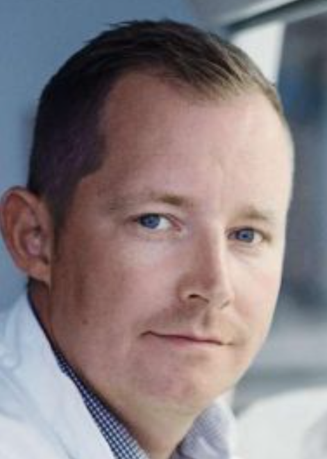 Martin Roelsgaard Jakobsen合作论文数Department of Biomedicine, Aarhus Universitet23
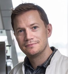 Bak Rasmus O合作论文数Dept Biomed, Aarhus Univ19
 Søren Riis Paludan合作论文数Department of Biomedicine, Aarhus University9
 Jacob Giehm Mikkelsen合作论文数9
 Christian K Holm合作论文数6
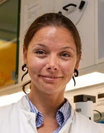 Renee M. Van Der Sluis合作论文数Department of Biomedicine, Aarhus University6Sun Chenglong合作论文数Department of Biomedicine, Aarhus University;Aarhus Research Center for Innate Immunology, Aarhus University;Aarhus University, Aarhus University6
 David Olagnier合作论文数6
 Søren B. Jensen合作论文数6
 Kathrine Hansen合作论文数5Albert Gris-Oliver合作论文数Vall dHebron Inst Oncol, Expt Therapeut Grp, Barcelona, Spain5
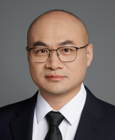 Yujia Cai (蔡宇伽)合作论文数Shanghai Center for Systems Biomedicine, Shanghai Jiao Tong University5per hallager合作论文数5Annika Andersena合作论文数4
 Yonglun Luo合作论文数4
 Trine Mogensen合作论文数Department of Biomedicine, Aarhus University;Department of Infectious Diseases, Aarhus University;Department of Clinical Medicine, Aarhus University;Aarhus University Hospital4Reinert Line S合作论文数4
 Thaneas Prabakaran合作论文数Department of Biomedicine, Aarhus University4
 Kjolby Mads合作论文数The Lundbeck Foundation Research Center, Aarhus University;Danish Research Institute of Translational Neuroscience DANDRITE, Aarhus University;Aarhus University, Aarhus University Hospital;Aarhus University, Aarhus University Hospital4
 Niels Uldbjerg合作论文数Department of Clinical Medicine, Aarhus University;Institut for Klinisk Medicin, Aarhus Universitet4
 Martin Tolstrup合作论文数4
 Tengchuan Jin (金腾川)合作论文数Laboratory of Structural Immunology, University of Science and Technology of China Life Sciences and Medicine;Institute of Health and Medicine, Hefei Comprehensive National Science Center;Key Laboratory of Innate Immunity and Chronic Disease, Chinese Academy of Sciences;Center for Excellence in Molecular Cell Science, Chinese Academy of Sciences4
 Lars Østergaard合作论文数John Innes Centre;Department of Biology, University of Oxford4Kristine R Gammelgaard合作论文数3
 Randi Berg合作论文数3Tobias W Bjerg合作论文数3Ena Cemalovic合作论文数3n h nielsen合作论文数3
 Yan Zhou合作论文数Lungene Technologies Co., Ltd.3
 Ulvi Ahmadov合作论文数Dept Biomed, Aarhus Univ3
 Jacob Thyrsted Jensen合作论文数Aarhus Univ, Dept Biomed, Aarhus Res Ctr Innate Immunol, Bartholin Alle 6, DK-8000 Aarhus, Denmark3
 Zongliang Gao (高宗良)合作论文数Guangzhou Medical University3
 Ole S Søgaard合作论文数Dept Infect Dis, Aarhus Univ Hosp3
 Raphaela T. Goldbach-Mansky合作论文数Translational Autoinflammatory Diseases Section, Laboratory of Clinical Immunology & Microbiology, National Institute of Allergy and Infectious Diseases, National Institutes of Health3
 Nielsen Asbjørn Haaning合作论文数Dept Civil Engn, Aalborg Univ3
 Tsan Sam Xiao (肖灿)合作论文数Department of Pathology, School of Medicine, Case Western Reserve University;Center for AIDS Research, School of Medicine, Case Western Reserve University;Case Comprehensive Cancer Center, Case Western Reserve University;Cleveland Center for Membrane and Structural Biology, Case Western Reserve University3
 Hans Brix合作论文数Department of Biology, Aarhus Universitet3
 Nadia Roan合作论文数Gladstone Institutes;Department of Urology, University of California, San Francisco3
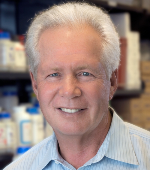 Warner Greene合作论文数Gladstone Center for HIV Cure Research, Gladstone Institutes;Gladstone Institute of Virology and Immunology, Gladstone Institutes;School of Medicine, University of California, San Francisco3
 Rune Hartmann合作论文数Department of Molecular Biology;Laboratory of Molecular Genetics;Institute for Virus Research;Kyoto University;DNA Technologies , Inc;Institute for Virus Research, Kyoto University3
 Jason Neidleman合作论文数Gladstone Institute of Virology and Immunology, University of California3Martin K. Thomsen合作论文数Section of Gene Function and Regulation, Institute of Cancer Research3
 Jes Vollertsen合作论文数Department of Life Sciences, Aalborg University3
 Thorkild Hvitved-Jacobsen合作论文数3
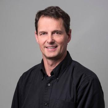 Matthew Porteus合作论文数Pediatrics Department, Stanford University School of Medicine;Division of Stem Cell Transplantation and Regenerative Medicine, Stanford University School of Medicine;Center for Definitive and Curative Medicine, Stanford University School of Medicine3Ramya Nandakumar合作论文数2M. Nyegaard合作论文数2Karen S. Jang合作论文数2Janni J Jørgensen合作论文数2




