Specific electroanalysis of neurotransmitters in the brain, bloodstream, cerebrospinal fluid (CSF), or at the cellular level critically depends on the availability of miniaturized electrodes for aptasensing. Yet, with electrode miniaturization, sensitivity of analysis and limits of detection (LOD) can be compromised. Here, we adapted the RNA-aptamer-based macroelectrode assay for dopamine to the microelectrode format, by using gold-plated carbon fiber microelectrodes (CFE), modified via thiol chemistry with cysteamine and an RNA aptamer, for specific dopamine detection. The sensitivity of analysis with gold-plated cylindrical microelectrodes improved 90-fold, to 9.75 μA μM-1 cm-2 (at +0.100 V) vs. 108 nA μM-1 cm-2 (at optimal +0.185 V) shown with gold disk macroelectrodes, with LOD being 60 and 100 nM, in PBS and in artificial CSF, respectively. Yet, epinephrine interfered at 0.1 V. At 0.05 V, the sensitivity dropped to 4.62 μA μM-1 cm-2 but the RNA-aptamer/cysteamine-modified CFEs demonstrated excellent selectivity for dopamine over epinephrine, norepinephrine, L-DOPA, DOPAC, and uric and ascorbic acids. These findings suggest a straightforward strategy for constructing biospecific aptamer-based microelectrodes. However, in matrices more complex than CSF, such as serum, dopamine oxidation was inhibited. Therefore, effective monitoring of dopamine levels in blood using aptamer microelectrodes will likely require the use of protective membranes.
Widespread human exposure to aluminum (Al) has raised increasing concerns about its effects on neuronal functions since Al3+ can cross the blood-brain barrier and accumulate in neuronal cells, especially considering its ionic similarity to ferric iron (Fe3+), a metal strongly associated with neurodegenerative diseases. In this study, we demonstrate that Al3+ affects vesicles in an iron-status-dependent manner in pheochromocytoma (PC12) cells, causing distinct changes under iron-normal, deficient, and overloaded conditions. Al3+ competes with Fe3+ for transferrin binding and enters cells via transferrin receptor (TfR)-mediated endocytosis, ultimately leading to cellular iron deficiency. This disruption alters dopamine-related proteins and molecular pathways, impairing exocytotic dynamics and reducing the average vesicular transmitter storage. When iron level is overloaded, TfR expression is downregulated to avoid Al3+ affecting vesicles. However, supplementation with ferric ammonium citrate (FAC) fails to reverse Al3+-induced iron deficiency due to Al3+ simultaneously upregulating TfR, allowing excess Fe3+ to further decrease neurotransmitter release. Furthermore, deferoxamine (DFOM), an Fe chelator, destabilizes exocytotic fusion pores, suggesting a potential drawback of chelation therapy. These findings highlight iron dysregulation as a key pathway through which Al3+ impairs vesicular function, providing a new insight into how metal ion interactions modulate neurotransmission.
The electrochemical activities of biomolecular condensates represent a new fundamental functioning mechanism in biochemistry and cell biology. However, our understanding of the underlying molecular mechanism and the interfacial field-dependent chemical activities remains limited. This is due to the lack of technology to probe such activities in real time and at a single-condensate level. Stress granules (SGs) are membraneless organelles that form in the cytoplasm to adapt to cell stress, which are found to encapsulate reactive oxygen species (ROS) in our lab. Here, we design and implement a collision-based electrochemical nanosensor that enables probing of the redox activities of SGs at a single-condensate level in live cells. We show that ex-vivo separated SGs drive the redox reactions depending on their own interfacial potentials and the constituents of the solution system. Surprisingly, we found that water molecules, rather than solvated oxygen (the main source of ROS produced by a conventional enzyme reaction in cells), are the main chemical origin of the redox activity of SGs. Finally, we demonstrate the application of this electrochemical nanosensor in real-time probing of the generation of hydrogen peroxide from SGs in mammalian cells and show that the electrochemical environment of the cells can regulate the redox activity of SGs. This work uncovers the likely mechanisms encoding nonenzymatic redox activities of SGs and demonstrates a key fundamental technological capability that can be highly useful in exploring the intracellular electroactive pathways of macroscale assemblies.
Insulin secretion from vesicles within pancreatic beta cells occurs through the rapid (≤10 ms) process of exocytosis. A crucial final step in this process is the formation of a fusion pore, which connects the insulin vesicle interior to the extracellular space and insulin secretion rate. We employed real-time single-cell amperometry (SCA) with microsensors to quantitatively and dynamically monitor serotonin secretion (used as a proxy for insulin) and alterations in the dynamics of fusion pore formation, including opening, duration, and closing times, during single exocytosis events. Additionally, total internal reflection fluorescence (TIRF) microscopy was used to track docked vesicles and optically measure insulin release. In beta cells from human organ donors with type 2 diabetes (T2D), the fusion pore opening was prematurely aborted, leading to diminished cargo release. Furthermore, the number of docked vesicles per beta cell differed between healthy donors and individuals with T2D. In conclusion, our combined super-sensitive optical and electrochemical analyses from a new perspective highlight a link between defective fusion pore dynamics and reduced insulin secretion (a hallmark of T2D) that has not been previously reported. These findings reveal the role of impaired fusion pore dynamics in T2D, independent of its underlying etiology, with potential therapeutic implications.
The aim of this paper is to overview the meeting on New horizons in nanoelectrochemistry held at Nanjing University in China in October 2024 and to give some perspective to the work presented. This paper is based on my summary talk and breaks down the subjects in the following areas of nanoelectrochemistry presented at the meeting: nanowires, nanonets, and nanoarrays; nanopores; nanopipettes; spectroelectrochemistry, scanning ion-conductance microscopy and light-active processes at nanointerfaces; scanning electrochemical microscopy and scanning electrochemical cell microscopy; and nanosensors. I end with some discussion of online meetings and where the field might go including artificial intelligence and by asking AI to define the challenges and future of nanoelectrochemistry.
The two essential fatty acids, alpha-linolenic acid and linoleic acid, and the higher unsaturated fatty acids synthesized from them are critical for the development and maintenance of normal brain functions. Deficiencies of these fatty acids have been shown to cause damage to the neuronal development, cognition, and locomotor function. We combined electrochemistry and imaging techniques to examine the effects of the two essential fatty acids on catecholamine release dynamics and the vesicle content as well as on the cell membrane phospholipid composition to understand how they impact exocytosis and by extension neurotransmission at the single-cell level. Incubation of either of the two fatty acids reduces the size of secretory vesicles and enables the incorporation of more double bonds into the cell membrane structure, resulting in higher membrane flexibility. This subsequently affects proteins regulating the dynamics of the exocytotic fusion pore and thereby affects exocytosis. Our data suggest a possible pathway whereby the two essential fatty acids affect the membrane structure to impact exocytosis and provide a potential treatment for diseases and impairments related to catecholamine signaling.
The microtubule-associated protein tau participates in neurotransmission regulation via its interaction with synaptic vesicles (SVs). The precise nature and mechanics of tau's engagement with SVs, especially regarding alterations in vesicle dynamics, remain a matter of discussion. We report an electrochemical method using a synapse-mimicking nanopipette to monitor vesicle dynamics induced by tau. A model vesicle of ~30 nm is confined within a lipid-modified nanopipette orifice with a comparable diameter to mimic the synaptic lipid environment. Both tau and phosphorylated tau (p-tau) present two-state dynamic behavior in this biomimetic system, showing typical ionic current oscillation, induced by lipid-tau interaction. The results indicate that p-tau has a stronger affinity to the lipid vesicles in the confined environment, blocking the vesicle movement to a higher degree. Taken together, this method bridges a gap for sensing synaptic vesicle dynamics in a confined lipid environment, mimicking vesicle movement near the synaptic membrane. These findings contribute to understanding how different types of tau protein regulate synaptic vesicle motility and to underlying its functional and pathological behaviours in disease.
Single-entity electrochemistry is a powerful tool that enables the study of electrochemical processes at interfaces and provides insights into the intrinsic chemical and structural heterogeneities of individual entities. Signal processing is a critical aspect of single-entity electrochemical measurements and can be used for data recognition, classification, and interpretation. In this review, we summarize the recent five-year advances in signal processing techniques for single-entity electrochemistry and highlight their importance in obtaining high-quality data and extracting effective features from electrochemical signals, which are generally applicable in single-entity electrochemistry. Moreover, we shed light on electrochemical noise analysis to obtain single-molecule frequency fingerprint spectra that can provide rich information about the ion networks at the interface. By incorporating advanced data analysis tools and artificial intelligence algorithms, single-entity electrochemical measurements would revolutionize the field of single-entity analysis, leading to new fundamental discoveries.
Serotonin, a monoamine neurotransmitter, is important in both the central nervous system (CNS) and the peripheral nervous system. Malfunction of serotonin signaling leads to various disorders. We studied serotonin signaling from serotonergic neurons inside the ventral nerve cord of Drosophila melanogaster . Serotonergic neurons and stimulated release were visualized and achieved with mCherry and channelrhodopsin-2 (an optogenetically transfected ion channel), respectively, and two electrochemical techniques quantified serotonin release and vesicular content. Mean vesicular serotonin content released during exocytosis from these neurons was 84 %, considerably higher than reported in previous studies regarding octopamine (4.5 %) and glutamate release (31 %). Serotonin content within all vesicles is uniformly changed when serotonin concentration is inhibited or enhanced. However, serotonin release exhibits two Gaussian distributions: higher frequency of small release events, and similar or slightly higher frequency of large events, resulting in differential release fractions ranging from partial (13–18 %) to full (100 %) release after treatment with agents to either enhance or diminish release. This is the first example of consistent full exocytotic release events we have observed in any system. We suggest one pool of vesicles can release significantly diverse fractions of transmitter load during exocytosis, a potentially novel pathway to regulate exocytosis and neuronal signaling.
Long COVID, as currently defined by the World Health Organization (WHO) and other authorities, is a symptomatic condition that has been shown to affect an estimated 10-30% of non-hospitalized patients after one infection. However, COVID-19 can also cause organ damage in individuals without symptoms, who would not fall under the current definition of Long COVID. This organ damage, whether symptomatic or not, can lead to various health impacts such as heart attacks and strokes. Given these observations, it is necessary to either expand the definition of Long COVID to include organ damage or recognize COVID-19-induced organ damage as a distinct condition affecting many symptomatic and asymptomatic individuals after COVID-19 infections. It is important to consider that many known adverse health outcomes, including heart conditions and cancers, can be asymptomatic until harm thresholds are reached. Many more medical conditions can be identified by testing than those that are recognized through reported symptoms. It is therefore important to similarly recognize that while Long COVID symptoms are associated with organ damage, there are many individuals that have organ damage without displaying recognized symptoms and to include this harm in the characterization of COVID-19 and in the monitoring of individuals after COVID-19 infections.
Long COVID, as currently defined by the World Health Organization (WHO) and other authorities, is a symptomatic condition that has been shown to affect an estimated 10 %-30 % of non-hospitalized patients after one infection. However, COVID-19 can also cause organ damage in individuals without symptoms, who would not fall under the current definition of Long COVID. This organ damage, whether symptomatic or not, can lead to various health impacts such as heart attacks and strokes. Given these observations, it is necessary to either expand the definition of Long COVID to include organ damage or recognize COVID-19-induced organ damage as a distinct condition affecting many symptomatic and asymptomatic individuals after COVID-19 infections. It is important to consider that many known adverse health outcomes, including heart conditions and cancers, can be asymptomatic until harm thresholds are reached. Many more medical conditions can be identified by testing than those that are recognized through reported symptoms. It is therefore important to similarly recognize that while Long COVID symptoms are associated with organ damage, there are many individuals that have organ damage without displaying recognized symptoms and to include this harm in the characterization of COVID-19 and in the monitoring of individuals after COVID-19 infections.
Elucidating exocytosis processes provide insights into cellular neurotransmission mechanisms, and may have potential in neurodegenerative diseases research. Amperometry is an established electrochemical method for the detection of neurotransmitters released from and stored inside cells. An important aspect of the amperometry method is the sub-millisecond temporal resolution of the current recordings which leads to several hundreds of gigabytes of high-quality data. In this study, we present a universal method for the classification with respect to diverse amperometric datasets using data-driven approaches in computational science. We demonstrate a very high prediction accuracy (greater than or equal to 95%). This includes an end-to-end systematic machine learning workflow for amperometric time series datasets consisting of pre-processing; feature extraction; model identification; training and testing; followed by feature importance evaluation - all implemented. We tested the method on heterogeneous amperometric time series datasets generated using different experimental approaches, chemical stimulations, electrode types, and varying recording times. We identified a certain overarching set of common features across these datasets which enables accurate predictions. Further, we showed that information relevant for the classification of amperometric traces are neither in the spiky segments alone, nor can it be retrieved from just the temporal structure of spikes. In fact, the transients between spikes and the trace baselines carry essential information for a successful classification, thereby strongly demonstrating that an effective feature representation of amperometric time series requires the full time series. To our knowledge, this is one of the first studies that propose a scheme for machine learning, and in particular, supervised learning on full amperometry time series data.
An overreactive stress granule (SG) pathway and long-lived, stable SGs formation are thought to participate in the progress of neurodegenerative diseases (NDs). To understand if and how SGs contribute to disorders of neurotransmitter release in NDs, we examined the interaction between extracellular isolated SGs and vesicles. Amperometry shows that the vesicular content increases and dynamics of vesicle opening slow down after vesicles are treated with SGs, suggesting larger vesicles are formed. Data from transmission electron microscopy (TEM) clearly shows that a portion of large dense-core vesicles (LDCVs) with double/multiple cores appear, thus confirming that SGs induce homotypic fusion between LDCVs. This might be a protective step to help cells to survive following high oxidative stress. A hypothetical mechanism is proposed whereby enriched mRNA or protein in the shell of SGs is likely to bind intrinsically disordered protein (IDP) regions of vesicle associated membrane protein (VAMP) driving a disrupted membrane between two closely buddled vesicles to fuse with each other to form double-core vesicles. Our results show that SGs induce homotypic fusion of LDCVs, providing better understanding of how SGs intervene in pathological processes and opening a new direction to investigations of SGs involved neurodegenerative disease.
Amperometry is a commonly used electrochemical method for studying the process of exocytosis in real-time. Given the high precision of recording that amperometry procedures offer, the volume of data generated can span over several hundreds of megabytes to a few gigabytes and therefore necessitates systematic and reproducible methods for analysis. Though the spike characteristics of amperometry traces in the time domain hold information about the dynamics of exocytosis, these biochemical signals are, more often than not, characterized by time-varying signal properties. Such signals with time-variant properties may occur at different frequencies and therefore analyzing them in the frequency domain may provide statistical validation for observations already established in the time domain. This necessitates the use of time-variant, frequency-selective signal processing methods as well, which can adeptly quantify the dominant or mean frequencies in the signal. The Fast Fourier Transform (FFT) is a well-established computational tool that is commonly used to find the frequency components of a signal buried in noise. In this work, we outline a method for spike-based frequency analysis of amperometry traces using FFT that also provides statistical validation of observations on spike characteristics in the time domain. We demonstrate the method by utilizing simulated signals and by subsequently testing it on diverse amperometry datasets generated from different experiments with various chemical stimulations. To our knowledge, this is the first fully automated open-source tool available dedicated to the analysis of spikes extracted from amperometry signals in the frequency domain.
Single cell Amperometry (SCA) is a powerful, sensitive, high temporal resolution electrochemical technique used to quantify secreted molecular messengers from individual cells and vesicles. This technique has been extensively applied to study the process of exocytosis, and it has also been applied, albeit less frequently, to investigate insulin exocytosis from single pancreatic beta cells. Insufficient insulin release can lead to diabetes, a chronic lifestyle disorder that affects millions of people worldwide. This review aims to summarize and highlight electrochemical measurements of insulin via monitoring its secretion from beta cells by SCA with micro- and nanoelectrodes since the 1990s and to explain how and why serotonin is used as a proxy for monitoring insulin during exocytosis from single beta cells. Finally, we describe how the combination of SCA measurements with the intracellular vesicle impact electrochemical cytometry (IVIEC) technique has led to important findings regarding fractional release types in beta cells. These findings, reported recently, have opened a new window in the study of pore formation, exocytosis from single vesicles, and the mechanisms of insulin secretion. This sensitive cellular electroanalysis approach should help in the development of novel therapeutic strategies targeting diabetes in the future.
Stress granules (SGs) are stress-induced biomolecular condensates which originate primarily from inactivated RNA translation machinery and translation initiation factors. SG formation is an important defensive mechanism for cell survival, while its dysfunction has been linked to neurodegenerative diseases. However, the molecular mechanisms of SG assembly and disassembly, as well as their impacts on cellular recovery, are not fully understood. More thorough investigations into the molecular dynamics of SG pathways are required to understand the pathophysiological roles of SGs in cellular systems. Here, we characterize the SG and cytoplasmic protein turnover in neuronal progenitor cells (NPCs) under stressed and non-stressed conditions using correlative STED and NanoSIMS imaging. We incubate NPCs with isotopically labelled (15N) leucine and stress them with the ER stressor thapsigargin (TG). A correlation of STED and NanoSIMS allows the localization of individual SGs (using STED), and their protein turnover can then be extracted based on the 15N/14N ratio (using NanoSIMS). We found that TG-induced SGs, which are highly dynamic domains, recruit their constituents predominantly from the cytoplasm. Moreover, ER stress impairs the total cellular protein turnover regimen, and this impairment is not restored after the commonly proceeded stress recovery period.
In multicellular organisms, cells typically communicate by sending and receiving chemical signals. Chemical messengers involved in the exocytosis of neuroendocrine cells or neurons are generally assumed to only originate from the fusing of intracellular large dense core vesicles (LDCVs) or synaptic vesicles with the cellular membrane following stimulation. Accumulated evidence suggests that exosomes─one of the main extracellular vesicles (EVs)─carrying cell-dependent DNA, mRNA, proteins, etc., play an essential role in cellular communication. Due to experimental limitations, it has been difficult to monitor the real-time release of individual exosomes; this restricts a comprehensive understanding of the basic molecular mechanisms and the functions of exosomes. In this work, we introduce amperometry with microelectrodes to capture the dynamic release of single exosomes from a single living cell, distinguish them from other EVs, and differentiate the molecules inside exosomes and those secreted from LDCVs. We show that, similar to many LDCVs and synaptic vesicles, exosomes released by neuroendocrine cells also contain catecholamine transmitters. This finding reveals a different mode of chemical communication via exosome-encapsulated chemical messengers and a potential interconnection between the two release pathways, changing the canonical view of exocytosis of neuroendocrine cells and possibly neurons. This defines a new mechanism for chemical communication at the fundamental level and opens new avenues in the research of the molecular biology of exosomes in the neuroendocrine and central nervous systems.
Aberrant functioning of the proteasome has been associated with crucial pathologic conditions including neurodegeneration. Yet, the complex underlying causes at the cellular level remain unclear and there are conflicting reports of neuroprotective to neurodegenerative effects of proteasomal inhibitors such as lactacystin that are utilised as models for neurodegenerative diseases. The conflicting results may be associated with different dose regimes of lactacystin and hence we have performed a dose dependent study of the effects of lactacystin to identify concurrent changes in the cell membrane lipid profile and the dynamics of exocytosis using a combination of surface sensitive mass spectrometry and single cell amperometry. Significant changes of negatively charged lipids were associated with different lactacystin doses that showed a weak correlation with exocytosis while changes in PE and PE-O lipids showed dose dependent changes correlated with initial pore formation and total release of vesicle content respectively.
We review the methods of vesicle impact electrochemical cytometry, intracellular impact electrochemical cytometry, and single cell amperometry and their application to measuring the storage of neurotransmitters in cellular vesicles. We provide protocols to measure vesicle content, the release of catecholamines, and from there the fraction of transmitter released in each exocytosis event. The focus here has been a combination of methods to evaluate factors related to neuronal function at the cellular level and implications in, for example, cognition.
 Nicholas Winograd合作论文数Department of Materials Science and Engineering, College of Earth and Mineral Sciences, Pennsylvania State University41
 Dunevall Johan合作论文数†Department of Chemistry and Chemical Engineering, Chalmers University of Technology29
 Michael L. Heien合作论文数Department of Chemistry, The University of North Carolina27
 Nhu T N Phan合作论文数Department of Chemistry and Molecular Biology, University of Gothenburg24
 Kurczy Michael E合作论文数Dept Chem & Biol Engn, Chalmers Univ Technol24
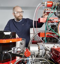 Per Malmberg合作论文数Wageningen22
 Ann-Sofie Cans合作论文数Chalmers University of Technology22
 Chaoyi Gu合作论文数Univ Gothenburg, Dept Chem & Mol Biol, Kemivagen 10, S-41296 Gothenburg, Sweden18
 Soodabeh Majdi合作论文数Department of Chemistry and Molecular Biology, University of Gothenburg18
 R. Mark Wightman合作论文数Department of Chemistry, The University of North Carolina at Chapel Hill18
 John S Fletcher合作论文数Department of Chemistry & Molecular Biology, University of Gothenburg18
 Xianchan Li (李鲜婵)合作论文数Department of Pharmaceutical Analysis, School of Pharmaceutical Sciences, Peking University16
 Lin Ren合作论文数Dept Chem & Chem Engn, Chalmers Univ Technol14
 Trouillon Raphaël合作论文数Dept Chem & Mol Biol, Univ Gothenburg14
 Jelena Lovric合作论文数13
 Nathan J. Wittenberg合作论文数Department of Chemistry;Pennsylvania State University;Department of Chemistry, Pennsylvania State University12Thomas P. Roddy合作论文数Department of Atherosclerosis Exploratory Biomarkers, Merck Research Laboratories12Mai H Philipsen合作论文数11Sombers Leslie A合作论文数Dept Chem, N Carolina State Univ11Ra Wallingford合作论文数10
 Xiulan He (何秀兰)合作论文数College of Chemistry, Beijing Normal University10
 Neda Najafinobar合作论文数10Y. Y. Lau合作论文数10D. M. Cannon合作论文数10Peter F. Gavin合作论文数10
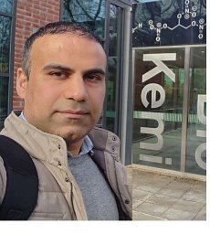 Amir Hatamie合作论文数Faculty of Sciences, Shahid Chamran Universitry9
 Lisa Mellander合作论文数Department of Chemistry and Molecular Biology, University of Gothenburg9
 Jorg Hanrieder合作论文数Department of Physical and Analytical Chemistry, Uppsala University9
 Carina Berglund合作论文数9
 Piehowski Paul D合作论文数Biol Sci Div, Pacific Northwest Natl Lab8Larsson Anna合作论文数Oral &The Sahlgrenska Academy, University of Gothenburg;The Sahlgrenska Academy, University of Gothenburg8
 Masoumeh Dowlatshahi Pour合作论文数8
 Xinwei Zhang (张欣伟)合作论文数College of Chemistry and Molecular Sciences, Wuhan University8
 Christian A. Amatore (克里斯汀·阿芒托)合作论文数Xiamen University;Centre National de la Recherche Scientifique;École Normale Supérieure8Reginaldo A. Saraceno合作论文数8Timothy G. Strein合作论文数8Kelly L. Adams合作论文数8Guangyao Chen合作论文数8Franklin D. Swanek合作论文数8Tm Olefirowicz合作论文数7
 Sara Ostrowski合作论文数7Tho Nguyen Duc Khanh合作论文数Goteborgs Universitet7
 F. Kanoufi合作论文数Laboratoire de Chimie et Electrochimie des Matériaux Moléculaires, ESPCI, 10, rue Vauquelin, 75231 Paris Cedex 05, France7Makos Monique A合作论文数7
 Amir Saeid Mohammadi合作论文数7
 Keke Hu (胡可可)合作论文数College of Chemistry and Chemical Engineering, Xiamen University7
 Royce W. Murray合作论文数University of North Carolina at Chapel Hill7Roger Karlsson合作论文数7
 Danny K Y Wong合作论文数Division of Environmental and Life Sciences;Centre for Professional Development;Macquarie University;Centre for Professional Development, Macquarie University7




