BACKGROUND:Extracorporeal life support (ECLS) can cause bleeding via excessive cleavage of multimeric protein von Willebrand Factor (VWF) by protease ADAMTS-13, a bleeding diathesis termed acquired von Willebrand syndrome. VWF high-molecular-weight multimers (HMWM) are more procoagulant than smaller multimers. Acute-phase responses generate high levels of VWF-HMWM and reduced ADAMTS-13 activity. OBJECTIVES:Assess whether VWF-HMWM degradation is less pronounced and VWF:collagen binding activity (CB)/VWF:antigen (Ag) ratios are lower during ECLS in acute trauma vs noninjury settings. METHODS:Anesthetized, mechanically ventilated swine (45-60 kg) were randomized to polytrauma (INJ CTRL, n = 14) managed with mechanical ventilation, uninjured receiving veno-venous extracorporeal carbon dioxide removal (ECLS CTRL, n = 20), or polytrauma managed with extracorporeal carbon dioxide removal (ECLS INJ, n = 22). In ECLS groups, 50% of animals received heparin anticoagulation, while 50% received no systemic anticoagulation. VWF:Ag, VWF:CB, VWF multimer size distribution, and ADAMTS-13 activity were evaluated at baseline, post-injury/intervention, and at 6, 24, 48, and 72 hours. RESULTS:VWF-HMWM were depleted at 72 hours in ECLS CTRL (-19 ± 3%, P < .0001), but not in ECLS INJ (-10 ± 5%, P = .09). VWF:CB/VWF:Ag ratios relative to baseline were reduced at 48 to 72 hours in ECLS CTRL (0.84 ± 0.04, P = .002) and ECLS INJ (0.69 ± 0.05, P < .001) but not INJ CTRL. ADAMTS-13 activity decreased in all groups. Animals treated with heparin showed less pronounced VWF-HMWM degradation, higher VWF:CB/VWF:Ag ratios, and higher ADAMTS-13 activity vs matched non-anticoagulated animals. CONCLUSION:Trauma managed with ECLS was associated with reduced VWF:CB/VWF:Ag ratios and lower ADAMTS-13 activity, but VWF-HMWM depletion was minimal. Thus, potential for acquired von Willebrand syndrome is a concern for trauma patients undergoing ECLS. Heparin partially countered effects of ECLS on VWF and ADAMTS-13.
Solutions to reduce the need for systemic anticoagulation during extracorporeal life support would improve safety and utility. The study objective was to evaluate the safety and efficacy of a nitric oxide-generating extracorporeal carbon dioxide removal (ECCO 2 R) system without systemic anticoagulation in a translational swine model. We hypothesized that nitric oxide reduces circuit thrombosis, without untoward systemic effects. Anesthetized, mechanically ventilated swine (50–60 kg) received bicaval jugular cannulation for 72 hour venovenous ECCO 2 R. Control (n = 6) received a standard ECCO 2 R device with systemic heparinization. Treatment (n = 6) received the same device, but with nitric oxide–generating circuitry and 80 ppm nitric oxide added to sweep gas, without systemic heparinization. No between-group differences in vitals, ventilator settings, blood gases, extracorporeal gas exchange, or fluid balance occurred. In both groups, ECCO 2 R enabled reduction in tidal volume. Oxygenator thrombus area, quantified following dissection and imaging, was reduced in treatment (10.2 ± 1.2%) versus control (15.2 ± 1.6%) ( p = 0.03). One control oxygenator occluded. No nitric oxide–related adverse effects were observed, including methemoglobinemia. Nitric oxide–enhanced ECCO 2 R enabled 72 hours of support without systemic anticoagulation, and without altering oxygenator performance or causing untoward systemic effects. Future studies are needed to investigate efficacy in subjects with underlying coagulopathy and/or contraindications for systemic anticoagulation.
INTRODUCTION:Absence of pre-hospital coagulation tests challenges prompt management of hemostasis after trauma. The Viscoelastic Coagulation Monitor (VCM, Entegrion, Durham, NC) is a hand-held coagulation test for point-of-care. We evaluated VCM in a translational swine polytrauma model, hypothesizing that VCM correlates with a laboratory reference method, the TEG 5000 (Haemonetics, Boston, MA), and can identify coagulopathic phenotypes relevant to trauma. Our secondary hypothesis was that pre-warming of VCM disposable test cartridges using a heating plate versus pre-warming of cartridges by carrying the cartridge in the user's pocket does not significantly alter results. MATERIALS AND METHODS:This study was conducted in tandem with a parent study involving anesthetized, mechanically ventilated swine (n = 20; 54 ± 5 kg) that encountered traumatic brain injury, pulmonary contusion and hemorrhage, or combination/polytrauma injury. Blood was collected at baseline, post-injury, post-shock, post-transfusion, and 6-, 24-, and 48 h post-injury to perform VCM at point-of-care. Within-group effect of time was assessed. Spearman correlation examined linear relations between VCM and standard laboratory-based coagulation tests; as well as lactate, ionized calcium, and body temperature. Logistic regression examined predictiveness of VCM to identify coagulopathic phenotypes, with receiver operator characteristic curves generated to assess diagnostic capability. At a subset of timepoints, necessity of pre-warming the VCM test cartridge using a heating plate versus pre-warming the cartridge by placement in the user's pocket was assessed by conducting simultaneous tests on two separate instruments, with results analyzed by paired t-test with crossover design. RESULTS:VCM revealed time-dependent changes in clotting time, clot formation time (CFT), alpha, maximum clot firmness (MCF), and lysis index (LI30). All VCM metrics correlated with the respective TEG 5000 metrics, with strongest correlation for VCM MCF with TEG MA (rhos = 0.77, P < .0001) and VCM LI30 with TEG LY30 (rhos = -0.76, P < .0001). VCM demonstrated good (area under the curve >0.70) to excellent (area under the curve >0.90) diagnostic accuracy in detection of low platelet count (MCF), low hematocrit (clotting time, clot formation time, alpha, and MCF), low fibrinogen (MCF), and high fibrinogen (alpha, MCF). There was no statistically or clinically relevant effect of cartridge warming method on results. CONCLUSIONS:In a trauma model, VCM detected significant changes in coagulation at point-of-care in a simplified portable form factor. VCM could enable informed hemostasis management in pre-hospital settings where coagulations tests are unavailable, pending further validation in clinical trials.
BACKGROUND:Aeromedical evacuation (AE) environments are characterized by hypobaria and may cause secondary insult to casualties with traumatic brain injury (TBI). As increased FiO2 is commonly administered to mechanically ventilated casualties during AE, it may exacerbate TBI. We hypothesized that hyperoxia at ground level and/or during simulated AE worsens neuroinflammation and neurodegeneration after mild-to-moderate blunt TBI. MATERIALS AND METHODS:Female Yorkshire swine were anesthetized, mechanically ventilated, and received blunt TBI via a modified humane stunner. Animals were randomized into 1 of 4 groups (n = 8/group): Ground Normoxia (G-Norm); Ground Hyperoxia (G-Hyper); AE Normoxia (AE-Norm); and AE Hyperoxia (AE-Hyper), with AE groups placed in a hypobaric chamber for 6 hours at 8,000 ft and then managed in an ICU for 24 hours. Neuroinflammation and neurodegeneration were assessed via histological injury scores (0 = no injury, and 4 = most severe injury), as well as measurements of systemic HMGB1, S100β, GFAP, and cytokines such as IL-1β, IL-6, and IL-10 levels. In addition, we assessed hemodynamics, intracranial pressure, blood gases and chemistry, and coagulation variables. RESULTS:TBI led to increased intracranial pressure, tachycardia, and transient hypertension with subsequent periodic fluctuations in heart rate and blood pressure. The G-Norm and G-Hyper groups had mild-to-moderate injuries (cerebrum: 2.0 and 1.75, respectively, NS; brainstem: 2.0 and 1.71, respectively, NS). The AE-Norm and AE-Hyper groups had numerically less injuries (cerebrum: 1.5 and 1.71, respectively, NS; brainstem: 0.86 and 1.25, respectively, NS). HMGB1 increased 2-fold after TBI in all 4 groups (P<.003). IL-6 increased after TBI and remained elevated relative to baseline in all groups except AE-Hyper group (P<.0001). No within or between-group differences were observed in any other variables. CONCLUSIONS:This 24-hour intent-to-treat study did not identify discernable differences in normoxic versus hyperoxic management of TBI at ground level or during AE. Future studies should evaluate exposures to hypobaria and hyperoxia over longer durations.
INTRODUCTION:Changes in blood lactate (Lac) carry prognostic value in pathologic processes and guide decision-making on therapeutic interventions. This feasibility study evaluated a new wearable continuous lactate monitor (CLM) currently undergoing preclinical validation in severe combat-relevant porcine trauma models. PURPOSE:Assess the performance of CLM compared to arterial blood gas (ABG) analysis. HYPOTHESIS:CLM-derived Lac values correlate with ABG Lac. METHODS:Female Yorkshire swine (50-60 kg, N = 6) were anesthetized, mechanically ventilated, subjected to a combination of the following injuries: bilateral pulmonary contusion (PC), traumatic brain injury (TBI), or hemorrhagic shock (HEM): (PC N = 3, PC + TBI N = 1; PC + HEM N = 1; PC + TBI + HEM N = 1). Animals were managed with immediate application of veno-venous extracorporeal life support. Two CLMs were placed in the skin of the lower abdomen. ABG analysis was performed using a GEM Premier 4000 [Werfen, Bedford, MA]. Raw CLM data were retrospectively calibrated using ABG values. Pearson Correlation Coefficient (P < 0.05) was calculated to compare CLM and ABG Lac values. Bland-Altman analysis was performed to assess device agreement before and after injury for up to 72 h. RESULTS:Three hundred ninety-four paired measurements were accepted for analysis. Retrospectively calibrated CLM Lac correlated with ABG Lac, Pearson Correlation Coefficient = 0.687, P < 0.0001; however, a Bland-Altman analysis showed moderate agreement between the methods. CONCLUSIONS:CLM positively correlated with ABG Lac values, with moderate agreement between devices. Future optimization and utilization of CLM may significantly enhance sensor performance and reliability, making it suitable for monitoring and decision support during patient management.
BACKGROUND:Trauma is a leading cause of mortality, but injury-specific molecular targets remain largely unknown. We hypothesized that distinctive yet unrecognized tissue targets accessible to circulating ligands might emerge during trauma, thereby underscoring a trauma-related proteome. METHODS:We screened a peptide library to discover targets in a porcine model of major trauma: compound femur fracture with hemorrhagic shock. Bioinformatics yielded conserved motifs, and candidate receptors were affinity purified. In silico and in vitro approaches served to investigate possible associations between candidate receptors and calcium, a major component of skeletal muscle and bone. In vivo homing and molecular imaging (PET/MRI and SPECT/CT) studies of the most promising ligand peptide candidate were performed in the porcine model and were also confirmed in a corresponding rat model of major trauma. Optical methodologies and molecular dynamics simulations served to explore the molecular attributes of the ligand-receptor binding. FINDINGS:Nearly all molecular targets of the selected ligand peptides were calcium-dependent proteins, which become accessible upon trauma. We validated specific binding of homing peptides to these receptors in injured tissues, including CLRGFPALVC:CASQ1, CSEIGVRAC:HSP27, and CRQRPASGC:CALR. Notably, we determined that ligand peptide CRQRPASGC targets an injury-specific calcium-facilitated conformation of calreticulin, enabling specific molecular imaging of trauma. CONCLUSIONS:We conceptually propose the term "traumome" for the functional receptor repertoire that becomes readily amenable for ligand-directed targeting upon major trauma. These preclinical findings pave the way toward clinic-ready targeted theragnostic approaches in the setting of trauma. FUNDING:Major funding was provided by the Defense Advanced Research Projects Agency (DARPA).
Abstract Background Optimizing resuscitation to reduce inflammation and organ dysfunction following human trauma-associated hemorrhagic shock is a major clinical hurdle. This is limited by the short duration of pre-clinical studies and the sparsity of early data in the clinical setting. Methods We sought to bridge this gap by linking preclinical data in a porcine model with clinical data from patients from the Prospective, Observational, Multicenter, Major Trauma Transfusion (PROMMTT) study via a three-compartment ordinary differential equation model of inflammation and coagulation. Results The mathematical model accurately predicts physiologic, inflammatory, and laboratory measures in both the porcine model and patients, as well as the outcome and time of death in the PROMMTT cohort. Model simulation suggests that resuscitation with plasma and red blood cells outperformed resuscitation with crystalloid or plasma alone, and that earlier plasma resuscitation reduced injury severity and increased survival time. Conclusions This workflow may serve as a translational bridge from pre-clinical to clinical studies in trauma-associated hemorrhagic shock and other complex disease settings.
BACKGROUND Awareness of ventilator-induced lung injury contributed to increased use of extracorporeal interventions, but not immediately after injury,before acute respiratory distress syndrome (ARDS) ensues. Our objective was to evaluate the role of venovenous extracorporeal carbon dioxide removal (ECCO2R) in management of mechanically ventilated swine with smoke inhalation injury and 40% body surface area burns. METHODS Yorkshire swine (n = 29, 43.2 ± 0.5 kg) underwent anesthesia, instrumentation, severe smoke inhalation, and 40% body surface area burns, followed by 72 hours of round-the-clock intensive care unit care with mechanical ventilation, fluids, pressors, bronchoscopic cast removal, computer tomography scans, and arterial blood assays. Within 1 hour after injury, animals received ECCO2R with either MiniLung (Xenios AG, Heilbronn, Germany; n = 10) or Hemolung (ALung Technologies, Pittsburgh, PA; n = 10), or no ECCO2R in injured controls (INJC, n = 12). RESULTS Immediate postinjury ECCO2R reduced minute ventilation (p < 0.001) and prevented ARDS in 37.5% of MiniLung and 11.1% of Hemolung animals. Time to ARDS (partial pressure of arterial oxygen to fraction of inspired oxygen ratio below 300) was shortest (14 ± 2.2 hours) in INJC, intermediate (21.6 ± 3.5 hours) in Hemolung (HEMO), and most delayed in MiniLung (31.1 ± 7.2 hours, p = 0.0121, log-rank test vs. INJC). Driving pressure was lower in MiniLung versus INJC (p < 0.0001) and HEMO versus INJC (p = 0.0005) at 48 hours. Extracorporeal CO2 removal reduced systemic levels of tumor necrosis factor α versus INJC. CONCLUSION In swine with severe smoke inhalation and burns, immediate postinjury ECCO2R reduced ventilator settings, delayed or prevented ARDS, and reduced its severity. Proactive early percutaneous ECCO2R initiation via simplified, purpose-built devices should be considered as a low-maintenance lung injury management approach with significant disease modifying clinical benefit potential.
Background Patients with kidney failure are at risk for lethal complications from hyperkalemia. Resuscitation, medications, and hemodialysis are used to mitigate increased potassium (K+) levels in circulating blood; however, these approaches may not always be readily available or effective, especially in a resource limited environment. We tested a sorbent cartridge (KC, K+ontrol CytoSorbents Medical Inc., Monmouth Junction, New Jersey) which contains a resin adsorber for K+. The objective of this study was to test the utility of KC in an ex vivo circulation system. We hypothesized that KC reduces K+ levels in extracorporeal circulation of donor swine whole blood infused with KCl. Methods A six-hour circulation study was carried out using KC, a NxStage (NxStage Medical, Inc., Lawrence, MA) membrane, blood bag containing heparinized whole blood with KCl infusion, 3/16-inch ID tubing, a peristaltic pump, and flow sensors. The NxStage permeate line was connected back to the main circuit in the Control group ( n = 6), creating a recirculation loop. For KC group ( n = 6), KC was added to the recirculation loop, and a continuous infusion of KCl at 10 mEq/hour was administered for two hours. Blood samples were acquired at baseline and every hour for 6 h. Results In the control group, K+ levels remained at ∼9 mmol/L; 9.1 ± 0.4 mmol/L at 6 h. In the KC group, significant decreases in K+ at hour 1 (4.3 ± 0.3 mmol/L) and were sustained for the experiment duration equilibrating at 4.6 ± 0.4 mmol/L after 6 h ( p = 0.042). Main loop blood flow was maintained under 400 mL/min; recirculation loop flow varied between 60 and 70 mL/min in the control group and 45–55 mL/min in the KC group. Decreases in recirculation loop flow in KC group required 7% increase of pump RPM. Conclusions During ex-vivo extracorporeal circulation using donor swine blood, KC removed approximately 50% of K+, normalizing circulating levels.
Microfluidic membrane oxygenators are designed to mimic branching vasculature of the native lung during extracorporeal lung support. To date, scaling of such devices to achieve clinically relevant blood flow and lung support has been a limitation. We evaluated a novel multilayer microfluidic blood oxygenator (BLOx) capable of supporting 750–800 ml/min blood flow versus a standard hollow fiber membrane oxygenator (HFMO) in vivo during veno-venous extracorporeal life support for 24 hours in anesthetized, mechanically ventilated uninjured swine (n = 3/group). The objective was to assess feasibility, safety, and biocompatibility. Circuits remained patent and operated with stable pressures throughout 24 hours. No group differences in vital signs or evidence of end-organ damage occurred. No change in plasma free hemoglobin and von Willebrand factor multimer size distribution were observed. Platelet count decreased in BLOx at 6 hours (37% dec, P = 0.03), but not in HFMO; however, thrombin generation potential was elevated in HFMO (596 ± 81 nM·min) versus BLOx (323 ± 39 nM·min) at 24 hours ( P = 0.04). Other coagulation and inflammatory mediator results were unremarkable. BLOx required higher mechanical ventilator settings and showed lower gas transfer efficiency versus HFMO, but the stable device performance indicates that this technology is ready for further performance scaling and testing in lung injury models and during longer use conditions.
INTRODUCTION:Clot formation, infection, and biofouling are unfortunate but frequent complications associated with the use of blood-contacting medical devices. The challenge of blood-foreign surface interactions is exacerbated during medical device applications involving substantial blood contact area and extended duration of use, such as extracorporeal life support (ECLS). We investigated a novel surface modification, a liquid-impregnated surface (LIS), designed to minimize protein adsorption and thrombus development on medical plastics. METHODS:The hemocompatibility and efficacy of LIS was investigated first in a low-shear model with LIS applied to the lumen of blood incubation vials and exposed to human whole blood. Additionally, LIS was evaluated in a 6 h ex vivo circulation model with swine blood using full-scale ECLS circuit tubing and centrifugal pumps with clinically relevant flow rate (1.5 L/min) and shear conditions for extracorporeal carbon dioxide removal. RESULTS:Under low-shear, LIS preserved fibrinogen concentration in blood relative to control polymers (+40 ± 6 mg/dL vs polyvinyl chloride, p < .0001), suggesting protein adsorption was minimized. A fibrinogen adhesion assay demonstrated a dramatic reduction in protein adsorption under low shear (87% decrease vs polyvinyl chloride, p = .01). Thrombus deposition and platelet adhesion visualized by scanning electron microscopy were drastically reduced. During the 6 h ex vivo circulation, platelets in blood exposed to LIS tubing did not become significantly activated or procoagulant, as occurred with control tubing; and again, thrombus deposition was visually reduced. CONCLUSIONS:A LIS coating demonstrated potential to reduce thrombus formation on medical devices. Further testing is needed specialized to clinical setting and duration of use for specific medical target applications.
Coating all portions of an extracorporeal membrane oxygenation (ECMO) circuit with materials exhibiting inherent, permanent antithrombotic properties is an essential step to prevent thrombus-induced complications. However, developing antithrombotic coatings for oxygenator fibers within membrane oxygenators of ECMO systems has proven challenging. We have used polydopamine (PDA) to coat oxygenator fibers and immobilize a Cu-based metal-organic framework (MOF) on the surface to act as a nitric oxide (NO) catalyst. Importantly, the PDA/MOF coating will produce NO indefinitely from endogenous S-nitrosothiols and it has not previously been applied to ECMO oxygenator fibers.
Recent global events such as COVID‐19 pandemic amid rising rates of chronic lung diseases highlight the need for safer, simpler, and more available treatments for respiratory failure, with increasing interest in extracorporeal membrane oxygenation (ECMO). A key factor limiting use of this technology is the complexity of the blood circuit, resulting in clotting and bleeding and necessitating treatment in specialized care centers. Microfluidic oxygenators represent a promising potential solution, but have not reached the scale or performance required for comparison with conventional hollow fiber membrane oxygenators (HFMOs). Here the development and demonstration of the first microfluidic respiratory assist device at a clinical scale is reported, demonstrating efficient oxygen transfer at blood flow rates of 750 mL min⁻1, the highest ever reported for a microfluidic device. The central innovation of this technology is a fully 3D branching network of blood channels mimicking key features of the physiological microcirculation by avoiding anomalous blood flows that lead to thrombus formation and blood damage in conventional oxygenators. Low, stable blood pressure drop, low hemolysis, and consistent oxygen transfer, in 24‐hour pilot large animal experiments are demonstrated – a key step toward translation of this technology to the clinic for treatment of a range of lung diseases.
Introduction: Post injury pfHb is associated with outcomes in trauma patients. The HemCheck (HemCheck Sweden AB, [Karlstad, Sweden]) is a CE-marked simplified point-of-care hemolysis testing device. Using Bland-Altman analysis, we evaluated the HemCheck versus the FDA-approved HemoCue® Plasma/Low Hb System (HemoCue, [Angelholm, Sweden]) reference device for measurement of pfHb in swine with ARDS due to smoke inhalation injury and 40% total body surface area burns managed with or without extracorporeal CO2 removal. Methods: EDTA plasma from anesthetized, mechanically ventilated, Female Yorkshire swine (n=20) subjected to injury (smoke inhalation and burn), with or without ECLS (750-1000 ml/min blood flow) for 72-h was analyzed. Samples for assessment of pfHb were collected at baseline, post-injury, post-ECLS, then 6, 24, 48 and at 72-hours. The HemoCue® was used per manufacturer guidelines. For HemCheck, 0.1 ml of plasma was added to a HemCheck s-Test. Bland-Altman was performed to compare HemCheck against HemoCue. Results: See Figure. HemoCue values were systematically higher than HemCheck. No agreement was found between the two devices for the measure of pfHb when considering all data points overall due to skewness of data in the above 300mg/dL range of values. pfHb values in the 0-300mg/dL range showed good agreement HemCheck and HemoCue. Conclusions: In this cohort of animals with smoke inhalation injury and burns managed with and without VV ECCO2R, HemCheck showed good agreement with HemoCue values in the pfHb range of 0-300mg/dL. More research is needed for accurate assessment of pfHb in subjects with different injury patterns.
Abstract ARDS is one of the leading causes of high morbidity and mortality in trauma patients. Previously our data demonstrated autologous MSCs treatment improved survival in a swine model of smoke inhalation and burn injury. However, the pathophysiological mechanisms are still largely unknown. Twenty anesthetized female swine underwent smoke inhalation injury and 40% TBSA burns, were then randomly assigned to either mock treatment (IC, n=10), or autologous MSCs treatment (MSCs, n=10), followed by ICU care up to 72 hours except in case of early death. Three doses of MSCs collected by bone marrow aspiration and concentrated using a bedside cell concentrator device were applied at 2, 24, and 48 hours of post-injury (PI). Blood and tissue samples were collected for ELISA and IHC analyses. In IC group, 10/10 injured pigs developed ARDS, but only 6/10 pigs in MSCs group developed ARDS. Serum analysis revealed that the HMGB1 level gradually increased after injury and reached a peak at 48h PI (5.7-fold increase vs. baseline). The SDC-1 and C3a levels also increased after injury but reached a peak at 24h (2.9-fold) and 72h PI (2.2-fold), respectively. The MSCs treatment significantly reduced the HMGB1 level in the serum, especially at 6h and 12h PI compared to the IC group but failed to inhibit the SDC-1 and C3a increases. IHC analyses showed that injury triggered a significantly higher expressions of HMGB1 and TLR4, as well as co-localization of HMGB1 and TLR4 in the lung; while MSCs treatment was able to disrupt the HMGB1-TLR4 interaction and significantly reduce their expressions. Our data indicate that the way MSCs treatment mitigates ARDS might be by reducing HMGB1 release and inhibiting the HMGB1-TLR4 signal pathway activation in pigs after smoke and burn injury. This work was supported by the US Army Medical Research and Development Command (USAMRDC) under Grant No. W81XWH-13-2-0005.
Introduction: Clinical evaluation of extracorporeal lung support (ECLS) device safety and efficacy across manufacturers is challenging due to center specific management practices and underlying patient conditions. In our translational research laboratory, we performed a cross-study comparison of CardioHELP HLS Advanced 7.0 system (Maquet/Getinge; Rastatt, Germany) versus the XLUNG system (Fresenius; Bad Homburg, Germany) in uninjured swine utilizing the same experimental conditions regarding anticoagulation and subject management. The objective of the study was to assess device specific differences in performance and hemocompatibility. Methods: Swine (50-60 kg) were anesthetized (TIVA), mechanically ventilated, and placed on ECLS with either the HLS 7.0 (n=6) or XLUNG (n=2) system for 72-hours. Flow rates were initiated at 1.5 L/min blood flow and 3 L/min sweep gas flow. Heparin (UFH) was administered to target ACT of 150% baseline value. Systemic blood samples were collected for arterial blood gas and coagulation assessment. Additionally, pre- and post- membrane oxygenator samples were collected for blood gas analysis. Statistical analyses were two-sided with p<0.05 for significance. Results: No difference in pre- and post-membrane oxygenator pO2 and pCO2 values were detected over time between devices (Figure 1). Systemic arterial blood pH, pO2, pCO2, lactate, base excess, bicarbonate, and hemoglobin levels were not different between groups. No group difference in aPTT, INR, platelet count and plasma free hemoglobin was observed (Figure 2). Conclusions: In this pilot assessment we did not observe a difference in gas exchange efficiency or hemocompatibility between the HLS 7.0 and XLUNG systems. Data collection is ongoing.
Device-induced thrombosis remains a major complication of extracorporeal life support (ECLS). To more thoroughly understand how blood components interact with the artificial surfaces of ECLS circuit components, assessment of clot deposition on these surfaces following clinical use is urgently needed. Scanning electron microscopy (SEM), which produces high-resolution images at nanoscale level, allows visualization and characterization of thrombotic deposits on ECLS circuitry. However, methodologies to increase the quantifiability of SEM analysis of ECLS circuit components have yet to be applied clinically. To address these issues, we developed a protocol to quantify clot deposition on ECLS membrane oxygenator gas transfer fiber sheets through digital and SEM imaging techniques. In this study, ECLS membrane oxygenator fiber sheets were obtained, fixed, and imaged after use. Following a standardized process, the percentage of clot deposition on both digital images and SEM images was quantified using ImageJ through blind reviews. The interrater reliability of quantitative analysis among reviewers was evaluated. Although this protocol focused on the analysis of ECLS membrane oxygenators, it is also adaptable to other components of the ECLS circuits such as catheters and tubing. Key features • Quantitative analysis of clot deposition using digital and scanning electron microscopy (SEM) techniques • High-resolution images at nanoscale level • Extracorporeal life support (ECLS) devices • Membrane oxygenators • Blood-contacting surfaces Graphical overview.
The effect of using donation after circulatory death (DCD) hearts on waitlist outcomes has not been substantiated. We retrospectively analyzed 184 heart transplant (HT) candidates at our institution from 2019 to 2021. Patients were stratified into 2 observation periods centered on September 12, 2020, when the adult DCD HT program officially began. The primary outcome was a comparison of transplant rate between period 1 (pre-DCD) and period 2 (post-DCD). Secondary outcomes included waitlist time-to-transplant, waitlist mortality rate, independent predictors of incidence of HT, and posttransplant outcomes. A total of 165 HTs (n = 92 in period 1 and n = 73 in period 2) were performed. The median waitlist time-to-transplant decreased from 47.5 to 19 days in periods 1 and 2, respectively (P = .004). The transplant rate increased from 181 per 100 patient-years in period 1 to 579 per 100 patient-years in period 2 (incidence rate ratio, 1.87; 95% CI, 1.04-3.38; P = .038). There were no statistical differences in waitlist mortality rate (P = .566) and 1-year survival (P = .699) between the 2 periods. DCD HTs (n = 36) contributed to 49.3% of overall HT activity in period 2. We concluded that utilization of DCD hearts significantly reduced waitlist time and increased transplant rate. Short-term posttransplant outcomes were comparable between the pre-DCD and post-DCD periods.
 Leopoldo Cancio合作论文数103
 Cancio Leopoldo C合作论文数53
 Kevin K. Chung合作论文数SeaStar Medical47Teryn R Roberts合作论文数44
 Josefina Salinas Tipiani合作论文数US Army Institute of Surgical Research44
 Corina Necsoiu合作论文数U.S. Army Institute of Surgical Research42
 Slava M. Belenkiy合作论文数the US Army Institute of Surgical Research;and Department of Surgery, San Antonio Military Medical Center;Uniformed Services University of the Health Sciences (J.C.P., K.K.C. J.W.C.),;From the US Army Institute of Surgical Research, Uniformed Services University of the Health Sciences40Bryan S. Jordan合作论文数MTR Branch, U.S. Army Institute of Surgical Research27George T Harea合作论文数25
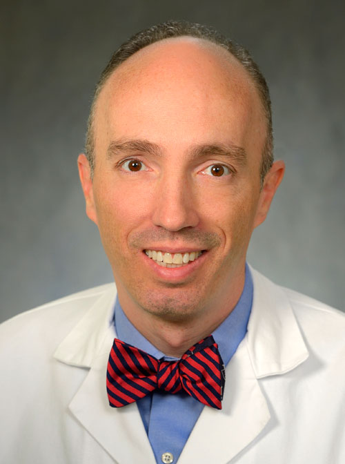 Jeremy W. Cannon合作论文数Surgery Faculty, Perelman School of Medicine, University of Pennsylvania25Daniel S Wendorff合作论文数22
 Brendan Michael Beely合作论文数Univ Incarnate Word, Sch Osteopath Med, Dept Translat Med, San Antonio, TX USA22William L. Baker合作论文数Department of Geography;University of Wyoming;Department of Geography, University of Wyoming19
 Michael A. Dubick合作论文数U.S. Army Institute of Surgical Research16Kerfoot P. Walker合作论文数15
 Andrew Cap合作论文数14
 Aden James K合作论文数14
 Antebi Ben合作论文数Faculty of Dental Medicine, Hebrew University14
 Valerie G Sams合作论文数Department of Surgery, University of Tennessee Medical Center13Liu Nehemiah T合作论文数13Vittorio Scaravilli合作论文数Department of Experimental Medicine, University of Milano-Bicocca13Jae Hyek Choi合作论文数12Yanyi Zang合作论文数12
 John B. Holcomb合作论文数Department of Surgery, Heersink School of Medicine, The University of Alabama at Birmingham;Decisio Health, Inc.12
 John Jones合作论文数Novartis Inst BioMed Res11
 Jordan Bryan合作论文数Broad Inst, Cambridge, MA USA11Linden Katharina合作论文数11
 Stefan Kreyer合作论文数11Ian J. Stewart合作论文数10
 Thomas Langer合作论文数Fondazione IRCCS Ca' Granda Ospedale Maggiore Policlinico di Milano, Università degli Studi10Tom Kuusela合作论文数10Kyle N Sieck合作论文数9Sine Christy R合作论文数9Vitali Karaliou合作论文数8
 Arezoo Mohammadipoor合作论文数Oak Ridge Inst Sci & Educ8
 Victor A. Convertino合作论文数US Army Institute of Surgical Research8dara regn合作论文数7Vittoria Vecchi合作论文数7
 Antonio Pesenti合作论文数Department of Pathophysiology and Transplantation, University of Milan7Henderson Jonathan L合作论文数7Park Timothy S合作论文数7Buel Allison R合作论文数7
 Charles E. Wade合作论文数Department of Surgery, McGovern Medical School, The University of Texas Health Science Center7Lydia C Piper合作论文数6Michael Morris合作论文数Department of Chemistry, University of Sheffield6
 Melissa M. Reynolds合作论文数Department of Chemistry, Department of Chemical and Biological Engineering, and School of Biomedical Engineering, Colorado State University6
 Hitesh Handa合作论文数College of Engineering, University of Georgia6Matt Brenner合作论文数6
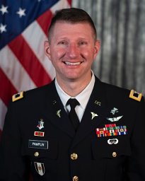 Jeremy C. Pamplin合作论文数6




