In 2024, it will be 140 years since the birth of Jan Karol Parnas, one of the most outstanding biochemists and one of the most outstanding Polish scientists of the first half of the 20th century. The article presents a synthetic biography of Prof. Parnas, his influence on the organization of scientific life in interwar Poland, and presents his students and collaborators who created the so-called "The School of Parnassus". It shows how this figure still influences the thinking of Polish biochemists about their Society and the attitude towards biochemical communities abroad, especially in Ukraine, which owes as much to Prof. Parnas as Poland does.
Artykuł przybliża postać jednego z najwybitniejszych biochemików II Rzeczypospolitej: Jakuba Karola Parnasa. Jego badania naukowe odegrały kluczową rolę w zrozumieniu fundamentalnego procesu biochemicznego, jakim jest glikoliza. Kariera naukowa Jakuba Karola Parnasa jest jednocześnie symbolem wyjątkowej atmosfery intelektualnej Galicji w 20. i 30. latach ubiegłego wieku. Dokonania naukowe Parnasa oraz prowadzone badania stanowią inspirującą historię sukcesu naukowego, osiągniętego w miejscu oddalonym od światowych centrów nauki. Ten sukces wynikał z talentu, determinacji i pasji naukowej, a także sprzyjającego środowiska Uniwersytetu Jana Kazimierza we Lwowie.
In 2024, it will be 140 years since the birth of Jan Karol Parnas, one of the most outstanding biochemists and one of the most outstanding Polish scientists of the first half of the 20th century. The article presents a synthetic biography of Prof. Parnas, his influence on the organization of scientific life in interwar Poland, and presents his students and collaborators who created the so-called "The School of Parnassus". It shows how this figure still influences the thinking of Polish biochemists about their Society and the attitude towards biochemical communities abroad, especially in Ukraine, which owes as much to Prof. Parnas as Poland does.
Muscle fructose-1,6-bisphosphatase (FBPase), which catalyzes the hydrolysis of fructose-1,6-bisphosphate (F1,6BP) to fructose-6-phosphate (F6P) and inorganic phosphate, regulates glucose homeostasis by controlling the glyconeogenic pathway. FBPase requires divalent cations, such as Mg2+, Mn2+, or Zn2+, for its catalytic activity; however, calcium ions inhibit the muscle isoform of FBPase by interrupting the movement of the catalytic loop. It has been shown that residue E69 in this loop plays a key role in the sensitivity of muscle FBPase towards calcium ions. The study presented here is based on five crystal structures of wild-type human muscle FBPase and its E69Q mutant in complexes with the substrate and product of the enzymatic reaction, namely F1,6BP and F6P. The ligands are bound in the active site of the studied proteins in the same manner and have excellent definition in the electron density maps. In all studied crystals, the homotetrameric enzyme assumes the same cruciform quaternary structure, with the κ angle, which describes the orientation of the upper dimer with respect to the lower dimer, of -85o. This unusual quaternary arrangement of the subunits, characteristic of the R-state of muscle FBPase, is also observed in solution by small-angle X-ray scattering (SAXS).
Fructose-1,6-bisphosphatase (FBPase) catalyzes the hydrolysis of fructose 1,6-bisphosphate to fructose 6-phosphate and is a key enzyme of gluconeogenesis and glyconeogenesis and, more generally, of the control of energy metabolism and glucose homeostasis. Vertebrates, and notably Homo sapiens, express two FBPase isoforms. The liver isozyme is expressed mainly in gluconeogenic organs, where it functions as a regulator of glucose synthesis. The muscle isoform is expressed in all cells, and recent studies have demonstrated that its role goes far beyond the enzymatic function, as it can interact with various nuclear and mitochondrial proteins. Even in its enzymatic function, the muscle enzyme is different from the liver isoform, as it is 100-fold more susceptible to allosteric inhibition by AMP and this effect can be abrogated by complex formation with aldolase. All FBPases are homotetramers composed of two intimate dimers: the upper dimer and the lower dimer. They oscillate between two conformational states: the inactive T form when in complex with AMP, and the active R form. Parenthetically, it is noted that bacterial FBPases behave somewhat differently, and in the absence of allosteric activators exist in a tetramer-dimer equilibrium even at relatively high concentrations. [Hines et al. (2007), J. Biol. Chem. 282, 11696-11704]. The T-to-R transition is correlated with the conformation of the key loop L2, which in the T form becomes `disengaged' and unable to participate in the catalytic mechanism. The T states of both isoforms are very similar, with a small twist of the upper dimer relative to the lower dimer. It is shown that at variance with the well studied R form of the liver enzyme, which is flat, the R form of the muscle enzyme is diametrically different, with a perpendicular orientation of the upper and lower dimers. The crystal structure of the muscle-isozyme R form shows that in this arrangement of the tetramer completely new protein surfaces are exposed that are most likely targets for the interactions with various cellular and enzymatic partners. The cruciform R structure is stabilized by a novel `leucine lock', which prevents the key residue, Asp187, from locking loop L2 in the disengaged conformation. In addition, the crystal structures of muscle FBPase in the T conformation with and without AMP strongly suggest that the T-to-R transition is a discrete jump rather than a shift of an equilibrium smooth transition through multiple intermediate states. Finally, using snapshots from three crystal structures of human muscle FBPase, it is conclusively demonstrated that the AMP-binding event is correlated with a β→α transition at the N-terminus of the protein and with the formation of a new helical structure.
X-Ray synchrotron diffraction data for human muscle fructose-1,6-bisphosphatase, crystallized in the presence of adenosine monophosphate in the T-state. Befor measurement crystals were soaked in mother liquor supplemented with 100 mM MgCl2 and 20% (v/v) glycerol to remove AMP. The diffraction images were collected to the resolution of 2.9 \xc5 at the BESSY beamline 14.2 in Berlin at 100K using a MX-225 detector. The data was recorded using X-ray wavelength 0.918 \xc5 with oscillation angle 0.1\xb0 and crystal-to-detector distances 280mm.
The mechanism by which calcium inhibits the activity of muscle fructose 1,6-bisphosphatase (FBPase) and destabilizes its interaction with aldolase, regulating glycogen synthesis from non-carbohydrates in skeletal muscle is poorly understood. In the current paper, we demonstrate evidence that Ca(2+) affects conformation of the catalytic loop 52-72 of muscle FBPase and inhibits its activity by competing with activatory divalent cations, e.g. Mg(2+) and Zn(2+). We also propose the molecular mechanism of Ca(2+)-induced destabilization of the aldolase-FBPase interaction, showing that aldolase associates with FBPase in its active form, i.e. with loop 52-72 in the engaged conformation, while Ca(2+) stabilizes the disengaged-like form of the loop.
The activity of fructose-1,6-bisphosphatase (FBPase; EC 3.1.3.11) isozymes is influenced by AMP, Ca2 + and by reversible interactions with subcellular structures. In contrast to mammalian and avian isozymes, the kinetic properties of FBPases from ectothermal vertebrates are not fully described. To get some insight into mechanism of glycogen resynthesis in ectothermal vertebrates we examined the features of FBPases isolated from Cyprinus carpio skeletal muscle and liver. To investigate the evolutionary origin of the sensitivity of FBPase to effectors, we performed a phylogenetic analysis of known animal amino acids sequences of the enzyme. Based on our findings, we hypothesize that the high, mammalian-like, sensitivity of FBPase to Ca2 + is not essential for controlling the stability of glyconeogenic complex in striated muscles, instead it ensures the precise regulation of mitochondrial metabolism during prolonged Ca2 + elevation in contracting muscle fibers. Comparison of the kinetic properties of vertebrate and insect FBPases suggests that the high sensitivity of muscle isozyme to inhibitors has arisen as an adaptation enabling coordination of energy metabolism in warm-blooded animals.
Human fructose-1,6-bisphosphatase is an allosteric enzyme that is regulated by different ligands. There are only two known isozymes in human tissues: the liver isozyme (the key enzyme of gluconeogenesis), which is regulated by fructose 2,6-bisphosphate, and its muscle counterpart (participating in glycogen synthesis), which is regulated by calcium ions. AMP, which is an allosteric inhibitor of both isozymes, inhibits the muscle isozyme with an I-0.5 that is 35-100 times lower than for the liver isozyme and the reason for this difference remains obscure. In studies aiming at an explanation of the main differences in the regulation of the two isozymes, it has been shown that only one residue, in position 69, regulates the sensitivity towards calcium ions. As a consequence of this finding, an E69Q mutant of the muscle isozyme, which is insensitive to calcium ions while retaining all other kinetic properties resembling the liver isozyme, has been prepared and crystallized. Here, two crystal structures of this mutant enzyme in complex with AMP with and without fructose 6-phosphate (the product of the catalytic reaction) are presented. The AMP binding pattern of the muscle isozyme is quite similar to that of the liver isozyme and the T conformations of the two isozymes are nearly the same.
Glyconeogenesis, the synthesis of glycogen from carbohydrate precursors like lactate, seems to be an important pathway participating in replenishing glycogen in cardiomyocytes. Fructose-1,6-bisphosphatase (FBPase), an indispensible enzyme of glyconeogenesis, has been found in cardiomyocytes on the Z-line, in the nuclei and in the intercalated discs. Glyconeogenesis may proceed only when FBPase accumulates on the Z-line. Searching for the mechanism of a FBPase regulation we investigated the effects of the calcium ionophore A23187, a muscle relaxant dantrolene, glucagon, insulin and medium without glucose on the subcellular localization of this enzyme in primary culture of neonatal rat cardiomyocytes. Immunofluorescence was used for protein localization and the intracellular calcium concentration was measured with Fura. We found that the concentration of calcium ions was the decisive factor determining the localization of muscle FBPase on the Z-line. Calcium ions had no effect on the localization of the enzyme in the intercalated discs or in the nuclei, but accumulation of FBPase in the nuclei was induced by insulin.
D-Fructose-1,6-bisphosphate 1-phosphohydrolase FBPase; [EC 3.1.3.11] was isolated from Pelophylax esculentus muscle in an electrophoretically homogeneous form with ca 30% yield. Its subunit molecular mass is ca 37 kDa. In this study, we determined the basic kinetic properties of the frog muscle enzyme. FBPase exhibited a maximum activity at pH 7.5. Like other FBPases the frog enzyme requires magnesium ions for its activity (K(a)=263 microM) and is activated by potassium ions (K(a)=63.6 microM). I(0.5) for calcium ion (91 microM) is 100 times higher than the corresponding value of mammalian muscle FBPase. K(s) for the substrate was 1.68 microM. Substrate excess inhibited the enzyme (K(si)=55 microM). AMP and fructose-2,6-bisphosphate (Fru-2,6P(2)) are potent inhibitors of frog muscle FBPase with I(0.5) of 0.2 microM and K(i) of 114 nM, respectively. Both inhibitors act synergistically on the frog muscle FBPase. In the presence of 0.05-0.5 microM of AMP, K(i) for Fru-2,6P(2) is 92 and 28 nM. I(0.5) for AMP for P. esculentus muscle FBPase is 55 times lower than the corresponding value for P. esculentus liver isozyme.
Antioxidant enzymes: catalase (CAT) [EC 1.11.1.6],peroxidase [EC 1.11.1.7],glutathione reductase (GSSGR) [EC 1.6.4.2] and glucose-6-phosphate dehydrogenase (G6PDH) [EC 1.1.1.49]were investigated in terrestrial Helix aspersa (O.F.Müll.) and aquatic Pomacea bridgesi (Reeve).The activity was determined in the heamolymph and homogenates of hepatopancreas and foot muscle.No CAT and peroxidase activity was detected in the hepatopancreas and foot muscle of P. bridgesi, and its haemolymph displayed a very low CAT activity (0.005 U/mg of protein).In H. aspersa the highest activities (U/g) observed for CAT and GSSGR in the hepatopancreas were 0.40, and 1.05, respectively; for peroxidase and G6PDH in the foot muscle the respective values were 1.22 and 0.22.The activities of antioxidant enzymes determined in both snail species are much lower than the corresponding values for mammalian tissues.
The contribution of Jakub Karol Parnas (1884-1949), a prominent Polish scientist, to the understanding of muscle biochemistry, including ammonia and carbohydrates sources and nucleotide metabolism, is described. Among his main achievements was the discovery of glycogen phosphorolysis, the first use of radioactive phosphorus in biological studies, and formulation and proof of phosphate transfer between glycolytic intermediates and ATP. The rewarding and successful life led by this man of great scientific and intellectual abilities up to the beginning of the Second World War, his dramatic fate during the war, and his tragic death in a Soviet prison reflects the turbulent 20th history of the region.
 Dariusz Rakus合作论文数27Daria Dziewulska-Szwajkowska合作论文数15
 Agnieszka Gizak合作论文数12
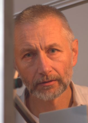 Mariusz Jaskólski合作论文数Department of Crystallography, Faculty of Chemistry, Adam Mickiewicz University, Poznań;Department of Structural Biology of Eukaryotes, Institute of Bioorganic Chemistry, Polish Academy of Sciences11Małgorzata Łozińska-Gabska合作论文数11Kołodziejczyk R合作论文数7Piotr Mamczur合作论文数7A Poloczek-Adamowicz合作论文数6K Skalecki合作论文数6J Wojtaszek合作论文数6M. Zarzycki合作论文数5Z Kozubek合作论文数5
 Marek Zarzycki合作论文数5Anna Adamowicz合作论文数5Jolanta Barańska合作论文数5J. Wisniewski合作论文数4J. Barciszewski合作论文数4Ha Eldorry合作论文数4Danuta Duś合作论文数4
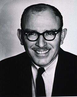 Bernard Horecker合作论文数Cornell University4Klaus Eschrich合作论文数4Marian Kochman合作论文数4S Pontremoli合作论文数3
 Adam Szewczyk合作论文数Nencki Institute of Experimental Biology3D Chu合作论文数3
 Robert Kolodziejczyk合作论文数3
 Robert Wysocki合作论文数3Stanislaw Ulaszewski合作论文数3E Maciaszczyk合作论文数2m przybylskawojtyszyn合作论文数2
 Janusz Wisniewski合作论文数Faculty of Biological Sciences, University of Wrocław2J Buczyłko合作论文数2
 Ewa Maciaszczyk-Dziubinska合作论文数2Barciszewski Jakub合作论文数2M. Pasek合作论文数2Ewa Maciaszczyk合作论文数2Monika Zmojdzian合作论文数2Janina Kwiatkowska-Korczak合作论文数2Orestes Tsolas合作论文数2Hubert Krotkiewski合作论文数2G J Xu合作论文数1a ogorzalek合作论文数1H Dihazi合作论文数1B Jungowska-Klin合作论文数1M. Majkowski合作论文数1Agostino Tafuri合作论文数1Maciej Zabel合作论文数1I Mroz合作论文数1Ellis W T Wong合作论文数1




