CD39 (ecto-nucleoside triphosphate diphosphohydrolase-1; E-NTPDase1) is a plasma membrane ecto-enzyme that regulates purinergic receptor signaling by controlling the levels of extracellular nucleotides. In blood vessels this enzyme exhibits a thromboregulatory role through the control of platelet aggregation. CD39 is localized in caveolae, which are plasma membrane invaginations with distinct lipid composition, similar to dynamic lipid microdomains, called rafts. Cholesterol is enriched together with sphingolipids in both rafts and caveolae, as well as in other specialized domains of the membrane, and plays a key role in their function. Here, we examine the potential role of cholesterol-enriched domains in CD39 function. Using polarized Madin-Darby canine kidney (MDCK) cells and caveolin-1 gene-disrupted mice, we show that caveolae are not essential either for the enzymatic activity of CD39 or for its targeting to plasma membrane. On the other hand, flotation experiments using detergent-free or detergent-based approaches indicate that CD39 associates, at least in part, with distinct lipid assemblies. In the apical membrane of MDCK cells, which lacks caveolae, CD39 is localized in microvilli, which are also cholesterol and raft-dependent membrane domains. Interfering with cholesterol levels using drugs that either deplete or sequester membrane cholesterol results in a strong inhibition of the enzymatic and anti-platelet activity of CD39. The effects of cholesterol depletion are completely reversed by replenishment of membranes with pure cholesterol, but not by cholestenone. These data suggest a functional link between the localization of CD39 in cholesterol-rich domains of the membrane and its role in thromboregulation.
Prothymosin α (ProTα) is a histone H1‐binding protein localized in sites of active transcription in the nucleus. We report here that ProTα physically interacts with the CREB‐binding protein (CBP), which is a versatile transcription co‐activator. Confocal laser scanning microscopy reveals that ProTα partially colocalizes with CBP in discrete subnuclear domains. Using transient transfections, we show that ProTα synergizes with CBP and stimulates AP1‐ and NF‐κB‐dependent transcription. Furthermore, overexpression of ProTα enhances the transactivation potential of CBP. These findings reveal a new function for ProTα in transcription activation, probably through CBP‐mediated recruitment to different promoters.
Capillary electrophoresis is a relatively new analytical technique that begins to have an impact on both routine and research in clinical laboratories. Recently, a fully automated system has become commercially available (Paragon CZE 2000, Beckman, USA) for the analysis of human serum proteins. Urine protein analysis, on the other hand, is currently accomplished by electrophoresis of concentrated urine specimens. The method is used to distinguish the glomerular from the tubular proteinuria and for the identification of Bence-Jones proteins. The procedure is labor-intensive and technically demanding. We developed a technique for the serum capillary electrophoresis instrument that can also be applied routinely to the differential diagnosis of proteinurias. Overriding the programmed dilution step of the instrument, we were able to distinguish different types of proteinurias without concentration of specimens with a total protein content of 150-200 mg/l as determined by sulfosalicylic acid. The different electrophoretic patterns obtained by the capillary electrophoresis system for various specimens correlated well with established techniques (Hydragel Proteinurie Kit, Sebia, France). The method is applicable for routine analysis of urinary proteins. It is reliable, less expensive and faster than the conventional methods (electrophoretic or immunonephelometric) used today for the differentiation of proteinurias, and it can be used as a quick screening test.
Decreased serum uric acid levels resulting from renal urate wasting have been occasionally encountered in jaundiced patients. However, in these cases, there are no data concerning the underlying renal tubular defects. In the present study, we investigated the renal tubular function in 35 patients with obstructive jaundice of various severity and causes (11 with lithiasis, 17 with carcinoma, and 7 with intrahepatic cholestasis). A detailed study of the renal tubular function was performed. Beyond the conventional methods,H-1-NMR spectroscopy of urine was used to evaluate noninvasively renal damage by the characteristic perturbation in the excretion pattern of low-molecular weight endogenous metabolites. On admission, patients with obstructive jaundice had significantly lot-ver serum uric acid and phosphate levels and higher bile acid concentrations compared with 40 age- and sex-matched controls. Serum uric acid levels presented a negative correlation with the total and direct bilirubin as well as the fractional excretion of uric acid. Furthermore, a great number of the patients studied developed one or more proximal tubular dysfunction manifestations beyond uricosuria, such as renal glucosuria, phosphaturia, and increased excretion of alpha (1)-microglobulin. H-1-NMR spectroscopy of the urine showed decreased levels of citrate and hippurate and increased levels of 3-hydroxybutyrate and acetate. In 12 patients partial or complete remission of jaundice was followed by an improvement of the proximal renal tubular damage. In conclusion, obstructive jaundice can cause a partially reversible generalized proximal tubular dysfunction.
OBJECTIVEVariations in thyroid function are known to be associated with changes in adrenocortical activity. Previous studies in animals have suggested that long-standing hyperthyroidism may be associated with diminished adrenal functional reserve despite a continuing hyperactivity of the hypothalamic-pituitary-adrenal (HPA) axis. In humans, there has been no direct assessment of adrenal secretory reserve in clinical thyrotoxicosis. This study aimed to assess adrenocortical reserve in response to low-dose ACTH, following dexamethasone suppression, in patients with severe thyrotoxicosis.DESIGN AND METHODSTen patients (four men and six women, 30-45 years) with severe long-standing thyrotoxicosis due to Graves' disease (n=6) or toxic nodular goitre (n=4) were studied at diagnosis and again when in a stable euthyroid state following drug therapy for 8-12 months. All patients underwent ACTH stimulation tests at 0800h with ACTH(1-24) (Cortrosyn; 0.1microg/kg body weight, i.v.) following overnight suppression of the HPA axis with dexamethasone (1mg per os at 2300h). Serum cortisol was assayed at -15, 0, 15, 30, 60 and 90min after the administration of ACTH.RESULTSThe mean (+/-s.d.) peak and delta cortisol responses to ACTH (634.5+/-164nmol/l and 618+/- 196nmol/l respectively), as well as the net area under the response curve (36769+/-12188nmol/lx min) in the hyperthyroid patients were significantly lower compared with the values when the same patients were euthyroid (911+/-157nmol/l, 905+/-160nmol/l and 57652+/-10128nmol/lxmin respectively; P<0.005). Subnormal peak cortisol responses (<500nmol/l) were observed in two severely toxic patients. The findings were independent of the cause of thyrotoxicosis.CONCLUSIONIn patients with severe thyrotoxicosis, cortisol secretion in response to low-dose ACTH stimulation, following dexamethasone suppression, is lower in the hyperthyroid than in the euthyroid state. It appears that thyrotoxicosis is associated with subtle impairment of adrenocortical reserve.
OBJECTIVES:The plasma apolipoprotein B (apo B) concentrations have been considered to be a more accurate representation of atherogenic particles and it has been proposed that the formula LDL-C (mmol/L) = 0.41TC - 0.32TG + 1.70apo B - 0.27 is reliable for the estimation of LDL-C (Clin Chem 1997; 43: 808-15). We undertook the present study to investigate the reliability of this formula in a large number of hyperlipidemic patients. DESIGN AND METHODS:1) The Friedewald formula (LDL-F) and the apo B-based formula (LDL-B) were compared with the beta-quantification reference procedure in 130 individuals with a wide range of total cholesterol (TC) and triglyceride (TG) levels, and 2) the LDL-C levels obtained by the Friedewald formula were compared with those calculated by the apo B-based formula in 1010 individuals attending our outpatient lipid clinic. RESULTS:The LDL-F and the LDL-B formulae for LDL-C estimation were found to be in good agreement with the beta-quantification (r = 0.96 and 0.97, respectively). The bias of each method plotted as a function of TG (up to 4.52 mmol/L) was found positive for the LDL-F, whereas the LDL-B was independent of the concentrations of TG. When a large number of individuals were examined, a good correlation between the two equations was found (n = 1010, r = 0.98). The difference between the two methods was not correlated with serum TG levels. However, it was correlated to serum TC, and apo B levels. CONCLUSIONS:The LDL-B formula is a more reliable and accurate method than the LDL-F formula, especially at TG levels >2.26 mmol/L, although it underestimates LDL-C concentrations. Furthermore, this equation can be used in hypertriglyceridemic patients (TG >4.52 mmol/L) in whom the Friedewald equation is inaccurate.
Objectives: To evaluate the analytical performance of a new homogeneous HDL-cholesterol assay (Olympus Diagnostica). To investigate possibly discrepant results in chronic hemodialysis patients who commonly exhibit quantitative and qualitative lipoprotein abnormalities, responsible for atherogenic complications in these patients.Design and methods: Serum samples were collected from 50 healthy subjects and 65 chronic hemodialysis patients. HDL-C levels measured by the homogeneous assay were compared with the routine dextran sulfate-Mg2+ precipitation method and the ultracentrifugation/dextran sulfate-Mg2+ precipitation as reference method.Results: The homogeneous assay was linear up to at least 220 mg/dL The analytical precision was estimated with three different sets of commercial controls and one set of human pooled serum control. The within-day CV ranged between 1.7% and 3.8% and the between-day CV ranged between 1.0% and 2.3%. HDL-C values in both populations correlated highly with the dextran sulfate-Mg2+ precipitation method and the ultracentrifugation/dextran sulfate-Mg2+ precipitation method (r greater than or equal to 0.96, bias between -0.9 and 2.3 mg/dL). Lipemia up to triglyceride concentration of 600 mg/dL did not alter the HDL-C value.Conclusions: The homogeneous assay for HDL-C (Olympus) uses much less sample. is accurate and convenient to handle, and allows full automation. The test should considerably facilitate the screening of individuals at an increased risk of cardiovascular disease, including hemodialysis patients. Copyright (C) 1999 The Canadian Society of Clinical Chemists.
Northwestern Greece was identified in the 1960s for its high prevalence of endemic goiter and iodine deficiency. Although iodized salt has been commercially available since then, a recent epidemiological survey of 3916 schoolchildren found that low-grade goiter is still prevalent in endemic proportions (21%). The aim of this study was to further assess the cause of goiter and the severity of iodine deficiency in children from this endemic area of Greece. Of the 800 children with clinically detectable goiter, 97 children (60 girls and 37 boys, 8-15 years) were recruited for determination of urinary iodine excretion, as well as assessment of thyroid volume and function and detection of antithyroid antibodies. The median urinary iodine concentration was 8.4 microg/dL, indicative of a mild iodine deficiency. Thyroid function was normal in all but 11 children who had subclinical hypothyroidism. Sixteen children (16.5%), including all those with subclinical hypothyroidism, were positive for antithyroid antibodies. Their median urinary iodine concentration (20.6 microg/dL) was higher compared to children who were negative for antibodies (7.4 microg/dL; p<0.001). The mean thyroid volume by ultrasonography (12.2+/-4.1 mL) was above the upper limit of normal for this age group. Thyroid volume was inversely related to the urinary iodine content in the children with negative antithyroid antibodies. Iodine deficiency is still prevalent in northwestern Greece although of mild severity and constitutes the primary cause of goiter among schoolchildren. However, it appears that autoimmune thyroiditis is emerging as a frequent cause of goiter in those children with sufficient iodine intake.
DNA of lymphocytes from human peripheral blood was analyzed by using the single cell gel electrophoresis technique (comet assay). The cells were used either as received from the donors or after treatment with various concentrations of the H2O2-generating enzyme glucose oxidase, in order to achieve a continuous flow of H2O2. The formation of single strand breaks (SSB) was dose-related but the time course of the induction of SSB by relatively low concentrations of glucose oxidase was of a biphasic mode with a fast increase 2 to 5 min after the addition of glucose oxidase followed by a gradual decrease toward the original base level during the next 35 to 60 min. This response of the cells appears to be based on the activation of already existing defense system(s) because it was shown that H2O2 is continuously released during the reaction time and the inhibition of protein synthesis does not affect the observed pattern. Supplementation of the growth medium with various antioxidants resulted in substantial protection only when the agents were taken up by the cells. The presence of the intracellular calcium chelator BAPTA protected the cells from H2O2-induced DNA damage in a dose-dependent manner. Only at the higher rate of H2O2-generation considerable DNA damage was observed in the presence of BAPTA.These results suggest that H2O2, at low concentrations induces DNA damage through intracellular Ca2+-mediated processes, which lead to DNA strand breaks possibly by endonuclease activation.
It has been reported that cumulative carnitine losses through dialysis membranes may worsen hyperlipidemia during long-term hemodialysis. However, carnitine supplementation has not shown a consistent beneficial response. We undertook the present study to determine if there is any hypolipidemic effect of L-carnitine on Greek dialysis patients in concert with the dialysate buffer composition (acetate or bicarbonate). A total of 28 patients (16 male, 12 female), mean age 43 years (range 21-61), with end-stage renal disease on maintenance hemodialysis for a mean period of 25 months (range 7-84) were studied. The dialysis schedule was 4 h, 3 times/week using cuprophane hollow-fiber dialyzers and acetate (n = 14) or bicarbonate (n = 14) dialysate. In all patients L-carnitine (5 mg/kg body weight) was infused intravenously 3 times/week at the end of each hemodialysis session. Blood samples for carnitine and lipid determinations were obtained before treatment, and 3 and 6 months following treatment. Even though L-carnitine did not modify most of the serum lipid levels, a significant decrease in serum triglycerides was evident in the whole group of patients (from 225 +/- 76 to 201 +/- 75 mg/dl, p = 0.03). Furthermore, L-carnitine could decrease serum triglycerides only in hypertriglyceridemic patients (from 260 +/- 64 to 226 +/- 82 mg/dl, p < 0.05). L-Carnitine resulted in a reduction of serum triglycerides in both patients on bicarbonate and on acetate dialysis, while there were no significant differences in the changes of lipid parameters after L-carnitine between the two groups of hemodialysis patients. We conclude that relatively low doses of L-carnitine supplementation could contribute to the management of some hypertriglyceridemic hemodialysis patients.
The herbicide paraquat (1,1'-dimethyl-4,4'-bipyridylium dichloride; PQ), is a poison known to cause delayed mortality due to lung and kidney injuries. High-resolution proton nuclear magnetic resonance (1H NMR) spectroscopy has been extensively applied in evaluating nephrotoxicity by the characteristic perturbations in the excretion pattern of low molecular weight endogenous metabolites. The application of the method allows the rapid localization of the renal injury noninvasively. In this study, we report 1H NMR and conventional clinical chemistry urinalysis in two patients suffering from paraquat intoxication after overdose with suicidal intent. The alterations in the urine NMR spectrum suggest necrosis of the pars recta of the proximal renal tubules. The molecule of paraquat is also clearly detected in the same spectrum. In conclusion, the rapid screening of urine by NMR spectroscopy provides information about both the identity of the poison and the abnormal pattern of endogenous metabolites that characterize the location of the injury in renal tubules and reveals alterations in unusual metabolites that are not commonly measured.
Prothymosin alpha (ProTalpha) is an abundant acidic nuclear protein that may be involved in cell proliferation. In our search for its cellular partners, we have recently found that ProTalpha binds to linker histone H1. We now provide further evidence for the physiological relevance of this interaction by immunoisolation of a histone H1-ProTalpha complex from NIH 3T3 cell extracts. A detailed analysis of the interaction between the two proteins suggests contacts between the acidic region of ProTalpha and histone H1. In the context of a physiological chromatin reconstitution reaction, the presence of ProTalpha does not affect incorporation of an amount of histone H1 sufficient to increase the nucleosome repeat length by 20 bp, but prevents association of all further H1. Consistent with this finding, a fraction of histone H1 is released when H1-containing chromatin is challenged with ProTalpha. These results imply at least two different interaction modes of H1 with chromatin, which can be distinguished by their sensitivity to ProTalpha. The properties of ProTalpha suggest a role in fine tuning the stoichiometry and/or mode of interaction of H1 with chromatin.
Background and Aim: As a continuation of our investigational program we carried out the present study to evaluate and compare the impact of LH-RH analogues and tamoxifen on serum lipid parameters in premenopausal women with breast cancer. Methods and Results: The study group consisted of 17 fully ambulatory women aged 36-48 years with histologically proven breast cancer randomly selected to receive an LH-RH analogue. Additionally a group of 17 premenopausal women matched for age and body mass index chosen from a pool of women treated with tamoxifen, acted as control. A complete lipid profile was determined before treatment and after 1, 3, 6 and 9 months. A decrease in serum total cholesterol, LDL cholesterol, Apo B, and Lp(alpha) levels was observed in women treated with tamoxifen. On the contrary no changes in measured lipid parameters were found in women treated with luteinizing hormone-releasing hormone (LH-RH) analogue. Patients treated with tamoxifen exhibited lower total cholesterol, LDL cholesterol, and Apo B levels, as well as lower levels of the atherogenic risk ratio total cholesterol:HDL cholesterol than patients treated with LH-RH analogue throughout the study These changes depended upon both treatment and time as well as their interaction Conclusions: We conclude that while tamoxifen has a beneficial effect on serum lipid profile, LH-RH analogues have rather a neutral effect, possibly as a result of the contrasting effects of estrogens and both progestins and androgens on lipid metabolism. (C) 1998, Medikal Press.
Dear Sir,Markedly elevated Lp(a) concentrations have consistently been reported in patients with renal diseases and specifically in proteinuric patients and in patients receiving hemodialysis or chronic ambulatory peritoneal dialysis [1, 2, 3, 4]. Even though predialysis patients exhibit high serum Lp(a) levels [5, 6], there are sparse data with regard to serum Lp(a) concentrations in patients with mild/moderate renal failure [7, 8].Lipid parameters including Lp(a) were measured in the serum of 106 patients with mild/moderate renal failure (creatinine clearance 10–60 ml/min) of various causes. Patients with proteinuria (>0.5 g/24 h), severe secondary hyperparathyroidism (PTH levels >250 pg/ml), diabetes mellitus, endocrine or liver disease, malignancy, as well as patients treated with drugs that influence lipoprotein metabolism (lipid lowering agents, steroids, cyclosporin, and β-blockers) were excluded. One hundred and two healthy individuals matched for age and sex with the patients were used as controls. The serum lipid profile of both patients and controls is shown in table 1. Compared to the controls, patients with moderate renal failure exhibited increased serum concentration of triglycerides and decreased serum HDL cholesterol and ApoA1 levels. Serum Lp(a) concentration was also significantly increased in patients compared to controls. Additionally, the percentage of patients with increased (>30 mg/dl) serum Lp(a) levels was significantly higher in uremic patients compared to the control population (35 vs. 11.8%, p < 0.01). Serum Lp(a) levels were correlated with total cholesterol (r = 0.29, p < 0.02), LDL cholesterol (r = 0.35, p < 0.005) and ApoB (r = 0.32, p < 0.01) levels. On the other hand, no correlation was found between serum Lp(a) concentration and creatinine, albumin, PTH, as well as creatinine clearance, though there was a trend towards an inverse correlation between Lp(a) concentration and creatinine clearance (r = –0.24, p = 0.08).Our study showed that patients with mild-to-moderate renal failure devoid of factors that influence lipoprotein metabolism, such as hypoalbuminemia, glucose intolerance, and severe hyperparathyroidism exhibit increased serum Lp(a) concentration, along with the other more common lipid abnormalities observed in these patients. These results, as well as those previously found [7, 8], suggest that uremia per se could have influenced Lp(a) metabolism. It has been suggested that the kidney may play a direct role in the catabolism of Lp(a), and therefore the underlying renal failure, even though of modest degree, might result in elevated serum Lp(a) levels [1]. Alternatively, or synergically, the kidney may have an indirect influence on Lp(a) metabolism through either the secretion of a factor that regulates Lp(a) synthesis [9] or the presence of uremic toxins that can influence the regulatory mechanisms of Lp(a) synthesis in the liver and/or of the catabolic pathways [1].It should be mentioned that the weak positive correlation between total and LDL cholesterol as well as ApoB and Lp(a) is likely to reflect the possible, even though disputable, role of the LDL receptor in the Lp(a) catabolism.
The dysfunction of the immune system has been implicated in the cause of essential hypertension (EH). On the other hand, interleukin- 1beta (IL-1beta) has strongly been involved in the pathogenesis of atheromatosis, whereas our preliminary experiments in serum samples from hypertensive patients before any drug therapy have shown the presence of high concentrations of IL-1beta and the absence of interleukin-2 (IL-2). The aim of this study was first to confirm our preliminary findings and second to investigate the possible interrelation(s) among the parameters studied, particularly between the immunologic markers and the blood pressure or the lipid parameters, because so far there are no data regarding the possible participation of IL-1beta in the cascade phenomena presented during the process of EH such as atherogenesis. Serum samples from 28 consecutive unselected patients with EH before any drug administration or after discontinuation of the antihypertensive therapy for at least 4 weeks, 31 normotensive patients with familial hypercholesterolemia (FH, disease control group), and 35 healthy individuals In a control group matched for age and sex were investigated for the presence of IL-1beta (commercial enzyme immunoassay), soluble IL-2 receptors (slL-2Rs, sandwich enzyme-linked immunosorbent assay set up in our laboratory), and some of the acute phase proteins by nephelometry. In addition, total cholesterol, triglycerides, high-density lipoprotein cholesterol, low-density lipoprotein cholesterol, apolipoproteins A1 and B, and lipoprotein (a) were determined by standard methods. The data were analyzed by unpaired t test, Mann Whitney-U, chi-squared analysis after Yate's correction, analysis of variance, or Kruskal-Wallis where applicable. Correlation coefficient was calculated by simple regression analysis (r) or nonparametric Spearman correlation coefficient (rs). We found that (1) none of the patients had increased concentrations of sIL-2Rs, and (2) the IL-1beta levels significantly differed in the three groups (p = 0.0001). In more detail, the concentrations of IL-1beta were significantly higher in patients with EH compared with those in patients with FH (p < 0.0005) and the healthy control group (p = 0.0001). By contrast, the IL-1beta concentrations did not differ between patients with FH and the healthy control group. (3) Sixteen (57.1%) patients with EH and only 6 (19.4%) patients with FH (p < 0.01) had increased levels of IL-1beta, and (4) the IL-1beta was not correlated with the acute phase reactants or the lipid parameters in the groups studied. However, the group of patients with EH and increased IL-1beta levels had significantly higher mean concentrations of triglycerides (p < 0.05) and significantly lower mean concentrations of high-density lipoprotein cholesterol (p < 0.05) than those who had IL-1beta levels lower than the cutoff point. (5) The IL-1beta concentrations were positively though slightly correlated with the mean blood pressure only in the group of patients with EH (r = 0.38, p < 0.05). This study demonstrated the presence of high concentrations of IL-1beta and the absence of indicators of cellular immune activation in the systemic circulation of patients with EH, suggesting that this cytokine may be involved in the pathogenesis of EH. In addition, this study showed that the high levels of IL-1beta were associated with lipid indicators of atheromatosis only in the group of patients with EH. More studies are required in an attempt to address whether IL-1beta could have a pathogenetic importance in EH. Taking into account these findings, however, it can be suggested that the presence of high IL-1beta levels may be an additional and perhaps independent risk factor for atheromatosis in patients with EH.
Conference Abstract| November 01 1996 REGULATION OF PROTHYMOSIN a DURING THE CELL CYCLE. EVIDENCE FOR E2F INVOLVEMENT K. Varell; K. Varell 1Laboratory of Biological Chemistry, Medical School University of Ioannina, GR 451 10 Ioannina, Greece Search for other works by this author on: This Site PubMed Google Scholar O. Tsolas; O. Tsolas 1Laboratory of Biological Chemistry, Medical School University of Ioannina, GR 451 10 Ioannina, Greece Search for other works by this author on: This Site PubMed Google Scholar M. Frangou-Lazaridis M. Frangou-Lazaridis 1Laboratory of Biological Chemistry, Medical School University of Ioannina, GR 451 10 Ioannina, Greece Search for other works by this author on: This Site PubMed Google Scholar Biochem Soc Trans (1996) 24 (4): 516S. https://doi.org/10.1042/bst024516s Views Icon Views Article contents Figures & tables Video Audio Supplementary Data Peer Review Share Icon Share Twitter LinkedIn Cite Icon Cite Get Permissions Citation K. Varell, O. Tsolas, M. Frangou-Lazaridis; REGULATION OF PROTHYMOSIN a DURING THE CELL CYCLE. EVIDENCE FOR E2F INVOLVEMENT. Biochem Soc Trans 1 November 1996; 24 (4): 516S. doi: https://doi.org/10.1042/bst024516s Download citation file: Ris (Zotero) Reference Manager EasyBib Bookends Mendeley Papers EndNote RefWorks BibTex toolbar search Search Dropdown Menu toolbar search search input Search input auto suggest filter your search All ContentAll JournalsBiochemical Society Transactions Search Advanced Search This content is only available as a PDF. © 1996 Biochemical Society1996 Article PDF first page preview Close Modal You do not currently have access to this content.
We have studied the molecular associations of parathymosin, an acidic polypeptide with a wide tissue distribution, by means of three approaches; ligand blotting; native electrophoresis; and immunoprecipitation. We report here that parathymosin binds specifically to the linker histone H1. This binding is enhanced by Zn2+ and is dependent on the concentration of parathymosin. Poly(glutamic acid) is able to compete fully with parathymosin for binding to histone H1, suggesting that this interaction is mediated by the acidic domain of the protein. Moreover, we demonstrate that parathymosin interacts with the globular domain of histone H1 under native conditions. Based on these data, we postulate that parathymosin may belong to a group of nuclear acidic proteins that affect histone H1 function.
 Eleni Th Bairaktari合作论文数Medical School, University of Ioannina17
 Moses Elisaf合作论文数Atherosclerosis and Obesity Department, University Hospital of Ioannina;Department of Internal Medicine, Medical School, University of Ioannina16Kc Siamopoulos合作论文数14
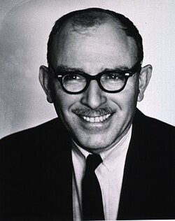 Bernard Horecker合作论文数Cornell University14
 Konstantin Seferiadis合作论文数9Ch Tzallas合作论文数9S Pontremoli合作论文数8T Papamarcaki合作论文数7Haralampos M Moutsopoulos合作论文数7
 Athanasios G. Tzioufas合作论文数School of Medicine, University of Athens6M Pappas合作论文数5Maria Frangou-Lazaridis合作论文数5H Bairaktari合作论文数4s c sun合作论文数4
 Vassiliki A Boumba合作论文数Medical School, University of Ioannina4Pranvera Lazo合作论文数4Dimitrios Lolis合作论文数4G Sferopoulos合作论文数3
 Omar Crivellaro合作论文数3George Fountzilas合作论文数Aristotle University of Thessaloniki3Bernard L Horecker合作论文数3
 Kostas C. Siamopoulos, MSc, MD, FRSH, FERA合作论文数3Louis Joris合作论文数3
 Edon Melloni合作论文数3
 Stathis Frillingos合作论文数Laboratory of Biological Chemistry, University of Ioannina Medical School3Savvas Christoforidis合作论文数Medical School, University of Ioannina3
 Ioannis Georgiou合作论文数Laboratory of Reproductive Genetics, University of Ioannina3E KOULOURIDIS合作论文数2Sc Sun合作论文数2P. Loizou合作论文数2Nicolaides C合作论文数2N Pavlidis合作论文数2
 Carol Murphy合作论文数Institute of Molecular Biology and Biotechnology, Foundation of Research and Technology-Hellas2
 Konstantinos Katopodis合作论文数Department of Nephrology, University Hospital of Ioannina2Afroditi Katsaraki合作论文数2Alexandros D Tselepis合作论文数2
 Andrzej Dzugaj合作论文数2P. D. Lapatsanis合作论文数2
 Dimitrios Galaris合作论文数Laboratory of Biological Chemistry, University of Ioannina Medical School2A.A. Haritos合作论文数2Z. Karetsou合作论文数2Vishwa Nath Singh合作论文数2
 Haralampos Milionis合作论文数Department of Medicine, School of Health Sciences, Faculty of Medicine, University of Ioannina2
 Panayiotis Vlachoyiannopoulos合作论文数School of Health Sciences, National and Kapodistrian University of Athens;Medical School, National and Kapodistrian University of Athens2George Makrydimas合作论文数Department of Obstetrics and Gynecology, University Hospital of Ioannina2
 Elizabeth O. Johnson合作论文数School of Medicine, University of Ioannina2E B Tsianos合作论文数2AK MAVRIDIS合作论文数2
 Agathocles Tsatsoulis合作论文数Department of Endocrinology, School of Health Sciences, University of Ioannina2




