Pathogenic variants in GDAP1 cause Charcot-Marie-Tooth disease (CMT), an inherited peripheral neuropathy characterized by progressive axonal degeneration. Although GDAP1 is an atypical glutathione S-transferase localized to the outer mitochondrial membrane, it has been proposed to function as a redox sensor that likely maintains inter-organelle communication in neurons. However, the mechanisms by which GDAP1 performs these functions remain unclear. To address this question, we here used a robust multi-tier approach that combines high-resolution and live-cell imaging with pH-sensitive probes, membrane contact sites (MCSs) analysis, lipid studies, transcriptomics, and nerve ultrastructural studies in both patient-derived fibroblasts and Gdap1-/- mice. We find that deletion of the GDAP1 gene induces localized pH and redox imbalances at mitochondria-lysosomes contact sites, which propagate to defective mitochondria-peroxisome interactions, impaired peroxisome biogenesis and morphology, leading to altered lipid homeostasis. These defects are accompanied by axonal organelle mislocalization, disruption of nodes of Ranvier, and structural abnormalities in peripheral nerves. Investigations on the potential reversibility of these processes, reveal that restoration of redox balance rescues MCS organization, identifying a therapeutically tractable MCS-peroxisome axis downstream of GDAP1. Together, our findings position GDAP1 as a redox-sensing organizer of mitochondrial membrane contact sites whose dysfunction triggers a cascade of organelle and axonal defects underlying CMT pathogenesis. Thus, this new knowledge should be taken into consideration in the future design of therapeutic interventions that can ameliorate the symptoms of this dismal disease.
Background/Objectives: Pancreatic ductal adenocarcinoma (PDAC) arises predominantly from activating KRAS mutations, yet individual genetic variants differ markedly in signaling output and clinical impact. G12D, the most prevalent variant, strongly drives oncogenic programs, whereas G12R signals less efficiently through the AKT and ERK pathways and is associated with longer patient survival than G12D-driven PDAC. Methods: To elucidate how these differences influence early cellular transformation, we expressed a panel of KRAS mutants in non-cancerous pancreatic ductal epithelial cells as a model of early PDAC initiation and profiled transcriptional and phospho-proteomic responses. We next examined whether epigenetic differences translate into mutation-specific changes in nuclear organization using quantitative imaging of G12D- and G12R-expressing nuclei at 24 and 48 h. Results: Each variant established a unique regulatory program enriched for chromatin remodelers, histone modifiers, and nuclear structural factors, indicating that variant-specific KRAS signaling rapidly develops divergent epigenetic states. Integrated transcriptomic and phospho-proteomic analyses identified G12D and G12R as the most divergent variants. G12D induced pronounced nuclear remodeling, including increased nuclear size, irregular morphology, and reorganization of the nucleolus and spliceosome, consistent with extensive chromatin and transcriptional reprogramming. In contrast, G12R elicited a weaker response, with minimal or delayed structural changes. Conclusions: Together, these findings demonstrate that KRAS mutational context in pancreatic ductal epithelial cells shapes early transcriptional reprogramming that actively remodels nuclear architecture and nuclear sub-compartments. This work establishes nuclear structural remodeling as a structural state of KRAS-driven epigenetic dysregulation during PDAC initiation.
Background and Aims:Metabolic dysfunction-associated steatotic liver disease affects about a third of adults worldwide and is projected soon to be the leading cause of liver cirrhosis. It occurs when fat accumulates in hepatocytes and can progress to metabolic dysfunction-associated steatohepatitis, liver cirrhosis, and HCC. Metabolic dysfunction-associated steatotic liver disease pathogenesis is believed to involve a combination of genetic and environmental risk factors. Single nucleotide polymorphisms have been implicated, but non-syndromic monogenic causes are lacking. Approach and Results:We identified a novel genetic variant in a familial case of metabolic dysfunction-associated steatohepatitis and performed deep variant functional analysis, including protein modeling, dynamics, and cell-based assays to assess molecular mechanisms of dysfunction and altered cellular signaling. We analyzed exome sequencing data of 3904 individuals with steatotic liver disease (SLD) to identify additional cases and establish the link between specific gene variants and SLD diagnosis. We discovered and functionally validated the NM_000245.4:c.3505A>T; p.(Ile1169Phe) variant in the MET (mesenchymal-epithelial transition) kinase domain as a monogenic cause of SLD. Subsequently, we detected additional ultra-rare, previously uninterpreted, and likely deleterious variants in MET from screening sequencing data. Among individuals with confirmed SLD based on electronic record review, 1.1% (45/3904) had rare predicted deleterious MET variants. Eight of 45 (17.7%) individuals had predicted deleterious variants in the MET kinase domain confirmed to be functionally like the familial case variant. Conclusions:We report the first germline nonmalignant rare MET-driven disease, a monogenic form of SLD.
Human Papillomavirus (HPV)-negative head and neck squamous cell carcinoma (HNSCC) remains a challenging malignancy, with radiotherapy, alone or combined with immune checkpoint inhibitors, often failing to achieve durable disease control. Here, by conducting longitudinal multi-omic analyses of pre- and post-radiation biopsies from patients receiving a pre-operative hypofractionated radiation regimen, we uncover that radiation rapidly depletes a subpopulation of tumor-infiltrating lymphocytes (TIL), characterized by a proliferative, cytotoxic, and tissue-resident gene signature (TProlif_Tox). We provide multi-dimensional evidence for tumor antigen-specificity of TProlif_Tox clonotypes and show that post-radiation tumors are instead repopulated by regulatory and non-specific clones. Finally, TIL depletion correlates with radiorecurrent disease after conventional radiation, emphasizing the potential impact of radiation-induced TIL loss regardless of fractionation. Thus, this study provides key insights into radiotherapy-induced alterations in the immune microenvironment that drive immunologic radioresistance and proposes restoring tumor antigen-specific T cell clonotypes as a strategy to improve radioimmunotherapy responses in HNSCC.
Pancreatic ductal adenocarcinoma (PDAC) is one of the deadliest forms of human malignancy, and there is an urgency to develop more effective therapy. We previously showed that Metavert, a dual inhibitor of glycogen synthase kinase 3-beta (GSK-3β) and histone deacetylases (HDACs) prevents pancreatic ductal adenocarcinoma (PDAC) metastasis. In this study, we investigated the mechanisms that mediate metastasis and the roles of GSK-3β, HDACs, and Yes-associated protein (YAP) in this process. We found that HDAC4 and YAP are highly expressed in PDAC from patients with rapid disease progression and metastasis compared to those with prolonged recurrence-free survival. Pan-HDAC inhibition decreases metastasis in the splenic PDAC metastatic mouse model. Inhibition of HDAC4 reduces migration of cancer cells and decreases the mRNA and protein levels of transcription factor MYB Proto-Oncogene Like 1 (MybL1) and YAP. Mechanistic studies show that HDAC4 regulates transcription of YAP through up-regulating MybL1 expression. Comparable results were observed in colon and prostate cancers. ATAC-seq studies show that inhibition of HDAC4 causes chromatin remodeling in the YAP promoter region and reduces accessibility to the binding sites of multiple transcription factors, including those of MybL1. Pharmacological or molecular inhibition of YAP significantly decreases PDAC metastasis in vivo. Imaging Mass Cytometry (IMC) reveals no significant changes in immune cells, but a notable shift in the distribution patterns of cancer-associated hepatic stellate cells in the metastatic niche, when YAP is ablated in the cancer cells. The results demonstrate a novel metastasis-driving cell signaling pathway mediated by the functional interaction between HDAC4 and MybL1, which regulates YAP expression and metastasis.
Background:Pulmonary arteriovenous malformations (PAVMs) are vascular complications that universally develop in patients with single ventricle congenital heart disease after Glenn surgery. However, the pathophysiological mechanisms underlying single ventricle PAVMs are poorly understood. To comprehensively evaluate molecular changes post-Glenn, we performed single-cell RNA sequencing (scRNAseq) on rat lung samples after Glenn surgery. Methods:Using adult Sprague Dawley rats, we performed scRNAseq on unfiltered lung samples 3 weeks after left-sided Glenn or sham surgery. We compared endothelial cell (EC) differentially expressed genes (DEGs) in our model to two mouse models of hereditary hemorrhagic telangiectasia (HHT), a hereditary condition characterized by visceral AVMs. Finally, we modified the vitamin A (Vit A) content of Glenn and sham rat diets and re-assessed PAVM shunting and EC transcriptional differences. Results:Using scRNAseq (n=4 Glenn, n=4 sham), we identified 13 transcriptionally distinct lung cell clusters, including 3 EC clusters (general, capillary, lymphatic), with pronounced differences between Glenn and sham in the general EC cluster (~17% of genes). Comparison to HHT mouse models confirmed overlap of ~18% of DEGs, including identification of significantly downregulated genes involved in and regulated by all-trans retinoic acid (ATRA) signaling in all 3 models. Dietary deficiency of Vit A intake, a precursor of ATRA, caused increased PAVM shunting (p<0.01) that was mitigated with excess dietary Vit A intake. Lastly, EC-specific RNAseq identified Vit A diet-induced gene expression differences, including regulation of PI3K signaling. Conclusions:In this study, we report the novel application of scRNAseq to study mechanisms underlying single ventricle PAVMs in a surgical rat model. We identified multiple dysregulated biological processes in rat lung ECs post-Glenn, including decreased ATRA signaling and conserved gene expression patterns with HHT. Dietary modification of Vit A intake altered post-Glenn shunting and represents a novel potential therapeutic strategy for single ventricle PAVMs and HHT AVMs.
Gastroesophageal reflux disease (GERD) is associated with inflammatory and neoplastic changes in the esophageal epithelium. Despite widespread PPI use, esophageal adenocarcinoma (EAC) incidence continues to rise, implicating non-acidic reflux components such as pepsin in disease progression. We performed transcriptomic profiling to assess pepsin-induced changes and the protective effect of amprenavir in vitro. Het-1A (normal) and BAR-T (Barrett’s) cells (n = 3) were treated at pH 7.0 with pepsin and/or 10 μM amprenavir for 1 h. RNA-seq identified DEGs (FDR ≤ 0.05, |log₂FC| ≥ 0.375), and Ingenuity Pathway Analysis revealed enriched pathways. Pepsin exposure altered mitochondrial function, oxidative phosphorylation, epithelial integrity, signaling, and inflammatory pathways in both cell lines. Amprenavir attenuated these transcriptomic perturbations, preserving mitochondrial and stress-response pathways. Notably, BAR-T cells exhibited heightened activation of wound-healing and epithelial repair pathways, whereas Het-1A cells showed greater mitochondrial and systemic stress pathway alterations. Pepsin drives transcriptomic dysregulation in esophageal epithelial cells under non-acidic conditions, and amprenavir shows potential to counteract peptic injury. Further studies are needed to validate these findings and explore amprenavir’s therapeutic utility in GERD management and EAC prevention.
Pathogenic variants of GDAP1 cause Charcot-Marie-Tooth disease (CMT), an inherited neuropathy characterized by axonal degeneration. GDAP1, an atypical glutathione S-transferase, localizes to the outer mitochondrial membrane (OMM), regulating this organelle's dynamics, transport, and membrane contact sites (MCSs). It has been proposed that GDAP1 functions as a cellular redox sensor. However, its precise contribution to redox homeostasis remains poorly understood, as does the possible redox regulation at mitochondrial MCSs. Given the relationship between the peroxisomal redox state and overall cellular redox balance, we investigated the role of GDAP1 in peroxisomal function and mitochondrial MCSs maintenance by using high-resolution microscopy, live cell imaging with pH-sensitive fluorescent probes, and transcriptomic and lipidomic analyses in the Gdap1-/- mice and patient-derived fibroblasts. We demonstrate that GDAP1 deficiency disrupts mitochondria-peroxisome MCSs and leads to peroxisomal abnormalities, which are reversible upon pharmacological activation of PPARγ or glutathione supplementation. These results identify GDAP1 as a new tether of mitochondria-peroxisome MCSs that maintain peroxisomal number and integrity. The supply of glutathione (GSH-MEE) or GDAP1 overexpression suffices to rescue these MCSs. Furthermore, GDAP1 may regulate the redox state within the microdomain of mitochondrial MCSs, as suggested by decreased pH at mitochondria-lysosome contacts in patient-derived fibroblasts, highlighting the relationship between GDAP1 and redox-sensitive targets. Finally, in vivo analysis of sciatic nerve tissue in Gdap1-/- mice revealed significant axonal structural abnormalities, including nodes of Ranvier disruption and defects in the distribution and morphology of mitochondria, lysosomes, and peroxisomes, emphasizing the importance of GDAP1 in sustaining axon integrity in the peripheral nervous system. Taken together, this study positions GDAP1 as a multifunctional protein that mediates mitochondrial interaction with cellular organelles of diverse functions, contributes to redox state sensing, and helps maintain axonal homeostasis. In addition, we identify PPAR as a novel therapeutic target, based on knowledge of the underlying pathogenetic mechanisms.
Introduction: The Euchromatic Histone Methyl Transferase Protein 2 (EHMT2), also known as G9a, deposits transcriptionally repressive chromatin marks that play pivotal roles in the maturation and homeostasis of multiple organs. Recently, we have shown that Ehmt2 inactivation in the mouse pancreas alters growth and immune gene expression networks, antagonizing Kras-mediated pancreatic cancer initiation and promotion. Here, we elucidate the essential role of Ehmt2 in maintaining a transcriptional landscape that protects organs from inflammation.Methods: Comparative RNA-seq studies between normal postnatal and young adult pancreatic tissue from Ehmt2 conditional knockout animals (Ehmt2fl/fl) targeted to the exocrine pancreatic epithelial cells (Pdx1-Cre and P48Cre/+), reveal alterations in gene expression networks in the whole organ related to injury-inflammation-repair, suggesting an increased predisposition to damage. Thus, we induced an inflammation repair response in the Ehmt2fl/fl pancreas and used a data science-based approach to integrate RNA-seq-derived pathways and networks, deconvolution digital cytology, and spatial transcriptomics. We also analyzed the tissue response to damage at the morphological, biochemical, and molecular pathology levels.Results and discussion: The Ehmt2fl/fl pancreas displays an enhanced injury-inflammation-repair response, offering insights into fundamental molecular and cellular mechanisms involved in this process. More importantly, these data show that conditional Ehmt2 inactivation in exocrine cells reprograms the local environment to recruit mesenchymal and immunological cells needed to mount an increased inflammatory response. Mechanistically, this response is an enhanced injury-inflammation-repair reaction with a small contribution of specific Ehmt2-regulated transcripts. Thus, this new knowledge extends the mechanisms underlying the role of the Ehmt2-mediated pathway in suppressing pancreatic cancer initiation and modulating inflammatory pancreatic diseases.
PurposeChanges in quantitative magnetic resonance imaging (qMRI) are frequently observed during chemotherapy or radiation therapy (RT). It is hypothesized that qMRI features are reflective of underlying tissue responses. It's unknown what underlying genomic characteristics underly qMRI changes. We hypothesized that qMRI changes may correlate with DNA damage response (DDR) capacity within human tumors. Therefore, we designed the current study to correlate qMRI changes from daily RT treatment with underlying tumor transcriptomic profiles.Methods and MaterialsStudy participants were prospectively enrolled (National Clinical Trial 03500081). RNA expression levels for 757 genes from pretreatment biopsies were obtained using a custom panel that included signatures of radiation sensitivity and DDR. Daily qMRI data were obtained from a 1.5 Tesla MR linear accelerator. Using these images, d-slow, d-star, perfusion, and apparent diffusion coefficient-mean values in tumors were plotted per-fraction, over time, and associated with genomic pathways.ResultsA total of 1022 qMRIs were obtained from 39 patients and both genomic data and qMRI data from 27 total patients. For 20 of those patients, we also generated normal tissue transcriptomic data. Radio sensitivity index values most closely associated with tissue of origin. Multiple genomic pathways including DNA repair, peroxisome, late estrogen receptor responses, KRAS signaling, and UV response were significantly associated with qMRI feature changes (P < .001).ConclusionsGenomic pathway associations across metabolic, RT sensitivity, and DDR pathways indicate common tumor biology that may correlate with qMRI changes during a course of treatment. Such data provide hypothesis-generating novel mechanistic insight into the biologic meaning of qMRI changes during treatment and enable optimal selection of imaging biomarkers for biologically MR-guided RT.
AimsEpigenomics has significantly advanced through the incorporation of Systems Biology approaches. This study aims to investigate the human lysine methylome as a system, using a data-science approach to reveal its emergent properties, particularly focusing on histone mimicry and the broader implications of lysine methylation across the proteome.MethodsWe employed a data-science-driven OMICS approach, leveraging high-dimensional proteomic data to study the lysine methylome. The analysis focused on identifying sequence-based recognition motifs of lysine methyltransferases and evaluating the prevalence and distribution of lysine methylation across the human proteome.ResultsOur analysis revealed that lysine methylation impacts 15% of the known proteome, with a notable bias toward mono-methylation. We identified sequence-based recognition motifs of 13 lysine methyltransferases, highlighting candidates for histone mimicry. These findings suggest that the selective inhibition of individual lysine methyltransferases could have systemic effects rather than merely targeting histone methylation.ConclusionsThe lysine methylome has significant mechanistic value and should be considered in the design and testing of therapeutic strategies, particularly in precision oncology. The study underscores the importance of considering non-histone proteins involved in DNA damage and repair, cell signaling, metabolism, and cell cycle pathways when targeting lysine methyltransferases.
Abstract In pancreatic ductal adenocarcinoma (PDAC), cancer associated fibroblasts (CAFs) play critical and complex roles in the tumor microenvironment. CAFs are also a major cell type in the desmoplastic stroma in PDAC and may account for half of the entire tumor tissue. Here we aimed to investigate the origin, diversification, and function of CAFs. We constructed a dual-DNA-recombinase mouse genetic model carrying KrasG12D/+; Trp53frt/+; Pdx1Flpo/+; Isl1creER/+; R26Tomato/+ alleles, referred to as KPFIT. The DNA recombinase FlpO directs expression of an oncogene Kras (G12D mutation) and loss of a tumor suppressor p53 in pancreatic epithelial cells, while creER recombines the Tomato reporter in the splanchnic mesenchyme and its descendants. The splanchnic mesenchyme is a layer of fetal tissue surrounding the endoderm where the pancreatic epithelium arises. This approach capitalized on the independent functions of FlpO/Frt and CreER/LoxP systems, allowing us to lineage trace Tomato labeled splanchnic progenies (via IT) in a spontaneous pancreatic cancer model (via KPF) within the same mouse. Our study identified the splanchnic mesenchyme as the fetal origin of pancreatic fibroblasts during homeostasis and tumorigenesis. Notably, the other two postulated origins, bone marrow and epithelial cells, have minimal contributions to fibroblasts. Importantly, single cell transcriptomic analysis indicated persistent and dynamic gene expressions along the pancreatic mesenchymal trajectory during development, homeostasis, precancer lesion and cancer. Intriguingly, certain splanchnic factors are expressed in only subtypes of adult pancreatic fibroblasts in temporally and spatially distinct patterns. Furthermore, we constructed mouse genetic models to delete one of the splanchnic factors, Gata6, specifically in CAFs, which resulted in increased tumor burden in the pancreas. This suggests a non-cell autonomous function of GATA6 in CAFs to restrain pancreatic cancer progression. In summary, this study delineated a continuous cell trajectory of the mesenchymal lineage in the pancreas across different life stages. Moreover, persistent gene expressions along the mesenchymal trajectory contributes to pancreatic CAF heterogeneity. Importantly, such persistence may constitute an inherent mechanism to suppress pancreatic cancer. The enhancement of this mechanism could be explored further for therapeutic benefits. Citation Format: Lu Han, Tom Walter, Joseph Beaudet, Caroline Everett, Kun Fang, Michael Zimmermann, Angela Mathison, Raul Urrutia, Victor Jin, Gustavo Leone, Michael Ostrowski. Persistence of fetal gene signatures along the mesenchymal lineage trajectory defines heterogeneity and function of pancreatic cancer associated fibroblasts [abstract]. In: Proceedings of the American Association for Cancer Research Annual Meeting 2024; Part 1 (Regular Abstracts); 2024 Apr 5-10; San Diego, CA. Philadelphia (PA): AACR; Cancer Res 2024;84(6_Suppl):Abstract nr 1595.
Abstract ID 97246Poster Board 457Pancreatic ductal adenocarcinoma (PDAC) remains a highly lethal cancer with an urgent need for more effective therapeutic strategies. Genomic analyses suggest that upwards of 25% of PDAC patients have mutations in DNA damage response (DDR) pathway genes, including ATM, BRCA1/2, and PALB2. Mutation in ataxia telangiectasia mutated (ATM) kinase, a major kinase involved in recognizing double-strand breaks, occurs in upwards of 6% of PDAC patients. If ATM is mutated, then the cells must rely on the related ATR pathway for sensing DNA damage. G9a, a lysine methyltransferase, has been localized to DNA breaks via its ATM-mediated phosphorylation at S569 or binding to RPA, an ATR target. G9a is upregulated in pancreatic cancer and its inhibition abrogates cell growth, suggesting G9a as a potential therapeutic target. Previously, our lab has performed experiments demonstrating that combined targeting of G9a and a downstream effector of ATR, CHK1, synergistically inhibit PDAC growth. However, the functional interaction between G9a and the ATM or ATR proteins remains unclear. Using linear motif analysis, we identified two potential phosphorylation sites, namely S525 and S579, in addition to the previously described S569, that may be involved in ATM/ATR signaling. Indeed, in vitro kinase assays demonstrated ATM- and ATR-mediated phosphorylation of G9a at these 3 sites. We also found increased G9a automethylation in the presence of ATM or ATR, suggesting that phosphorylation stimulates the catalytic activity of this methyltransferase. Furthermore, affinity purification of G9a followed by mass spectrometry showed interaction with ATM, ATR and ATRIP in cells. To better understand the role of G9a in both ATM and ATR signaling, we evaluated the impact of G9a knockout on DDR effector signals upon induction of DNA damage through single-stranded (ATR) or double-stranded (ATM) breaks. To determine therapeutic relevance of targeting this pathway in PDAC, we found that inactivation of both G9a and ATM reduces PDAC cell proliferation rates and colony formation more than disruption of the individual pathways. Thus, collectively, our results suggest that dual G9a-ATM inhibition may serve as a novel therapeutic strategy for PDAC and that leveraging G9a inhibition in ATM-deficient PDAC holds potential as a precision medicine approach in this patient.
Objective: The adverse effects of ischemia-reperfusion injury (IRI) remain a principal barrier to a successful outcome after lifesaving orthotopic liver transplantation (OLT). Gene expression during different phases of IRI is dynamic and modified by individual exposures, making it attractive for identifying potential therapeutic targets for improving the number of suitable organs for transplantation and patient outcomes. However, data remain limited on the functional landscape of gene expression during liver graft IRI, spanning procurement to reperfusion and recovery. Therefore, we sought to characterize transcriptomic profiles of IRI during multiple phases in human OLT. Methods: We conducted clinical data analyses, histologic evaluation, and RNA sequencing of 17 consecutive human primary OLT. We performed liver allograft biopsies at 4 time points: baseline (B, before donor cross-clamp), at the end of cold ischemia (CI), during early reperfusion (ER, after revascularization), and during late reperfusion (LR). Data were generated and then recipients grouped by post-OLT outcomes categories: immediate allograft function (IAF; n = 11) versus early allograft dysfunction (EAD; n = 6) groups. Results: We observed that CI (vs B) modified a transcriptomic landscape enriched for a metabolic and immune process. Expression levels of hallmark inflammatory response genes were higher transitioning from CI to ER and decreased from ER to LR. IAF group predominantly showed higher bile and fatty acid metabolism activity during LR compared with EAD group, while EAD group maintained more immunomodulatory activities. Throughout all time points, EAD specimens exhibited decreased metabolic activity in both bile and fatty acid pathways. Conclusions: We report transcriptomic profiles of human liver allograft IRI from prepreservation in the donor to posttransplantation in the recipient. Immunomodulatory and metabolic landscapes across ER and LR phases were different between IAF and EAD allografts. Our study also highlights marker genes for these biological processes that we plan to explore as novel therapeutic targets or surrogate markers for severe allograft injury in clinical OLT.
Enhancer of zeste homologue 2 (EZH2) is part of the Polycomb Repressor Complex 2, which promotes trimethylation of lysine 27 on histone 3 (H3K27me3) and gene repression. EZH2 is overexpressed in many cancers, and studies in mice attributed both prooncogenic and tumor suppressive functions to EZH2 in pancreatic ductal adenocarcinoma (PDAC). EZH2 deletion enhances de novo KRAS-driven neoplasia following pancreatic injury, while increased EZH2 expression in patients with PDAC is correlated to poor prognosis, suggesting a context-dependant effect for EZH2 in PDAC progression. In this study, we examined EZH2 in pre- and early neoplastic stages of PDAC. Using an inducible model to delete the SET domain of EZH2 in adult acinar cells (EZH2ΔSET), we showed that loss of EZH2 activity did not prevent acinar cell regeneration in the absence of oncogenic KRAS (KRASG12D) nor did it increase PanIN formation following KRASG12D activation in adult mice. Loss of EZH2 did reduce recruitment of inflammatory cells and, when combined with a more aggressive PDAC model, promoted widespread PDAC progression and remodeling of the tumor microenvironment. This study suggests that expression of EZH2 in adult acinar cells restricts PDAC initiation and progression by affecting both the tumor microenvironment and acinar cell differentiation.
Abstract In pancreatic ductal adenocarcinoma (PDAC), cancer associated fibroblasts (CAFs) play critical and complex roles in the tumor microenvironment. While fibroblasts are sparse in the normal adult pancreas, CAFs are a major component in the desmoplastic stroma of PDAC and may account for half of the entire tumor tissue. Here we aimed to investigate the origin, diversification, and function of CAFs. We constructed a dual-DNA-recombinase mouse genetic model, which allowed for genetic alternations in pancreatic epithelial cells and fibroblasts independently and simultaneously. The splanchnic mesenchyme is a layer of tissue adjacent to the pancreatic endoderm during fetal development. Our lineage tracing studies demonstrated that the splanchnic mesenchyme is the fetal origin of adult pancreatic fibroblasts during homeostasis and tumorigenesis. Notably, the other two postulated CAF sources, bone marrow and epithelial cells, have minimal contributions to fibroblasts. We further compared mice carrying no genetic mutations, only Kras mutation, and Kras/p53 dual mutations in pancreatic epithelial cells. We found that fibroblasts in all three conditions are derived from the splanchnic mesenchyme, suggesting a continuous mesenchymal trajectory in coordination with the pancreatic epithelium. Additionally, single cell transcriptomic analysis indicated persistent and dynamic gene expressions along the pancreatic mesenchymal trajectory during development, homeostasis, precancer lesion and cancer. Intriguingly, certain splanchnic factors are expressed in adult pancreatic fibroblasts in temporally and spatially distinct patterns. To determine the function of splanchnic factors in fibroblasts, we constructed mouse genetic models to delete GATA6 specifically in CAFs, which resulted in increased tumor burden in the pancreas. This suggests a non-cell autonomous function of GATA6 in CAFs to restrain pancreatic cancer progression. In summary, this study delineated a continuous cell trajectory of the mesenchymal lineage in the pancreas across different life stages. Furthermore, persistent gene expressions along the mesenchymal trajectory contributes to pancreatic CAF heterogeneity. Importantly, such persistence may constitute an inherent mechanism to restrain pancreatic cancer. Citation Format: Lu Han, Tom Walter, Joseph Beaudet, Caroline Everett, Julia Piermattei, Alex Adams, Kun Fang, Michael Zimmermann, Angela Mathison, Raul Urrutia, Victor Jin, Gustavo Leone, Michael Ostrowski. Persistence of the splanchnic gene signature along the mesenchymal cell trajectory during pancreatic cancer progression [abstract]. In: Proceedings of the AACR Special Conference in Cancer Research: Pancreatic Cancer; 2023 Sep 27-30; Boston, Massachusetts. Philadelphia (PA): AACR; Cancer Res 2024;84(2 Suppl):Abstract nr A045.
Despite the promise of concurrent radiotherapy (RT) and immunotherapy in head and neck cancer (HNC), multiple randomized trials of this combination have had disappointing results. To evaluate potential immunologic mechanisms of RT resistance, we compared pre-treatment HNCs that developed RT resistance to a matched cohort that achieved curative status. Gene set enrichment analysis demonstrated that a pre-treatment pro-immunogenic tumor microenvironment (TME), including type II interferon [interferon gamma (IFNγ)] and tumor necrosis factor alpha (TNFα) signaling, predicted cure while type I interferon [interferon alpha (IFNα)] enrichment was associated with an immunosuppressive TME found in tumors that went on to recur. We then used immune deconvolution of RNA sequencing datasets to evaluate immunologic cell subset enrichment. This identified M2 macrophage signaling associated with type I IFN pathway expression in RT-recurrent disease. To further dissect mechanism, we then evaluated differential gene expression between pre-treatment and RT-resistant HNCs from sampled from the same patients at the same anatomical location in the oral cavity. Here, recurrent samples exhibited upregulation of type I IFN-stimulated genes (ISGs) including members of the IFN-induced protein with tetratricopeptide repeats (IFIT) and IFN-induced transmembrane (IFITM) gene families. While several ISGs were upregulated in each recurrent cancer, IFIT2 was significantly upregulated in all recurrent tumors when compared with the matched pre-RT specimens. Based on these observations, we hypothesized sustained type I IFN signaling through ISGs, such as IFIT2, may suppress the intra-tumoral immune response thereby promoting radiation resistance.
Breakthrough symptoms are thought to occur in roughly half of all gastroesophageal reflux disease (GERD) patients despite maximal acid suppression (proton pump inhibitor, PPI) therapy. Topical alginates have recently been shown to enhance mucosal defense against acid-pepsin insult during GERD. We aimed to examine potential alginate protection of transcriptomic changes in a cell culture model of PPI-recalcitrant GERD. Immortalized normal-derived human esophageal epithelial cells underwent pretreatment with commercial alginate-based anti-reflux medications (Gaviscon Advance or Gaviscon Double Action), a matched-viscosity placebo control, or pH 7.4 buffer (sham) alone for 1 min, followed by exposure to pH 6.0 + pepsin or buffer alone for 3 min. RNA sequencing was conducted, and Ingenuity Pathway Analysis was performed with a false discovery rate of ≤0.01 and absolute fold-change of ≥1.3. Pepsin-acid exposure disrupted gene expressions associated with epithelial barrier function, chromatin structure, carcinogenesis, and inflammation. Alginate formulations demonstrated protection by mitigating these changes and promoting extracellular matrix repair, downregulating proto-oncogenes, and enhancing tumor suppressor expression. These data suggest molecular mechanisms by which alginates provide topical protection against injury during weakly acidic reflux and support a potential role for alginates in the prevention of GERD-related carcinogenesis.
 Gwen Lomberk合作论文数GI Research Unit;Division of Gastroenterology;Mayo Clinic;GI Research Unit, Mayo Clinic85
 Urrutia Raul A合作论文数83
 Michael Thomas Zimmermann合作论文数31Iovanna Juan L合作论文数31de Assuncao Thiago M合作论文数31Ann Salmonson合作论文数17Navtej Buttar合作论文数14
 Guillermo Urrutia合作论文数12
 Ezequiel Calvo合作论文数12
 Grzenda Adrienne合作论文数Laboratory of Epigenetics and Chromatin Dynamics, Mayo Clinic11
 Vijay H. Shah合作论文数Department of Physiology & Biomedical Engineering, Mayo Clinic;Division of Gastroenterology and Hepatology, Department of Internal Medicine, Mayo Clinic11Mckenna Missfeldt合作论文数10Robert C. Huebert合作论文数10
 Brian F Volkman合作论文数9
 Asha Nair合作论文数9
 Smith Brian C合作论文数Department of Biomolecular Chemistry, University of Wisconsin-Madison9Elise N Levrence合作论文数8Trace Christensen合作论文数Mayo Clinic8Raul Urrutia合作论文数8
 Young-In Chi合作论文数Joslin Diabetes Center and Department of Medicine, Harvard Medical School8Timothy J Stodola合作论文数7Sarah Kossak合作论文数7Jennifer Bintz合作论文数7Monique Williams合作论文数7Jenica L Abrudan合作论文数6
 Seo Seungmae合作论文数Mayo Graduate School, Mayo Clinic6
 Usman Yaqoob合作论文数GI Research Unit and Cancer Cell Biology Program, Mayo Clinic6
 William A. Faubion合作论文数Department of Medicine, Mayo Clinic Rochester6
 Victor X. Jin合作论文数Comprehensive Cancer Center, The Ohio State University6
 Martin E. Fernandez-Zapico合作论文数Department of Oncology, Mayo Clinic;Division of Gastroenterology and Hepatology, Department of Internal Medicine, Mayo Clinic6
 Stephen J. Pandol合作论文数Pandol Lab, Cedars-Sinai5Gareth Pollin合作论文数4
 Nikki Johnston合作论文数4Atefeh Zeighami合作论文数4N. S. Buttar合作论文数4Nikita R Dsouza合作论文数Med Coll Wisconsin, Genom Sci & Precis Med Ctr, Bioinformat Res & Dev Lab, Milwaukee, WI 53226 USA4Svingen Phyllis A合作论文数4
 Tina Samuels合作论文数4
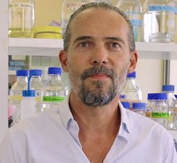 Nelson Dusetti合作论文数Cancrer Research Center of Marseille4
 Eric W. Klee合作论文数Mayo Clinic;2 In - vitrogen Corporation;Mayo Clinic, Mayo Clinic4Yuning Xiong合作论文数4Tom Walter合作论文数3Cathrine J DeMars合作论文数3Romica Kerketta合作论文数3Jorge A. Toro-Zapata合作论文数3Wendy M Demos合作论文数3A. Liebl合作论文数3
 Gustavo Leone合作论文数Dept Biochem, Med Coll Wisconsin3Michael Tschannen合作论文数3




