Plasmodium replicates within the liver prior to reaching the bloodstream and infecting red blood cells. Because clinical manifestations of malaria only arise during the blood stage of infection, a perception exists that liver infection does not impact disease pathology. By developing a murine model where the liver and blood stages of infection are uncoupled, we showed that the integration of signals from both stages dictated mortality outcomes. This dichotomy relied on liver stage-dependent activation of Vγ4+ γδ T cells. Subsequent blood stage parasite loads dictated their cytokine profiles, where low parasite loads preferentially expanded IL-17-producing γδ T cells. IL-17 drove extra-medullary erythropoiesis and concomitant reticulocytosis, which protected mice from lethal experimental cerebral malaria (ECM). Adoptive transfer of erythroid precursors could rescue mice from ECM. Modeling of γδ T cell dynamics suggests that this protective mechanism may be key for the establishment of naturally acquired malaria immunity among frequently exposed individuals.
Plasmodium replicates within the liver prior to reaching the bloodstream and infecting red blood cells. Because clinical manifestations of malaria only arise during the blood stage of infection, a perception exists that liver infection does not impact disease pathology. By developing a murine model where the liver and blood stages of infection are uncoupled, we showed that the integration of signals from both stages dictated mortality outcomes. This dichotomy relied on liver stage-dependent activation of Vy4+ y8 T cells. Subsequent blood stage parasite loads dictated their cytokine profiles, where low parasite loads preferentially expanded IL-17-producing y8 T cells. IL-17 drove extra-medullary erythropoiesis and concomitant reticulocytosis, which protected mice from lethal experimental cerebral malaria (ECM). Adoptive transfer of erythroid precur-sors could rescue mice from ECM. Modeling of y8 T cell dynamics suggests that this protective mechanism may be key for the establishment of naturally acquired malaria immunity among frequently exposed indi-viduals.
Severe malaria can manifest itself with a variety of well-recognized clinical phenotypes that are highly predictive of death – severe anaemia, coma (cerebral malaria), multiple organ failure, and respiratory distress. The reasons why an infected individual develops one pathology rather than another remain poorly understood. Here we use distinct rodent models of infection to show that the host microbiota is a contributing factor for the development of respiratory distress syndrome and host mortality in the context of malaria infections (malaria-associated acute respiratory distress syndrome, MA-ARDS). We show that parasite sequestration in the lung results in sustained immune activation. Subsequent production of the anti-inflammatory cytokine IL-10 by T cells compromises microbial control, leading to severe lung disease. Notably, bacterial clearance with linezolid, an antibiotic commonly used in the clinical setting to control lung-associated bacterial infections, prevents MA-ARDS-associated lethality. Thus, we propose that the host’s anti-inflammatory response to limit tissue damage can result in loss of microbial control, which promotes MA-ARDS. This must be considered when intervening against life-threatening respiratory complications.
While the liver and blood stages of the Plasmodium life cycle are commonly regarded as two separate fields of malaria research, several studies have pointed towards the existence of a bidirectional crosstalk, where one stage of mammalian infection may impact the establishment and progression of the other. Despite the constraints in experimentally addressing concurrent liver and blood stage Plasmodium infections, animal models and clinical studies have unveiled a plethora of molecular interactions between the two. Here, we review the current knowledge on the reciprocal influence of hepatic and erythrocytic infection by malaria parasites, and discuss its impacts on immunity, pathology and vaccination against this deadly disease. (c) 2022 Published by Elsevier Ltd on behalf of Australian Society for Parasitology. This is an open access article under the CC BY license (http://creativecommons.org/licenses/by/4.0/).
Malaria infection involves an obligatory, yet clinically silent liver stage1,2. Hepatocytes operate in repeating units termed lobules, exhibiting heterogeneous gene expression patterns along the lobule axis3, but the effects of hepatocyte zonation on parasite development at the molecular level remain unknown. Here we combine single-cell RNA sequencing4 and single-molecule transcript imaging5 to characterize the host and parasite temporal expression programmes in a zonally controlled manner for the rodent malaria parasite Plasmodium berghei ANKA. We identify differences in parasite gene expression in distinct zones, including potentially co-adaptive programmes related to iron and fatty acid metabolism. We find that parasites develop more rapidly in the pericentral lobule zones and identify a subpopulation of periportally biased hepatocytes that harbour abortive infections, reduced levels of Plasmodium transcripts and parasitophorous vacuole breakdown. These 'abortive hepatocytes', which appear predominantly with high parasite inoculum, upregulate immune recruitment and key signalling programmes. Our study provides a resource for understanding the liver stage of Plasmodium infection at high spatial resolution and highlights the heterogeneous behaviour of both the parasite and the host hepatocyte.
Anthracyclines are among the most used and effective anticancer drugs. Their activity has been attributed to DNA double-strand breaks resulting from topoisomerase II poisoning and to eviction of histones from select sites in the genome. Here, we show that the extensively used anthracyclines Doxorubicin, Daunorubicin, and Epirubicin decrease the transcription of nuclear factor kappa B (NF-κB)-dependent gene targets, but not interferon-responsive genes in primary mouse (Mus musculus) macrophages. Using an NMR-based structural approach, we demonstrate that anthracyclines disturb the complexes formed between the NF-κB subunit RelA and its DNA-binding sites. The anthracycline variants Aclarubicin, Doxorubicinone, and the newly developed Dimethyl-doxorubicin, which share anticancer properties with the other anthracyclines but do not induce DNA damage, also suppressed inflammation, thus uncoupling DNA damage from the effects on inflammation. These findings have implications for anticancer therapy and for the development of novel anti-inflammatory drugs with limited side effects for life-threatening conditions such as sepsis.
Plasmodium parasites, causative agents of malaria, scavenge host nutrients to sustain their intracellular replication. Modulation of the host's nutritional status can potentially help control infection by limiting the parasite's access to nutrients, or by boosting the immune system. Here, we show that dietary supplementation of mice employing a combination of arginine (R) with two additional amino acids, lysine (K) and valine (V), termed RKV, significantly decreases Plasmodium liver infection. RKV supplementation results in the elimination of parasites at a late stage of their development in the liver. Our data employing genetic knockout mouse models and in vivo depletion of specific cell populations suggest that RKV supplementation boosts the host's overall innate immune response, and that parasite elimination is dependent on MyD88 signaling in immune cells. The immunostimulatory effect of RKV supplementation opens a potential role for dietary supplementation as an adjuvant for prophylaxis or immunization strategies against Plasmodium infection.
The malaria parasite Plasmodium obligatorily infects and replicates inside hepatocytes surrounded by a parasitophorous vacuole membrane (PVM), which is decorated by the host-cell derived autophagy protein LC3. We have previously shown that the parasite-derived, PVM-resident protein UIS3 sequesters LC3 to avoid parasite elimination by autophagy from hepatocytes. Here we show that a small molecule capable of disrupting this interaction triggers parasite elimination in a host cell autophagy-dependent manner. Molecular docking analysis of more than 20 million compounds combined with a phenotypic screen identified one molecule, C4 (4-{[4-(4-{5-[3-(trifluoromethyl) phenyl]-1,2,4-oxadiazol-3-yl}benzyl)piperazino]carbonyl}benzonitrile), capable of impairing infection. Using biophysical assays, we established that this impairment is due to the ability of C4 to disrupt UIS3–LC3 interaction, thus inhibiting the parasite’s ability to evade the host autophagy response. C4 impacts infection in autophagy-sufficient cells without harming the normal autophagy pathway of the host cell. This study, by revealing the disruption of a critical host–parasite interaction without affecting the host’s normal function, uncovers an efficient anti-malarial strategy to prevent this deadly disease.
Plasmodium, the causative agent of malaria, is responsible for more than 200 million new infections and 400 000 deaths yearly. While in recent years the influence of the microbiota in homeostasis and a wide variety of disorders has taken center stage, its contribution during malaria infections has only now started to emerge. The few published studies suggest two distinct but complementary directions. Plasmodium infections can cause significant alterations in host (atleast gut) microbiota, and host gut microbiota can influence the clinical outcome of malaria infections.In this opinion article, we highlight the most fundamental unanswered questions in the field that will, hopefully, point future research directions towards unveiling key mechanistic in-sights of the Plasmodium-host-microbiota axis.
Under Multiple Sclerosis pathological conditions, oligodendrocyte precursor cells (OPCs) present in the brain parenchyma or derived from subventricular zone neural stem cells (SVZ-NSCs) can differentiate into oligodendrocytes (OLs), which migrate and partially remyelinate the lesioned areas. Previous data from our group demonstrated that activation of adenosine A2A receptors (A2AR) modulated SVZ-NSCs oligodendroglial differentiation, both in vitro and in vivo under physiological conditions. Hence, we aimed at understanding the role of A2AR in adult oligodendrogenesis derived from SVZ-NSCs in an in vivo mouse model of MS. For this, the Experimental Autoimmune Encephalomyelitis (EAE) mouse model of MS was developed, and behavioural tests were performed to evaluate motor function. Cellular differentiation was assessed by immunohistochemistry assays for bromodeoxyuridine (BrdU) colocalization with oligodendrocytic markers in brain regions of interest. Western blot and ELISA assays were used for myelin protein levels and inflammatory cytokine quantification. Our results for EAE model characterization showed that motor impairment is proportional to the score of the disease and cellular and molecular data showed an increase in the levels of the pro-inflammatory cytokine TNFα (n=5, p<0.01). A significant increase in NG2+BrdU+ cells in the corpus callosum (CC) of EAE mice was observed (n=3, p<0.05), hinting at the migration of OPCs from the SVZ to the CC. Ongoing studies encompass the in vivo modulation of A2AR and assessing its effect on EAE phenotype and adult oligodendrogenesis, ultimately unveiling the modulation of adult oligodendrogenesis derived from SVZ-NSCs by A2AR as a putative therapy for MS.
Event Abstract Back to Event From neural stem cells to oligodendrocytes: postnatal oligodendrogenesis as a target for MS therapy Joana M. Mateus1, 2, Marta A. Gomes1, 2, Rita Soares1, 2, Sara L. Paulo1, 2, Ângelo F. Chora2, Ana M. Sebastião1, 2 and Sara Xapelli1, 2* 1 Institute of Pharmacology and Neurosciences, Faculty of Medicine, University of Lisbon, Portugal 2 Institute of Molecular Medicine, Faculty of Medicine, University of Lisbon, Portugal Oligodendrocytes (OLs), the myelinating cells of the Central Nervous System (CNS), are generated upon differentiation of oligodendrocyte precursor cells (OPCs), which possess proliferative and migratory capabilities and are present in the CNS postnatally. Under pathological conditions such as in Multiple Sclerosis (MS), there is a depletion of OLs, but the OPCs present in the brain parenchyma or derived from subventricular zone (SVZ) neural stem cells (NSCs) can differentiate, migrate and partially remyelinate the lesioned areas. Herein, we aimed at characterizing the MS mouse model, experimental autoimmune encephalomyelitis (EAE), studying animal behaviour as well as the process of adult oligodendrogenesis. For this, the EAE mouse model was developed, and behavioural tests (open field, rotarod, pole test) were performed to evaluate motor function. Cellular differentiation was assessed by immunohistochemistry assays for bromodeoxyuridine (BrdU) colocalization with oligodendrocytic markers in brain regions of interest. Western blot and ELISA assays were used for myelin protein levels and inflammatory cytokine quantification. Results for clinical scores and behavioural characterization of the EAE model suggested that motor impairment is proportional to the score of the disease. Moreover, an increase in the levels of the pro-inflammatory cytokine TNFα (n=5, p<0.01), and a tendency for increased IL-1β were observed in EAE mice. Importantly, a tendency for increased BrdU+ cells in the SVZ, corpus callosum (CC) and cerebral cortex (CT) was observed, accompanied by a significant increase in NG2+BrdU+ cells in the CC of EAE mice (n=3, p<0.05), hinting at the migration of precursor cells from the SVZ to the CC. Altogether, this work allowed the characterization of the oligodendrogenesis process and of the EAE model throughout time, supporting future studies involving the modulation of adult oligodendrogenesis as a putative therapy for MS. Keywords: subventricular zone (SVZ), Neural stem cells (NSCs), Oligodendrocytes (OLs), Multiple Sclerosis, EAE (experimental autoimmune encephalomyelitis) Conference: XVI Meeting of the Portuguese Society for Neuroscience (SPN2019), Lisboa, Portugal, 30 May - 1 Jun, 2019. Presentation Type: Pitch communication + Poster presentation Topic: Glia / Neuroinflammation Citation: Mateus JM, Gomes MA, Soares R, Paulo SL, Chora ÂF, Sebastião AM and Xapelli S (2019). From neural stem cells to oligodendrocytes: postnatal oligodendrogenesis as a target for MS therapy. Front. Cell. Neurosci. Conference Abstract: XVI Meeting of the Portuguese Society for Neuroscience (SPN2019). doi: 10.3389/conf.fncel.2019.01.00047 Copyright: The abstracts in this collection have not been subject to any Frontiers peer review or checks, and are not endorsed by Frontiers. They are made available through the Frontiers publishing platform as a service to conference organizers and presenters. The copyright in the individual abstracts is owned by the author of each abstract or his/her employer unless otherwise stated. Each abstract, as well as the collection of abstracts, are published under a Creative Commons CC-BY 4.0 (attribution) licence (https://creativecommons.org/licenses/by/4.0/) and may thus be reproduced, translated, adapted and be the subject of derivative works provided the authors and Frontiers are attributed. For Frontiers’ terms and conditions please see https://www.frontiersin.org/legal/terms-and-conditions. Received: 02 Mar 2019; Published Online: 27 Sep 2019. * Correspondence: Prof. Sara Xapelli, Institute of Pharmacology and Neurosciences, Faculty of Medicine, University of Lisbon, Lisbon, Portugal, sxapelli@medicina.ulisboa.pt Login Required This action requires you to be registered with Frontiers and logged in. To register or login click here. Abstract Info Abstract The Authors in Frontiers Joana M Mateus Marta A Gomes Rita Soares Sara L Paulo Ângelo F Chora Ana M Sebastião Sara Xapelli Google Joana M Mateus Marta A Gomes Rita Soares Sara L Paulo Ângelo F Chora Ana M Sebastião Sara Xapelli Google Scholar Joana M Mateus Marta A Gomes Rita Soares Sara L Paulo Ângelo F Chora Ana M Sebastião Sara Xapelli PubMed Joana M Mateus Marta A Gomes Rita Soares Sara L Paulo Ângelo F Chora Ana M Sebastião Sara Xapelli Related Article in Frontiers Google Scholar PubMed Abstract Close Back to top Javascript is disabled. Please enable Javascript in your browser settings in order to see all the content on this page.
The relevance of genetic factors in conferring protection to severe malaria has been demonstrated, as in the case of sickle cell trait and G6PD deficiency 1 . However, it remains unknown whether environmental components, such as dietary or metabolic variations, can contribute to the outcome of infection 2 . Here, we show that administration of a high-fat diet to mice for a period as short as 4 days impairs Plasmodium liver infection by over 90%. Plasmodium sporozoites can successfully invade and initiate replication but die inside hepatocytes, thereby are unable to cause severe disease. Transcriptional analyses combined with genetic and chemical approaches reveal that this impairment of infection is mediated by oxidative stress. We show that reactive oxygen species, probably spawned from fatty acid β-oxidation, directly impact Plasmodium survival inside hepatocytes, and parasite load can be rescued by exogenous administration of the antioxidant N-acetylcysteine or the β-oxidation inhibitor etomoxir. Together, these data reveal that acute and transient dietary alterations markedly impact the establishment of a Plasmodium infection and disease outcome.
Severe sepsis remains a poorly understood systemic inflammatory condition with high mortality rates and limited therapeutic options in addition to organ support measures. Here we show that the clinically approved group of anthracyclines acts therapeutically at a low dose regimen to confer robust protection against severe sepsis in mice. This salutary effect is strictly dependent on the activation of DNA damage response and autophagy pathways in the lung, as demonstrated by deletion of the ataxia telangiectasia mutated (Atm) or the autophagy-related protein 7 (Atg7) specifically in this organ. The protective effect of anthracyclines occurs irrespectively of pathogen burden, conferring disease tolerance to severe sepsis. These findings demonstrate that DNA damage responses, including the ATM and Fancony Anemia pathways, are important modulators of immune responses and might be exploited to confer protection to inflammation-driven conditions, including severe sepsis.
Before they infect red blood cells and cause malaria, Plasmodium parasites undergo an obligate and clinically silent expansion phase in the liver that is supposedly undetected by the host. Here, we demonstrate the engagement of a type I interferon (IFN) response during Plasmodium replication in the liver. We identified Plasmodium RNA as a previously unrecognized pathogen-associated molecular pattern (PAMP) capable of activating a type I IFN response via the cytosolic pattern recognition receptor Mda5. This response, initiated by liver-resident cells through the adaptor molecule for cytosolic RNA sensors, Mavs, and the transcription factors Irf3 and Irf7, is propagated by hepatocytes in an interferon-α/β receptor-dependent manner. This signaling pathway is critical for immune cell-mediated host resistance to liver-stage Plasmodium infection, which we find can be primed with other PAMPs, including hepatitis C virus RNA. Together, our results show that the liver has sensor mechanisms for Plasmodium that mediate a functional antiparasite response driven by type I IFN.
Effective CD8+ T‐cell responses against tumor or microbial antigens that are not directly expressed in antigen‐presenting cells (APCs) depend on the cross‐presentation of these antigens on MHC class I in APCs. To identify signaling molecules that regulate cross‐presentation, we used lentiviral‐based RNA interference to test the roles of hundreds of kinases and phosphatases in this process. Our study uncovered eight previously unknown genes, consisting of one positive and seven negative regulators of antigen cross‐presentation. Depletion of Acvr1c, a type I receptor for TGF‐β family of signaling molecules, led to an increase in CD80 and CD86 co‐stimulator surface expression and secreted IL‐12 in mouse bone marrow‐derived DCs, as well as antigen‐specific T‐cell proliferation.
Sickle human hemoglobin (Hb) confers a survival advantage to individuals living in endemic areas of malaria, the disease caused by Plasmodium infection. As demonstrated hereby, mice expressing sickle Hb do not succumb to experimental cerebral malaria (ECM). This protective effect is exerted irrespectively of parasite load, revealing that sickle Hb confers host tolerance to Plasmodium infection. Sickle Hb induces the expression of heme oxygenase-1 (HO-1) in hematopoietic cells, via a mechanism involving the transcription factor NF-E2-related factor 2 (Nrf2). Carbon monoxide (CO), a byproduct of heme catabolism by HO-1, prevents further accumulation of circulating free heme after Plasmodium infection, suppressing the pathogenesis of ECM. Moreover, sickle Hb inhibits activation and/or expansion of pathogenic CD8(+) T cells recognizing antigens expressed by Plasmodium, an immunoregulatory effect that does not involve Nrf2 and/or HO-1. Our findings provide insight into molecular mechanisms via which sickle Hb confers host tolerance to severe forms of malaria.
The generation of diversity and plasticity of transcriptional programs are key components of effective vertebrate immune responses. The role of Alternative Splicing has been recognized, but it is underappreciated and poorly understood as a critical mechanism for the regulation and fine-tuning of physiological immune responses. Here we report the generation of loss-of-function phenotypes for a large collection of genes known or predicted to be involved in the splicing reaction and the identification of 19 novel regulators of IL-1β secretion in response to E. coli challenge of THP-1 cells. Twelve of these genes are required for IL-1β secretion, while seven are negative regulators of this process. Silencing of SFRS3 increased IL-1β secretion due to elevation of IL-1β and caspase-1 mRNA in addition to active caspase-1 levels. This study points to the relevance of splicing in the regulation of auto-inflammatory diseases.
Low-grade polymicrobial infection induced by cecal ligation and puncture is lethal in heme oxygenase-1-deficient mice (Hmox1(-/-)), but not in wild-type (Hmox1(+/+)) mice. Here we demonstrate that the protective effect of this heme-catabolizing enzyme relies on its ability to prevent tissue damage caused by the circulating free heme released from hemoglobin during infection. Heme administration after low-grade infection in mice promoted tissue damage and severe sepsis. Free heme contributed to the pathogenesis of severe sepsis irrespective of pathogen load, revealing that it compromised host tolerance to infection. Development of lethal forms of severe sepsis after high-grade infection was associated with reduced serum concentrations of the heme sequestering protein hemopexin (HPX), whereas HPX administration after high-grade infection prevented tissue damage and lethality. Finally, the lethal outcome of septic shock in patients was also associated with reduced HPX serum concentrations. We propose that targeting free heme by HPX might be used therapeutically to treat severe sepsis.
 Miguel Che Parreira Soares合作论文数Instituto Gulbenkian de Ciencia12
 Maria M. Mota合作论文数Instituto de Medicina Molecular, Universidade de Lisboa12
 Gozzelino Raffaella合作论文数Inst Gulbenkian Ciencias5
 Sofia Marques合作论文数5
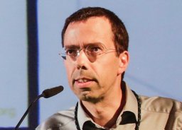 Moita Luis F合作论文数Inst Mol Med, Univ Lisbon4
 Gabriela Paiva-Silva合作论文数4
 Viktória Jeney合作论文数Medical and Health Science Center, University of Debrecen4
 Vanessa Zuzarte-Luis合作论文数Facultad de Medicina, Universidad de Cantabria4
 Karine Serre合作论文数4Ana Ferreira合作论文数3
 Tania Carvalho合作论文数3
 Maria Ines Marreiros合作论文数3Debanjan Mukherjee合作论文数Inst Med Mol, Univ Lisbon3
 Miguel Prudêncio合作论文数Fac Med, Univ Lisbon3
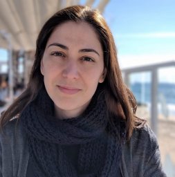 Silvia Cardoso合作论文数3Rodriguez Sonia合作论文数Indiana University Simon Cancer Center, Indiana University School of Medicine3
 Catarina Moita合作论文数Instituto Medicina Molecular, UBCSI Ed. Egas Moniz3
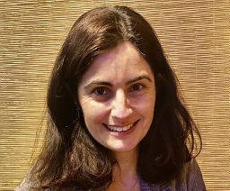 Ana Neves-Costa合作论文数Nuclear Dynamics and Function, Babraham Institute3Daniel Gomes da Costa合作论文数2
 Pedro Ruivo合作论文数2Isabel Pombo Grégoire合作论文数2Marta Gomes合作论文数2Pedro Pereira合作论文数2Ivo Marguti合作论文数2Luis Fernandez合作论文数2
 Aparajita Lahree合作论文数2Nadja Pejanovic合作论文数2
 Joana Mateus合作论文数2
 Sara L. Paulo合作论文数2Rita Soares合作论文数2
 Ann T. Tate合作论文数Department of Biological Sciences, Vanderbilt University;Department of Pathology, Microbiology, and Immunology, Vanderbilt University2
 William Heath合作论文数Department of Microbiology and Immunology, University of Melbourne2
 Fernandez-Ruiz Daniel合作论文数2
 Gama-Carvalho Margarida合作论文数BioFIG-Centre for Biodiversity, Functional and Integrative Genomics Lisbon, Universidade de Lisboa2Pedroso Dora合作论文数2
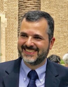 Ruy Ribeiro合作论文数Los Alamos National Laboratory;Center for Evolutionary & Theoretical Immunology, The University of New Mexico;Instituto de Medicina Molecular2Boyan C. Goumnerov合作论文数2
 Ricardo S. Ramiro合作论文数Centre for Immunity , Infection and Evolution;Institute of Immunology and Infection Research;School of Biological Sciences;University of ,;School of Biological Sciences, University of ,2
 Bruno Silva-Santos合作论文数Instituto de Medicina Molecular, Universidade de Lisboa2Peter Liehl合作论文数2
 Nadia Carlesso合作论文数Dana-Farber Cancer Institute and Department of, Medicine, Harvard Medical School2David Dombkowski合作论文数2Joao Alberto Ferreira合作论文数2
 Maria Helena Raquel合作论文数2
 Nir Hacohen合作论文数Center for Cancer Immunology, Massachusetts General Hospital;Mass General Research Institute;Broad Institute2
 Isabel Gordo合作论文数Gulbenkian Institute for Molecular Medicine2
 Laurence G. Rahme合作论文数Department of Surgery, Massachusetts General Hospital;Department of Microbiology, Blavatnik Institute, Harvard Medical School;Spero Therapeutics2
 Ana Sebastiao合作论文数Institute of Molecular Medicine, Faculty of Medicine, University of Lisbon2
 Jozsef Balla合作论文数Div. of Nephrology, Faculty of Medicine, University of Debrecen;Department of Internal Medicine, University of Debrecen;Department of Internal Medicine, Internal Medicine Clinic, University of Debrecen Clinical Centre2




