This editorial explores the novel The Secret of Secrets by Dan Brown as a gateway to longstanding debates in neuroscience, particularly surrounding consciousness and free will. Highlighting the novels references to real neurophotonic technologies, it examines where scientific evidence ends and speculative fiction begins.
Neuromodulatory transmitters have vasoactive properties. Therefore, the impulse response function (IRF) linking spontaneous neuronal activity with hemodynamics may depend on neuromodulation. To test this hypothesis, we used optical imaging to measure norepinephrine (NE) or acetylcholine (ACh), calcium (Ca2+) activity of cortical neurons and hemodynamics in cerebral cortex in awake mice. We show that modeling of hemodynamics as a weighted sum of Ca2+-specific and NE-specific IRFs (IRFCa2+ and IRFNE) convolved with the respective time courses dramatically improved the model performance compared to using IRFCa2+ alone. In contrast to NE, ACh was largely redundant with Ca2+ and, therefore, did not improve the hemodynamic estimation. Because NE covaried with arousal, we observed instances of the diminished hemodynamic coherence between cortical regions during high arousal despite coherent behavior of the underlying neuronal Ca2+ activity. We conclude that, without accounting for noradrenergic neuromodulation, diminished hemodynamic coherence can be falsely interpreted as neuronal desynchronizations in neuroimaging studies.
Sodium magnetic resonance imaging ( 23 Na MRI) provides a unique opportunity to probe ionic microenvironments in neural tissue because sodium ions play central roles in membrane electrophysiology, ion transport, and cellular homeostasis. Unlike conventional proton (¹H) MRI, which primarily reflects water distribution and tissue structure, ²³Na MRI is sensitive to ionic compartmentation and quadrupolar interactions arising from the spin-3/2 nature of the sodium nucleus. However, sodium MRI remains technically challenging due to intrinsically low signal sensitivity and rapid biexponential relaxation, particularly when imaging small biological systems. Here, we establish a high-field multinuclear MRI platform for imaging human cerebral organoids at 14 Tesla. Cerebral organoids derived from human induced pluripotent stem cells provide a simplified three-dimensional neural tissue model that enables investigation of ionic microenvironments without vascular or systemic confounds. Using a dual-tuned ¹H/²³Na radiofrequency coil, we performed co-registered structural, diffusion, and sodium imaging of individual fixed organoids. High-resolution ¹H MRI (33-100 μm) revealed pronounced microstructural heterogeneity, while multi-echo ²³Na MRI (300-400 μm) enabled voxel-wise characterization of quadrupolar relaxation behavior. Bi-exponential analysis of the sodium signal decay identified distinct relaxation components (T2* short ≈ 1 ms and T2* long ≈ 12 ms) and revealed spatial heterogeneity in sodium microenvironments across the organoid tissue. These results demonstrate the feasibility of quantitative sodium relaxometry in cortical organoids and establish a multinuclear imaging platform for investigating ionic microenvironment dynamics in three-dimensional neural tissue models.
Understanding distributed biological systems, particularly neural circuits, requires simultaneous cellular-resolution imaging across millimeter-scale fields of view (FOV). Existing miniature microscopes remain fundamentally constrained by trade-offs among FOV, spatial resolution, and optical complexity, limiting their ability to bridge cellular microscopy with cortex-scale imaging. Here we introduce distributed computational optics, a framework that distributes image formation across coordinated optical modules and computationally integrates their measurements into a unified image. We realize this framework in Bio-CM², a computational miniature mesoscope that partitions the imaging field across four optical modules while converging their measurements onto a common image sensor. This architecture overcomes the aberration-scaling limitations of conventional miniature optics while avoiding the hardware complexity of multi-camera systems and the contrast degradation associated with optical multiplexing. Bio-CM² achieves a 7.5 × 10 mm² FOV while enabling cellular-resolution in vivo imaging at video rates. We demonstrate its utility through two complementary imaging modalities in head-fixed mice: cortex-wide functional vascular imaging, enabling simultaneous quantification of pial arteriole vasomotion and mesoscale hemodynamic functional connectivity, and cellular-resolution calcium imaging, resolving the activity of over 3,000 neurons together with mesoscale neuronal functional connectivity. We further demonstrate the versatility of the platform through cellular-resolution imaging of entire coronal mouse brain sections, population-scale imaging of freely behaving Caenorhabditis elegans , and odor-evoked calcium imaging of the main olfactory bulb in head-fixed mice, highlighting its broad applicability across diverse biological systems and imaging modalities. By overcoming the conventional trade-off between FOV and spatial resolution in a compact miniature platform, Bio-CM² establishes distributed computational optics as a scalable framework for multiscale biological imaging.
Introduction Gamma entrainment using sensory stimuli (GENUS) uses 40Hz-pulsed sensory stimuli to entrain neural activity in the gamma band (30-150Hz). However, the effect of GENUS on low-frequency vascular oscillations has not been fully explored. Objectives The objective of this study is to elucidate the effect of GENUS on vasomotion in healthy mice and potential confounds for future application in disease studies. Methods Head-fixed, awake C57Bl/6 mice (n=18; 9M 9F) aged between 18 to 60 weeks were subjected to white light of either 40Hz visual flicker (GENUS), or constant stimulus (control). Blood flow was imaged using laser speckle contrast imaging (LSCI) before, during, immediately after 1 hour of stimulus, and 30min after the stimulus termination. Results A linear mixed-effects model showed that GENUS enhanced the magnitude of 0.2-0.4Hz blood flow oscillations by 38% during stimulation and by 30% at 30 minutes after stimulation compared to control when controlled for age, sex, and other factors. The effect on vasomotion was distributed across many cortical regions not limited to visual areas and lasted beyond 24 hours post-stimulus. Conclusion These results support the exploration of GENUS for increasing vasomotion in therapeutic contexts.
In this issue of Neuron, Sun, Peng, et al.1 identify two separate mechanisms that together provide a brain-wide noradrenergic control of cerebral blood flow in larval zebrafish.
During forebrain development, inhibitory interneurons and oligodendrocyte progenitor cells migrate long distances into the developing dorsal cortex. Human induced pluripotent stem cell-derived forebrain assembloids (FAs) provide direct experimental access to this migratory process in vitro. Using viral labeling to express yellow fluorescent protein (EYFP) and tandem-dimer tomato (tdTomato) driven by EF1α or SOX10 promoters, respectively, we tracked cells in FAs over 15–17 h using spinning disk confocal microscopy. We developed an end-to-end processing pipeline for 4D volumetric imaging data, consisting of background subtraction and drift correction, manual cell coordinate tracking, and an analysis workflow to describe migratory cell behavior. Image preprocessing significantly improved data quality for subsequent manual tracking in datasets with heterogeneous labeling density and brightness. Trajectory analysis of 336 EYFP- and 337 tdTomato-labeled cells from twelve FAs indicates that most cells show super-diffusive directed motility. Our pipeline represents a key resource for cell tracking in FAs and similar three-dimensional platforms. This pipeline represents the first open tracking resource for iPSC-derived FAs and can be used as a ground-truth resource for the development of automated cell detection and tracking algorithms.
Inducing apparent memory recall by tagging and optogenetically reactivating cells in the hippocampus was demonstrated over a decade ago. However, the hippocampal dynamics resulting from this reactivation remain largely unknown. While calcium imaging is commonly used as a measure of neuronal activity, GCaMP, the most common calcium indicator, cannot be used with optogenetic neuronal reactivation because both require blue light excitation. To resolve this overlap, we demonstrate optogenetic reactivation with a red-shifted opsin, ChrimsonR. We then conduct dual-color calcium imaging in CA1 during memory reactivation in DG. In addition to measuring population dynamics in CA1, CA1 cells tagged during the original experience were identified. In the fear-conditioned animals (FC+), nontagged cells in CA1 decreased their firing rate during stimulation, while tagged cells maintained their activity level. In the FC+ animals, as the behavioral effect of stimulation decreased across days, so did the changes in neural activity during stimulation. Our results both demonstrate the technical feasibility of calcium imaging during optogenetic reactivation of memory-associated neurons and advance our understanding of the dynamics underlying this reactivation.
Driven by a period of accelerated progress and recent technical breakthroughs, whole-brain functional neuroimaging in rodents offers exciting new possibilities for addressing basic questions about brain function and its alterations. In response to lessons learned from the human neuroimaging community, leading scientists and researchers in the field convened to address existing barriers and outline ambitious goals for the future. This article captures these discussions, highlighting a shared vision to advance rodent functional neuroimaging into an era of increased impact.
The leptomeninges, composed of the arachnoid mater, and pia mater, contains distinct subgroups of fibroblasts that differ in location and transcriptomic profiles. These fibroblasts contribute to the blood–cerebrospinal fluid barrier under physiological conditions, participate in fibrosis, and support blood–brain barrier integrity during injury and disease. However, their Ca2+ signaling profiles and underlying mechanisms in health and disease remain poorly understood. In this study, we divided leptomeningeal fibroblasts into three subgroups based on their locations: arachnoid fibroblasts, pia mater fibroblasts and perivascular fibroblasts. We employed two-photon microscopy in awake transgenic mice expressing Ca²⁺ indicators in leptomeningeal fibroblasts to investigate spontaneous and behaviorally evoked Ca²⁺ transients across different fibroblast subgroups. We found that each subgroup exhibits a distinct Ca²⁺ activity profile, with pia mater fibroblasts showing the highest-amplitude Ca²⁺ transients. Moreover, these fibroblasts displayed unique responses to both whisker air-puff stimulation and locomotion. We further demonstrated, using a chronically implanted cannula beneath the cranial window, that locomotion-associated vasodilation is followed by TRPV4 channel-mediated fibroblast Ca²⁺ elevations. Finally, systemic inflammation induced by lipopolysaccharide (LPS) reduced spontaneous Ca²⁺ transients in pia mater fibroblasts, likely due to macrophage infiltration following the inflammatory response. For the first time, this study characterizes spontaneous and behaviorally evoked Ca²⁺ dynamics in distinct leptomeningeal fibroblast subgroups in awake animals, providing novel insights into the functional roles of leptomeningeal fibroblasts in the healthy and diseased brain.
The editorial reflects on the secret ingredients that can elevate the flavor of our scientific ideas.
High-resolution awake mouse functional magnetic resonance imaging (fMRI) remains challenging despite extensive efforts to address motion-induced artifacts and stress. This study introduces an implantable radio frequency (RF) surface coil design that minimizes image distortion caused by the air/tissue interface of mouse brains while simultaneously serving as a headpost for fixation during scanning. Furthermore, this study provides a thorough acclimation method used to accustom animals to the MRI environment minimizing motion-induced artifacts. Using a 14 T scanner, high-resolution fMRI enabled brain-wide functional mapping of visual and vibrissa stimulation at 100 µm×100 µm×200 µm resolution with a 2 s per frame sampling rate. Besides activated ascending visual and vibrissa pathways, robust blood oxygen level-dependent (BOLD) responses were detected in the anterior cingulate cortex upon visual stimulation and spread through the ventral retrosplenial area (VRA) with vibrissa air-puff stimulation, demonstrating higher-order sensory processing in association cortices of awake mice. In particular, the rapid hemodynamic responses in VRA upon vibrissa stimulation showed a strong correlation with the hippocampus, thalamus, and prefrontal cortical areas. Cross-correlation analysis with designated VRA responses revealed early positive BOLD signals at the contralateral barrel cortex (BC) occurring 2 s prior to the air-puff in awake mice with repetitive stimulation, which was not detected using a randomized stimulation paradigm. This early BC activation indicated a learned anticipation through the vibrissa system and association cortices in awake mice under continuous exposure of repetitive air-puff stimulation. This work establishes a high-resolution awake mouse fMRI platform, enabling brain-wide functional mapping of sensory signal processing in higher association cortical areas.
Transient stoppages of red blood cell (RBC) flow through capillaries—termed capillary stalls—occur persistently in neurological disorders such as Alzheimer’s disease and ischemic stroke and can interrupt oxygen delivery and exacerbate neurological damage. Effective imaging tools and analyses are necessary to understand the nature, role, and prevention of stalls. In this study, we dissect differences in stalls measured by two-photon Bessel beam microscopy (Bessel-2PM) and optical coherence tomography (OCT) to gain insight into the temporal dynamics of stalls. Twenty-minute series of volumetric angiograms were obtained separately with Bessel-2PM and OCT on the same day in awake, head-fixed mice. The temporal dynamics of stalling in both methods revealed a minority population of susceptible capillaries that exhibited frequent stalls and a large majority of capillaries with infrequent stalls. Differences between OCT and Bessel-2PM in the repeatability and dynamics of stalls are explained by differences in their sensitivity to short or infrequent stalls based on scanning speed and detection off-time. Finally, stroke caused a shift toward the frequently stalling capillary subpopulation, lasting 1 week post-stroke. Dynamic stall analysis therefore enables examination of physiological and methodological contributions to the stalls measured in disease models and across studies.
Human cortical organoids (hCOs) are three-dimensional neural cell aggregates that recapitulate certain structural and functional aspects of the developing human cortex. Xenotransplantation of hCOs into the rodent brain enables human-centric modeling of neurodevelopmental processes in a physiologically relevant environment. Here, we present a neurorecording toolkit for longitudinal structural and functional assessment of hCO xenografts as they mature in vivo. Single hCOs were implanted into the retrosplenial cortex of adult immunodeficient mice and monitored for up to 8 months. Optical coherence tomography was used for label-free imaging of xenograft vascularization and structure, enabling quantitative assessments of capillary density and graft volume. To probe neuronal activity, human neurons were labeled with a calcium sensor before implantation using either adeno-associated or lentivirus for sparse or dense neuronal labeling, respectively. Fluorescent imaging was conducted using two-photon, widefield, and swept confocally-aligned planar excitation microscopy for single cell, whole-graft, and volumetric calcium imaging, respectively. Results from these modalities indicate an increase in neuronal activity and synchronicity over time during in vivo graft maturation. Further, we chronically implanted surface graphene microelectrode arrays (gMEAs) and performed recordings of xenograft and host local field potential signals simultaneously with 2P calcium imaging, confirming the spatial localization and human origin of electrical signals recorded at the xenograft surface.
Bessel beam two-photon microscopy has revealed shorter stalling events than previously measured. Even these brief disruptions in flow can result in hypoxia.
The editorial presents the two-part Special Section on Frontiers in Neurophotonics.
We introduce an ultrasound speckle decorrelation-based time-lagged functional ultrasound technique (tl-fUS) for the quantification of the relative changes in cerebral blood flow speed (rCBF [Formula: see text]), cerebral blood volume (rCBV) and cerebral blood flow (rCBF) during functional stimulations. Numerical simulations, phantom validations, and in vivo mouse brain experiments were performed to test the capability of tl-fUS to parse out and quantify the ratio change of these hemodynamic parameters. The blood volume change was found to be more prominent in arterioles compared to venules and the peak blood flow changes were around 2.5 times the peak blood volume change during brain activation, agreeing with previous observations in the literature. The tl-fUS shows the ability of distinguishing the relative changes of rCBFspeed, rCBV, and rCBF, which can inform specific physiological interpretations of the fUS measurements.
How transient hyperglycemia contributes to cerebro-vascular disease has been a challenge to study under controlled physiological conditions. We use amplified, ultrashort laser-pulses to physically disrupt brain-venule endothelium at targeted locations. This vessel disruption is performed in conjunction with transient hyperglycemia from a single injection of metabolically active D-glucose into healthy mice. The observed real-time responses to laser-induced disruption include rapid serum extravasation, platelet aggregation, and neutrophil recruitment. Thrombo-inflammation is pharmacologically ameliorated by a platelet inhibitor, by a scavenger of reactive oxygen species, and by a nitric oxide donor. As a control, vessel thrombo-inflammation is significantly reduced in mice injected with metabolically inert L-glucose. Venules in mice with diabetes show a similar response to laser-induced disruption and damage is reduced by restoration of normo-glycemia. Our approach provides a controlled method to probe synergies between transient metabolic and physical vascular perturbations and can reveal new aspects of brain pathophysiology.
Two-photon phosphorescence lifetime microscopy has been a key tool for studying cerebral oxygenation in mice. However, the accuracy of the partial pressure of oxygen (pO2) measurements is affected by out-of-focus signal. In this work, we applied reconfigurable differential aberration imaging to characterize and correct for out-of-focus signal contamination in intravascular pO2 imaging. Our results show that signal contamination is higher in more oxygenated vessels and that it could be effectively removed using the proposed method.
 David Boas合作论文数Boston University94
 Sava Sakadzic合作论文数Athinoula A Martinos Ctr Biomed Imaging, Massachusetts Gen Hosp66
 Anders Dale合作论文数Department of Neurosciences, School of Medicine, University of California, San Diego55
 Martin Thunemann合作论文数Interfakultäres Institut für Biochemie, University of Tübingen53
 Kiliç Kivilcim合作论文数37
 Mohammad A. Yaseen合作论文数Athinoula A Martinos Ctr Biomed Imaging, Massachusetts Gen Hosp28
 Payam A. Saisan合作论文数Christopher Glass Laboratory, School of Medicine, University of California, San Diego26
 Sergei A. Vinogradov合作论文数Department of Chemistry, College of Arts & Sciences, University of Pennsylvania;Department of Biochemistry and Biophysics, Perelman School of Medicine, University of Pennsylvania25
 Gaute Einevoll合作论文数University of Oslo;Department of Physics, Norwegian University of Life Sciences;International Neuroinformatics Coordinating Facility21
 Richard Bruce Buxton合作论文数Center for Functional MRI, Department of Radiology, School of Medicine, University of California San Diego19
 Eng Lo合作论文数Neuroprotection Research Lab, Massachusetts General Hospital;Harvard Medical School19Peifang Tian合作论文数Dept Neurosci, Univ Calif San Diego17
 Sencan Ikbal合作论文数Dept Elect Engn, Univ Calif Los Angeles16
 Vivek Jay Srinivasan合作论文数Biomedical Engineering Department, College of Engineering, University of California, Davis16
 Michele Desjardins合作论文数Laboratory of Optical and Molecular Imaging, Polytechnique Montreal16
 Louis Gagnon合作论文数Harvard Medical School, Massachusetts General Hospital16Emiri T Mandeville合作论文数15Natalie Fomin-Thunemann合作论文数14
 David Kleinfeld合作论文数Department of Neurobiology, School of Biological Sciences, University of California, San Diego14Buyin Fu合作论文数13
 Sanaz Sadegh合作论文数Colorado State Univ, Dept Elect & Comp Engn, Ft Collins, CO 80523 USA13Kura Sreekanth合作论文数Harvard Medical School, MHG/MIT/13Baoqiang Li (李宝强)合作论文数Shenzhen Institute of Advanced Technology, Chinese Academy of Sciences;University of Chinese Academy of Sciences13
 Frederic Lesage合作论文数Department of Electrical Engineering, Polytechnique Montréal13Mu-Han Yang合作论文数12
 Shaya Fainman合作论文数Department of Electrical and Computer Engineering, University of California, San Diego12
 Andrew K. Dunn合作论文数Department of Biomedical Engineering, Cockrell School of Engineering, The University of Texas at Austin;Center for Emerging Imaging Technologies, The University of Texas at Austin12
 Ole Andreassen合作论文数Centre for Precision Psychiatry, Institute of Clinical Medicine, Faculty of Medicine, University of Oslo12
 Cenk Ayata合作论文数Massachusetts General Hospital;Mass General Research Institute;Harvard Medical School12
 Elizabeth M. C. Hillman合作论文数Columbia University;Department of Imaging Sciences, St. Jude Children's Research Hospital11Jiang John合作论文数11
 Cheng Xiaojun合作论文数Department of Physics, Queens College of the City University of New York11Christopher G. L. Ferri合作论文数11
 Istvan Ulbert合作论文数Institute of Cognitive Neuroscience and Psychology, Hungarian Academy of Sciences;Faculty of Information Technology, Pázmány Péter Catholic University11Philipp Mächler合作论文数Dept Biomed Engn, Boston Univ10
 Emmanuel Rousakis合作论文数Wellman Center for Photomedicine, Massachusetts General Hospital, Harvard Medical School, Boston, MA, USA10
 Srdjan Djurovic合作论文数Department of Medical Genetics, Oslo University Hospital9
 Meryem Ayşe Yücel合作论文数Neurophotonics Center, Boston University;College of Engineering, Boston University9Lana Ruvinskaya合作论文数9joseph j musacchia合作论文数8Lei Tian合作论文数8
 Alysson Muotri合作论文数Department of Pediatrics, School of Medicine, University of California, San Diego8
 Lorraine Hossain合作论文数Univ Calif San Diego, Mat Sci & Engn Program, La Jolla, CA 92093 USA8Patrick R Doran合作论文数8
 Francesca Puppo合作论文数the Integrated Systems Laboratory, Ecole Polytechnique Federale de Lausanne;c;the Integrated Systems Laboratory, Ecole Polytechnique Federale de Lausanne8
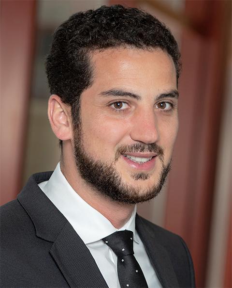 Shadi A. Dayeh合作论文数Integrated Electronics and Biointerfaces Laboratory, Department Electrical and Computer Engineering, UC San Diego8
 Uhlirova Hana合作论文数8Ivan C. Teng合作论文数7Matthieu Vandenberghe合作论文数UCSD, Dept Radiol, La Jolla, CA 92093 USA7




