Antibiotic resistance rapidly develops against almost all available therapeutics. Therefore, searching for new antibiotics to overcome the problem of antibiotic resistance alone is insufficient. Given that antibiotic resistance can be driven by mutagenesis, an avenue for preventing it is the inhibition of mutagenic processes. We previously showed that the DNA translocase Mfd is mutagenic and accelerates antibiotic resistance development. Here, we present our discovery of a small molecule that inhibits Mfd-dependent mutagenesis, ARM-1 (anti-resistance molecule 1). We found ARM-1 using a high-throughput, small molecule, in vivo screen. Using biochemical assays, we characterized the mechanism by which ARM-1 inhibits Mfd. Critically, we found that ARM-1 reduces mutagenesis and significantly delays antibiotic resistance development across highly divergent bacterial pathogens. These results demonstrate that the mutagenic proteins accelerating evolution can be directly inhibited. Furthermore, our findings suggest that Mfd inhibition, alongside antibiotics, is a potentially effective approach for prevention of antibiotic resistance development during treatment of infections.
An important step towards understanding the mechanistic basis of the central dogma is the quantitative characterization of the dynamics of nucleic-acid-bound molecular motors in the context of the living cell, where a crowded cytoplasm as well as competing and potentially antagonistic processes may significantly affect their rapidity and reliability. To capture these dynamics, we develop a novel method, lag-time analysis, for measuring in vivo dynamics. The approach uses exponential growth as the stopwatch to resolve dynamics in an asynchronous culture and therefore circumvents the difficulties and potential artifacts associated with synchronization or fluorescent labeling. Although lag-time analysis has the potential to be widely applicable to the quantitative analysis of in vivo dynamics, we focus on an important application: characterizing replication dynamics. To benchmark the approach, we analyze replication dynamics in three different species and a collection of mutants. We provide the first quantitative locus-specific measurements of fork velocity, in units of kb per second, as well as replisome-pause durations, some with the precision of seconds. The measured fork velocity is observed to be both locus and time dependent, even in wild-type cells. In addition to quantitatively characterizing known phenomena, we detect brief, locus-specific pauses at rDNA in wild-type cells for the first time. We also observe temporal fork velocity oscillations in three highly-divergent bacterial species. Lag-time analysis not only has great potential to offer new insights into replication, as demonstrated in the paper, but also has potential to provide quantitative insights into other important processes.
Transcriptional dysregulation is a key step in oncogenesis, but our understanding of transcriptional control has relied on genetic approaches that are slow and allow for compensation. Chemical-genetic approaches have shortened the time frame for the analysis of transcription factors from days or weeks to minutes. These studies show that while DNA-binding proteins bind to thousands of sites, they are directly required to regulate only a small cadre of genes. Moreover, these transcriptional control networks are far more distinct, with much less overlap and interconnectivity than predicted from DNA binding. The identified direct targets can then be used to dissect the mechanism of action of these factors, which could identify ways to therapeutically manipulate these oncogenic transcriptional control networks.
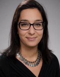 Merrikh Houra合作论文数Department of Microbiology, University of Washington2
 Brandon S. Sim合作论文数1Teresa W Lo合作论文数1
 Hillary M. Layden合作论文数Department of Biochemistry, Vanderbilt University School of Medicine1Dean Huang合作论文数Dept Phys, Univ Washington1Kwangho Kim合作论文数1
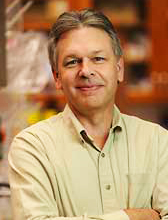 Scott W. Hiebert合作论文数Department of Biochemistry, Vanderbilt University1
 Martin Egli合作论文数Department of Biochemistry, School of Medicine, Vanderbilt University1
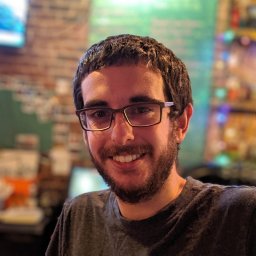 Carvajal-Garcia Juan合作论文数Lineberger Comprehensive Cancer Center, University of North Carolina1
 Paul Wiggins合作论文数Department of Physics, University of Washington1




