Multi-frequency EPR spectroscopy can provide high-level structural information on high-spin Fe3+ sites in proteins and enzymes. Unfortunately, analysis of the EPR spectra of these spin systems is hindered by the presence of broad distributions in the zero-field-splitting (ZFS) parameters, which reflect conformational heterogeneity of the iron sites. We present the analysis of EPR spectra of high-spin Fe3+ bound to human serum transferrin. We apply a method termed the grid-of-errors to extract the distributions of the individual ZFS parameters from EPR spectra recorded in the high-field limit at a microwave frequency of 275 GHz. Study of a series of transferrin variants shows that the ZFS distributions are as characteristic of the structure of a high-spin Fe3+ site as the ZFS parameters themselves. Simulations based on the extracted ZFS distributions reproduce spectra recorded at 34 GHz (Q band) and 9.7 GHz (X band), including subtle variations that were previously difficult to quantify. The X-band spectrum of transferrin shows a characteristic double peak, which has puzzled researchers for decades. We show that the double peak is uniquely related to the term B4-3O4-3(S) in the spin Hamiltonian. Our method is generally applicable in the analysis of spectra that arise from a broad distribution of parameters.
The transferrin (Tf) trafficking pathway is a promising mechanism for use in targeted cancer therapy due to the overexpression of transferrin receptors (TfRs) on cancerous cells. We have previously developed a mathematical model of the Tf/TfR trafficking pathway to improve the efficiency of Tf as a drug carrier. By using diphtheria toxin (DT) as a model toxin, we found that mutating the Tf protein to change its iron release rate improves cellular association and efficacy of the drug. Though this is an improvement upon using wild-type Tf as the targeting ligand, conjugated toxins like DT are unfortunately still highly cytotoxic at off-target sites. In this work, we address this hurdle in cancer research by developing a mathematical model to predict the efficacy and selectivity of Tf conjugates that use an alternative toxin. For this purpose, we have chosen to study a mutant of DT, cross-reacting material 107 (CRM107). First, we developed a mathematical model of the Tf-DT trafficking pathway by extending our Tf/TfR model to include intracellular trafficking via DT and DT receptors. Using this mathematical model, we subsequently investigated the efficacy of several conjugates in cancer cells: DT and CRM107 conjugated to wild-type Tf, as well as to our engineered mutant Tf proteins (K206E/R632A Tf and K206E/R534A Tf). We also investigated the selectivity of mutant Tf-CRM107 against non-neoplastic cells. Through the use of our mathematical model, we predicted that (i) mutant Tf-CRM107 exhibits a greater cytotoxicity than wild-type Tf-CRM107 against cancerous cells, (ii) this improvement was more drastic with CRM107 conjugates than with DT conjugates, and (iii) mutant Tf-CRM107 conjugates were selective against non-neoplastic cells. These predictions were validated with in vitro cytotoxicity experiments, demonstrating that mutant Tf-CRM107 conjugates is indeed a more suitable therapeutic agent. Validation from in vitro experiments also confirmed that such whole-cell kinetic models can be useful in cancer therapeutic design.
In erythroid cells, more than 90% of transferrin-derived iron enters mitochondria where ferrochelatase inserts Fe2+ into protoporphyrin IX. However, the path of iron from endosomes to mitochondrial ferrochelatase remains elusive. The prevailing opinion is that, after its export from endosomes, the redox-active metal spreads into the cytosol and mysteriously finds its way into mitochondria through passive diffusion. In contrast, this study supports the hypothesis that the highly efficient transport of iron toward ferrochelatase in erythroid cells requires a direct interaction between transferrin-endosomes and mitochondria (the "kiss-and-run" hypothesis). Using a novel method (flow sub-cytometry), we analyze lysates of reticulocytes after labeling these organelles with different fluorophores. We have identified a double-labeled population definitively representing endosomes interacting with mitochondria, as demonstrated by confocal microscopy. Moreover, we conclude that this endosome-mitochondrion association is reversible, since a "chase" with unlabeled holotransferrin causes a time-dependent decrease in the size of the double-labeled population. Importantly, the dissociation of endosomes from mitochondria does not occur in the absence of holotransferrin. Additionally, mutated recombinant holotransferrin, that cannot release iron, significantly decreases the uptake of 59Fe by reticulocytes and diminishes 59Fe incorporation into heme. This suggests that endosomes, which are unable to provide iron to mitochondria, cause a "traffic jam" leading to decreased endocytosis of holotransferrin. Altogether, our results suggest that a molecular mechanism exists to coordinate the iron status of endosomal transferrin with its trafficking. Besides its contribution to the field of iron metabolism, this study provides evidence for a new intracellular trafficking pathway of organelles.
Iron (Fe) acquisition by cells requires the binding of diferric transferrin to membrane transferrin receptors, followed by endocytosis. The path of Fe2+ from endosomes, after its released via DMT-1 to ferrochelatase for heme synthesis is not fully understood. By using 2D and 3D live confocal imaging, we have shown a transient interaction of endosomes with mitochondria (kiss-and-run) for efficient iron delivery to heme. We have demonstrated the interaction of these organelles after labeling them with Alexa Green Transferrin (AGTf) and MitoTracker Deep Red (MTDR) using novel method of flow subcytometry (FSC). The dynamic nature of these interactions showed time-dependent decrease of the double-labeled population when reticulocytes were washed and re-incubated with unlabeled Fe2-Tf. We have shown that the iron status of endosomes governs the efficacy of endosome-mediated iron delivery to mitochondria. In addition, experiments with mutants of fluorescence-labeled, recombinant human Tf, which either remain permanently bound to iron (L-Fe2-hTf) or cannot bind to iron (L-apo-hTf), in FSC studies showed failure of interaction of these two organelles. Experiments with 59Fe-Tf showed that L-Fe2-hTf significantly decreased incorporation of 59Fe into both cells and heme. These results suggest that L-Fe2-hTf remains associated with mitochondria for a longer time than WT-hTf, slows the transferrin cycle and thus blocks 59Fe incorporation into heme. Experiments with murine erythroleukemia and fetal liver cells show a significant increase in the magnitude of contacts between endosomes and mitochondria when these cells are induced for differentiation. Hence, we conclude that the mitochondria-endosome interaction is generally involved in iron delivery for heme biosynthesis, but that this process is dramatically augmented in erythroid cells. Taken together our results show that endosomes come in contact with mitochondria to deliver iron for heme biosynthesis Iron (Fe) acquisition by cells requires the binding of diferric transferrin to membrane transferrin receptors, followed by endocytosis. The path of Fe2+ from endosomes, after its released via DMT-1 to ferrochelatase for heme synthesis is not fully understood. By using 2D and 3D live confocal imaging, we have shown a transient interaction of endosomes with mitochondria (kiss-and-run) for efficient iron delivery to heme. We have demonstrated the interaction of these organelles after labeling them with Alexa Green Transferrin (AGTf) and MitoTracker Deep Red (MTDR) using novel method of flow subcytometry (FSC). The dynamic nature of these interactions showed time-dependent decrease of the double-labeled population when reticulocytes were washed and re-incubated with unlabeled Fe2-Tf. We have shown that the iron status of endosomes governs the efficacy of endosome-mediated iron delivery to mitochondria. In addition, experiments with mutants of fluorescence-labeled, recombinant human Tf, which either remain permanently bound to iron (L-Fe2-hTf) or cannot bind to iron (L-apo-hTf), in FSC studies showed failure of interaction of these two organelles. Experiments with 59Fe-Tf showed that L-Fe2-hTf significantly decreased incorporation of 59Fe into both cells and heme. These results suggest that L-Fe2-hTf remains associated with mitochondria for a longer time than WT-hTf, slows the transferrin cycle and thus blocks 59Fe incorporation into heme. Experiments with murine erythroleukemia and fetal liver cells show a significant increase in the magnitude of contacts between endosomes and mitochondria when these cells are induced for differentiation. Hence, we conclude that the mitochondria-endosome interaction is generally involved in iron delivery for heme biosynthesis, but that this process is dramatically augmented in erythroid cells. Taken together our results show that endosomes come in contact with mitochondria to deliver iron for heme biosynthesis
We report 275 GHz EPR spectra of human serum transferrin. At this high microwave frequency the zero-field splitting between the magnetic sublevels of the high-spin \(\text{Fe}^{3+}\) sites can be accurately determined. We find the zero-field splitting to be a sensitive probe of the structure of the transferrin iron-binding sites. Signals arising from iron bound to the transferrin N-lobe can clearly be distinguished from signals from iron bound to the C-lobe. Moreover, our spectra show that the structure of the iron site in the N-lobe is influenced by the presence and conformation of the C-lobe. The spectra of a series of N-lobe mutants altering the second-shell interaction of Arg124 with the synergistic anion carbonate reflect conformational changes induced at the iron site.
Following an internal contamination event, the transport of actinide (An) and lanthanide (Ln) metal ions through the body is facilitated by endogenous ligands such as the human iron-transport protein transferrin (Tf). The recognition of resulting metallo-transferrin complexes (M2Tf) by the cognate transferrin receptor (TfR) is therefore a critical step for cellular uptake of these metal ions. A high performance liquid chromatography-based method has been used to probe the binding of M2Tf with TfR, yielding a direct measurement of the successive thermodynamic constants that correspond to the dissociation of TfR(M2Tf)2 and TfR(M2Tf) complexes for Fe(3+), Ga(3+), La(3+), Nd(3+), Gd(3+), Yb(3+), Lu(3+), (232)Th(4+), (238)UO2(2+), and (242)Pu(4+). Important features of this method are (i) its ability to distinguish both 1 : 1 and 1 : 2 complexes formed between the receptor and the metal-bound transferrin, and (ii) the requirement for very small amounts of each binding partner (<1 nmol of protein per assay). Consistent with previous reports, the strongest receptor affinity is found for Fe2Tf (Kd1 = 5 nM and Kd2 = 20 nM), while the lowest affinity was measured for Pu2Tf (Kd1 = 0.28 μM and Kd2 = 1.8 μM) binding to the TfR. Other toxic metal ions such as Th(IV) and U(VI), when bound to Tf, are well recognized by the TfR. Under the described experimental conditions, the relative stabilities of TfR:(MxTf)y adducts follow the order Fe(3+) >> Th(4+) ~ UO2(2+) ~ Cm(3+) > Ln(3+) ~ Ga(3+) >>> Yb(3+) ~ Pu(4+). This study substantiates a role for Tf in binding lanthanide fission products and actinides, and transporting them into cells by receptor-mediated endocytosis.
Arenaviruses and hantaviruses cause severe human disease. Little is known regarding host proteins required for their propagation. We identified human proteins that interact with the glycoproteins (GPs) of a prototypic arenavirus and hantavirus and show that the lectin endoplasmic reticulum (ER)-Golgi intermediate compartment 53 kDa protein (ERGIC-53), a cargo receptor required for glycoprotein trafficking within the early exocytic pathway, associates with arenavirus, hantavirus, coronavirus, orthomyxovirus, and filovirus GPs. ERGIC-53 binds to arenavirus GPs through a lectin-independent mechanism, traffics to arenavirus budding sites, and is incorporated into virions. ERGIC-53 is required for arenavirus, coronavirus, and filovirus propagation; in its absence, GP-containing virus particles form but are noninfectious, due in part to their inability to attach to host cells. Thus, we have identified a class of pathogen-derived ERGIC-53 ligands, a lectin-independent basis for their association with ERGIC-53, and a role for ERGIC-53 in the propagation of several highly pathogenic RNA virus families.
It has been previously suggested that large amounts of oxalate in plasma could play a role in autism by binding to the bilobal iron transport protein transferrin (hTF), thereby interfering with iron metabolism by inhibiting the delivery of iron to cells. By examining the effect of the substitution of oxalate for the physiologically utilized synergistic carbonate anion in each lobe of hTF, we sought to provide a molecular basis for or against such a role. Our work clearly shows both qualitatively (6 M urea gels) and quantitatively (kinetic analysis by stopped-flow spectrofluorimetry) that the presence of oxalate in place of carbonate in each binding site of hTF does indeed greatly interfere with the removal of iron from each lobe (in the absence and presence of the specific hTF receptor). However, we also clearly demonstrate that once the iron is bound within each lobe of hTF, neither anion can displace the other. Additionally, as verified by urea gels and electrospray mass spectrometry, formation of completely homogeneous hTF-anion complexes requires that all iron must first be removed and hTF then reloaded with iron in the presence of either carbonate or oxalate. Significantly, experiments described here show that carbonate is the preferred binding partner; i.e., even if an equal amount of each anion is available during the iron loading process, the hTF-carbonate complex is formed.
Worldwide stocks of actinides and lanthanide fission products produced through conventional nuclear spent fuel are increasing continuously, resulting in a growing risk of environmental and human exposure to these toxic radioactive metal ions. Understanding the biomolecular pathways involved in mammalian uptake, transport and storage of these f-elements is crucial to the development of new decontamination strategies and could also be beneficial to the design of new containment and separation processes. To start unraveling these pathways, our approach takes advantage of the unique spectroscopic properties of trivalent curium. We clearly show that the human iron transporter transferrin acts as an antenna that sensitizes curium luminescence through intramolecular energy transfer. This behavior has been used to describe the coordination of curium within the two binding sites of the protein and to investigate the recognition of curium-transferrin complexes by the cognate transferrin receptor. In addition to providing the first protein-curium spectroscopic characterization, these studies prove that transferrin receptor-mediated endocytosis is a viable mechanism of intracellular entry for trivalent actinides such as curium and provide a new tool utilizing the specific luminescence of curium for the determination of other biological actinide transport mechanisms.
The Fe3+ binding protein human serum transferrin (hTF) is well known for its role in cellular iron delivery via the transferrin receptor (TFR). A new application is the use of hTF as a therapy and targeted drug delivery system for a number of diseases. Recently, production of hTF in plants has been reported; such systems provide a relatively inexpensive, animal-free (eliminating potential contamination by animal pathogens) method to produce large amounts of recombinant proteins for such biopharmaceutical applications. Specifically, the production of Optiferrin™ (hTF produced in rice, Oryza sativa, from InVitria) has been shown to yield large amounts of functional protein for use in culture medium for cellular iron delivery to promote growth. In the present work we describe further purification (by gel filtration) and characterization of hTF produced in rice (purified Optiferrin™) to determine its suitability in biopharmaceutical applications. The spectral, mass spectrometric, urea gel and kinetic analysis shows that purified Optiferrin™ is similar to recombinant nonglycosylated N-His tagged hTF expressed by baby hamster kidney cells and/or serum derived glycosylated hTF. Additionally, in a competitive immunoassay, iron-loaded Optiferrin™ is equivalent to iron-loaded N-His hTF in its ability to bind to the soluble portion of the TFR immobilized in an assay plate. As an essential requirement for any functional hTF, both lobes of purified Optiferrin™ bind Fe3+ tightly yet reversibly. Although previously shown to be capable of delivering Fe3+ to cells, the kinetics of iron release from iron-loaded Optiferrin™/sTFR and iron-loaded N-His hTF/sTFR complexes differ somewhat. We conclude that the purified Optiferrin™ might be suitable for consideration in biopharmaceutical applications.
Background: Not long after the Big Bang, iron began to play a central role in the Universe and soon became mired in the tangle of biochemistry that is the prima essentia of life. Since life's addiction to iron transcends the oxygenation of the Earth's atmosphere, living things must be protected from the potentially dangerous mix of iron and oxygen. The human being possesses grams of this potentially toxic transition metal, which is shuttling through his oxygen-rich humor. Since long before the birth of modern medicine, the blood-vibrant red from a massive abundance of hemoglobin iron has been a focus for health experts.Scope of review: We describe the current understanding of iron metabolism, highlight the many important discoveries that accreted this knowledge, and describe the perils of dysfunctional iron handling.General significance: Isaac Newton famously penned, "If I have seen further than others, it is by standing upon the shoulders of giants". We hope that this review will inspire future scientists to develop intellectual pursuits by understanding the research and ideas from many remarkable thinkers of the past.Major conclusions: The history of iron research is a long, rich story with early beginnings, and is far from being finished. This article is part of a Special Issue entitled Transferrins: Molecular mechanisms of iron transport and disorders. (C) 2011 Elsevier B.V. All rights reserved.
The recent crystal structure of two monoferric human serum transferrin (Fe(N)hTF) molecules bound to the soluble portion of the homodimeric transferrin receptor (sTFR) has provided new details about this binding interaction that dictates the delivery of iron to cells. Specifically, substantial rearrangements in the homodimer interface of the sTFR occur as a result of the binding of the two FeNhTF molecules. Mutagenesis of selected residues in the sTFR highlighted in the structure was undertaken to evaluate the effect on function. Elimination of Ca2+ binding in the sTFR by mutating two of four coordinating residues ([E465A,E468A]) results in low production of an unstable and aggregated sTFR. Mutagenesis of two histidines ([H475A,H684A]) at the dimer interface had little effect on the kinetics of release of iron at pH 5.6 from either lobe, reflecting the inaccessibility of this cluster to solvent. Creation of an H318A sTFR mutant allows assignment of a small pH-dependent initial decrease in the magnitude of the fluorescence signal to His318. Removal of the four C-terminal residues of the sTFR, Asp757-Asn758-Glu759-Phe760, eliminates pH-stimulated release of iron from the C-lobe of the Fe(2)hTF/sTFR Delta 757-760 complex. The inability of this sTFR mutant to bind and stabilize protonated hTF His349 (a pH-inducible switch) in the C-lobe of hTF accounts for the loss. Collectively, these studies support a model in which a series of pH-induced events involving both TFR residue His318 and hTF residue His349 occurs to promote receptor-stimulated release of iron from the C-lobe of hTF.
Efficient delivery of iron is critically dependent on the binding of diferric human serum transferrin (hTF) to its specific receptor (TFR) on the surface of actively dividing cells. Internalization of the complex into an endosome precedes iron removal. The return of hTF to the blood to continue the iron delivery cycle relies on the maintenance of the interaction between apohTF and the TFR after exposure to endosomal pH (≤6.0). Identification of the specific residues accounting for the pH-sensitive nanomolar affinity with which hTF binds to TFR throughout the cycle is important to fully understand the iron delivery process. Alanine substitution of 11 charged hTF residues identified by available structures and modeling studies allowed evaluation of the role of each in (1) binding of hTF to the TFR and (2) TFR-mediated iron release. Six hTF mutants (R50A, R352A, D356A, E357A, E367A, and K511A) competed poorly with biotinylated diferric hTF for binding to TFR. In particular, we show that Asp356 in the C-lobe of hTF is essential to the formation of a stable hTF-TFR complex: mutation of Asp356 in the monoferric C-lobe hTF background prevented the formation of the stoichiometric 2:2 (hTF:TFR monomer) complex. Moreover, mutation of three residues (Asp356, Glu367, and Lys511), whether in the diferric or monoferric C-lobe hTF, significantly affected iron release when in complex with the TFR. Thus, mutagenesis of charged hTF residues has allowed identification of a number of residues that are critical to formation of and release of iron from the hTF-TFR complex.
 Robert C Woodworth合作论文数58
 RTA MACGILLIVRAY合作论文数32
 Qingyu He (何庆瑜)合作论文数College of Life Science and Technology, Jinan University;Institute of Life and Health Engineering, Jinan University32and Ross T a Macgillivray合作论文数26
 N. Dennis Chasteen合作论文数College of Engineering and Physical Sciences, University of New Hampshire Durham18Halbrooks Peter J合作论文数16Byrne Shaina L合作论文数15Bm Tam合作论文数14
 Edward Baker合作论文数Faculty of Science, University of Auckland;School of Biological Sciences, University of Auckland14
 Peter Sadler合作论文数Department of Chemistry, University of Warwick13
 Igor A. Kaltashov合作论文数Center for Bioactive Delivery, Institute for Applied Life Sciences, University of Massachusetts Amherst12Steere Ashley N合作论文数11S.J. Everse合作论文数10Nicholas G. James合作论文数10Valerie C. Smith合作论文数9V.C. Smith合作论文数9Heather M. Baker合作论文数9walter d funk合作论文数8
 Daniel Kamei合作论文数Department of Bioengineering, School of Engineering, University of California, Los Angeles8John F. Brandts合作论文数7Brian C Monk合作论文数Department of Oral Sciences and Orthodontics, University of Otago7Stephen A. Brown合作论文数7Lung-Nan Lin合作论文数7Stephen J. Everse合作论文数7
 Maria C Bewley合作论文数7Wr Church合作论文数5
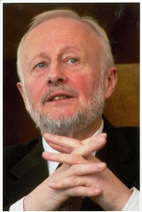 Ponka Prem合作论文数Jewish General Hospital, McGill University;Department of Physiology, Faculty of Medicine and Health Sciences, McGill University;Lady Davis Institute, McGill University5Bobst Cedric E合作论文数Dept Chem, Univ Massachusetts5
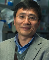 Hongzhe Sun (孙红哲)合作论文数Department of Chemistry, The University of Hong Kong;School of Chemistry, Sun Yat-sen University5
 Alex D Sheftel合作论文数McGill University5Sara K. Briggs合作论文数5T.E. Adams合作论文数4S.K. Briggs合作论文数4M.C. Cox合作论文数4Tb Kardos合作论文数4Yoon Dennis J合作论文数4Luck Ashley N合作论文数4
 Kerry Savage合作论文数Division of Medical Oncology, Faculty of Medicine, University of British Columbia4J.K. Grady合作论文数4
 Vinh Nguyen合作论文数Gippsland School of IT, Monash University4Dmitry R. Gumerov合作论文数4
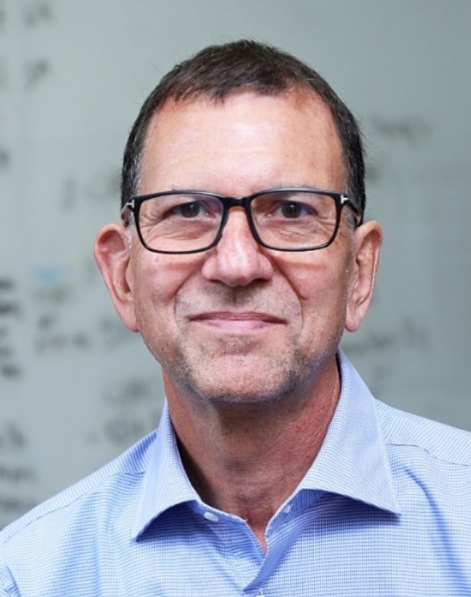 David S. Perlin合作论文数Hackensack Meridian Health Center for Discovery and Innovation;Department of Medical Sciences, Hackensack Meridian School of Medicine;Georgetown University School of Medicine;New Jersey Medical School, Rutgers, The State University of New Jersey4Emma J. Beatty合作论文数4
 Tariq Mahmood Roshan合作论文数School of Medical Sciences, University Sains Malaysia4Rwa Oliver合作论文数3G Kubal合作论文数3T.A. Frenkiel合作论文数3Jmb Smith合作论文数3Te Adams合作论文数3




