Positron emission tomography (PET) imaging has evolved over the last 20 years to a powerful tool for assessing vascular biology. The incidental uptake of PET tracers to the vessel wall provided the substrate for creating dedicated protocols and introducing novel agents for molecular imaging that targeted specific pathobiological mechanisms involved in the atherosclerotic process. Preliminary work with 18F-FDG underscored its efficacy to detect macrophage-rich inflammation, while, subsequent studies using 18F-NaF demonstrated its ability to detect active microcalcification and high-risk plaques. These encouraging results lead to the development of a new generation of tracers targeting macrophage receptors, chemokine signalling, angiogenesis, endothelial activation, and mitochondrial activity that have been validated against histology in animal models, endarterectomy, and autopsy tissue. This review provides an overview in the field describing the evolution of PET imaging in the study of atherosclerosis. It summarises the existing and emerging tracers, presents the biological processes that have been targeted by PET agents and collates the evidence from studies for vulnerable-plaque detection. Finally, it outlines technical hurdles and the current limitations of PET imaging that need to be addressed for clinical translation.
AIMS:Hybrid intravascular ultrasound-optical coherence tomography (IVUS-OCT) can enable more accurate plaque characterization than single-modality intravascular imaging, enhancing treatment planning and vulnerable plaque detection. However, image interpretation in IVUS-OCT is challenging and time-consuming. To overcome this limitation, we introduce a novel histology-trained deep learning (DL)-classifier for plaque component classification in IVUS-OCT images and compare its performance against single-modality DL and expert analysts. METHODS AND RESULTS:IVUS-OCT frames and matched histological sections from 10 cadaveric human hearts were included in this analysis. The histological data were used to define fibrotic, calcific, and necrotic core tissue regions of interest (ROIs) in IVUS-OCT and used to train three DL-classifiers for IVUS, OCT, or hybrid IVUS-OCT image analysis (992 frames) and test their performance (264 frames). The test set was additionally annotated by experts from three different core labs, and their estimations and those of the DL-classifiers were compared with histology.The IVUS-OCT DL-classifier had a superior performance to the IVUS-DL, OCT-DL, and the expert analysts in detecting plaque phenotypes (Kappa 0.60 vs. 0.19, 0.35, and 0.53, respectively) and accurately classified 68% of histologically defined fibroatheromas. The hybrid IVUS-OCT DL-classifier also had a better performance than single-modality DL-classifiers and the experts in assessing tissue types in ROIs annotated by histology (overall accuracy 86.7% compared with 73.2% for IVUS-DL, 66.6% for OCT-DL, and 70.6% for the experts). CONCLUSION:Plaque characterization using a histology-trained hybrid IVUS-OCT DL-classifier is feasible and enables more accurate detection of plaque components and phenotype classification than single-modality DL-classifiers and expert analysts.
AIMS:Left ventricular (LV) thrombus is a severe complication of acute myocardial infarction (AMI) and chronic heart failure. While current guidelines support the use of direct oral anticoagulants (DOACs) as alternatives to vitamin K antagonists (VKA), their benefit across different aetiologies remains uncertain. This study aimed to compare the efficacy and safety of DOAC vs. VKA across different aetiologies of LV dysfunction. METHODS AND RESULTS:We conducted a multi-centre observational study including 901 patients with confirmed LV thrombus treated with either a VKA or DOAC. The primary outcome was thrombus resolution, secondary outcomes included stroke and systemic embolization (SSE), major bleeding and mortality with analyses performed by aetiology. The principal aetiologies were AMI (38.3%), ischaemic cardiomyopathy (ICM) (38.0%) and non-ischaemic cardiomyopathy (NICM) (23.7%). Overall, thrombus resolution was significantly higher in DOAC treated patients, but this was driven by the AMI sub-group (P = 0.018). Direct oral anticoagulant use independently predicted thrombus resolution (OR 2.0, 95% Cl 1.29-3.24, P = 0.010). Major bleeding events (BARC ≥3) were more common with VKA use (P = 0.008). Non-ischaemic cardiomyopathy had the highest SSE rate (15.3%, P = 0.002), which were significantly raised in those treated with DOAC (P < 0.001). CONCLUSION:The underlying aetiology of LV dysfunction significantly influences both treatment response and outcomes in patients with LV thrombus. Direct oral anticoagulant were associated with superior efficacy and safety in AMI-related LV thrombus, but were linked to increased rates of SSE in NICM. These findings highlight the importance of aetiology on LV thrombus management and the potential need for tailored approaches.
BACKGROUND:The incorporation of side branches in vessel geometry influences wall shear stress (WSS) distribution. However, complete vessel reconstruction is time-consuming, and there is no evidence that its WSS estimations better predict atherosclerotic disease progression compared with the output of the conventional single-vessel reconstruction (SVR). METHODS:Patients who had baseline and 1-year follow-up intravascular ultrasound imaging (n=40 vessels), and patients with neoatherosclerotic lesions (n=13 vessels) on optical coherence tomography were included. All the studied vessels had at least one side branch with a diameter >1 mm; 3-dimensional complete vessel reconstruction and SVR were performed, and the time-averaged WSS and multidirectional WSS were computed. The performance of both methods in predicting disease progression in intravascular ultrasound and optical coherence tomography models was assessed. RESULTS:The incorporation of side branches in 3-dimensional geometry resulted in lower minimum predominant time-averaged WSS in the intravascular ultrasound (1.09 versus 1.58 Pa, P<0.001) and optical coherence tomography-based reconstructions (0.68 versus 1.33 Pa, P<0.001) and influenced the multidirectional WSS distribution. In native segments, complete vessel reconstruction-derived WSS metrics demonstrated superior predictive performance for disease progression-defined as lumen area reduction and plaque burden increase-compared with SVR, as evidenced by improved out-of-sample accuracy (leave-one-out information criterion: 429 versus 551), discrimination (C statistic: 0.725 versus 0.651), calibration (Brier score: 0.172 versus 0.226), and explained variance (27.8% versus 20.7%). Consistent findings were observed in stented segments, where complete vessel reconstruction-derived WSS metrics more accurately predicted neointimal proliferation than SVR-derived metrics. CONCLUSIONS:Incorporating side branches into vessel reconstruction influences WSS distribution and enables more accurate prediction of atherosclerotic disease progression in native and stented segments than SVR.
Cardiogenic shock is a life-threatening condition caused by the heart’s sudden inability to pump sufficient blood to maintain adequate tissue perfusion, most commonly occurring following a myocardial infarction or acute decompensated heart failure. The resultant hypoperfusion can quickly progress to end-organ failure and ultimately death if not treated urgently. This review explores the management of cardiogenic shock, highlighting current treatments, their effectiveness, and the challenges faced by healthcare providers. It looks at both pharmacological therapies and devices used for cardiac support, including mechanical circulatory support and emergency revascularisation procedures to restore blood flow. We also examine how different stages of shock affect survival and how new technologies including artificial intelligence and wearable monitors could help detect and treat this condition earlier. In addition, this review discusses the significant pressure that cardiogenic shock places on healthcare provision, including the typical financial cost of treatment in the UK, resource utilisation and regional disparities. Finally, we outline future directions for trial design, better prevention, more rapid diagnosis and improved treatments that could improve morbidity and mortality.
As transcatheter aortic valve implantation (TAVI) expands to younger populations, durability has become a concern, requiring a lifetime rather than a single-procedure perspective. While clinical trials suggest comparable mid-term performance to surgical bioprostheses, data beyond 10 years remain limited, particularly for bicuspid valves, valve-in-valve procedures, and complex anatomies. Computational modelling combines patient anatomy and device design in computer-based simulations to study valve performance under physiological loading. Applied to TAVI, these models can reproduce implantation, evaluate mechanical stresses, and simulate blood flow, providing mechanistic insights into deterioration processes, including altered leaflet loading, stent deformation, and thrombosis-prone flow. Although these simulations do not directly assess durability, they use surrogate metrics linked with these mechanisms, helping identify factors that may influence longevity and guide design and procedural refinements. Clinically, modelling could support patient-specific planning and reintervention strategies, informing decisions across the valve-replacement pathway, an important consideration as younger patients are likely to undergo multiple lifetime procedures. Integrating these tools into pre-procedural planning may help anticipate challenges such as coronary access, annular geometry, and redo feasibility. However, current studies report elements of verification and field-level validation, but none complete a pre-specified, calibrated surrogate-to-outcome validation with uncertainty/sensitivity analysis; thus, durability predictions remain exploratory. Progress needs transparent verification, field checks vs. bench or imaging, surrogate calibration to data, outcome testing in independent cohorts, and routine uncertainty/sensitivity reporting, with close clinician-engineer collaboration. This review underscores the need for a multidisciplinary approach and provides a critical analysis of the available tools and their potential to advance long-term outcomes.
Cardiovascular biomarkers are central to the diagnosis and management of a range of acute and chronic disease states, yet current approaches remain dependent on episodic venous blood sampling. Dermal interstitial fluid (ISF), a dynamic and accessible biofluid, offers a promising alternative for continuous and minimally invasive biomarker monitoring. In this Review, we explore the emerging convergence of ISF biosensing technologies with cardiovascular diagnostics. We discuss the physiological basis of ISF sampling and highlight engineering advances in microneedle arrays, hydrogel implants and electrokinetic extraction methods. We examine state-of-the-art biosensing platforms—ranging from enzymatic electrochemical sensors to aptamer- and antibody-based systems—and evaluate their applicability to key cardiovascular biomarkers, including troponin, natriuretic peptides, CRP and microRNAs. Although early studies demonstrate proof-of-concept for ISF-based detection of several cardiac biomarkers, clinical translation remains limited. While still in proof of concept stage, continuous ISF monitoring could disrupt traditional models of care by enabling earlier detection, personalised risk stratification and remote disease management. However, it also presents challenges related to clinical workflow integration, data interpretation and regulatory oversight. Bridging these gaps will require multidisciplinary innovation across materials science, device engineering and clinical validation. By transitioning from episodic blood sampling to continuous dermal monitoring, this technology promises to redefine cardiovascular care; shifting the paradigm from reactive acute management to proactive, personalised, and predictive precision medicine.
AbstractPatients who have previously undergone coronary artery bypass grafting (CABG) may present with non-ST-elevation acute coronary syndromes (NSTE-ACSs). In this setting, guidelines recommend routine invasive angiography. However, patients with CABG were often excluded from key trials that informed contemporary guidelines and invasive angiography is known to be of higher risk in these patients. In this article, the authors review the evidence and propose a trial to address the question, "Should patients with prior CABG who present with NSTE-ACSs routinely undergo invasive angiography?"
Thrombolysis in myocardial infarction frame count enables assessment of coronary flow but cannot measure coronary flow velocity (CFV), which is needed to examine microvascular function. To overcome this limitation, we introduce a semi-automated software for fast CFV computation using contrast bolus tracking techniques in angiography and compare its performance against experts. The study included patients undergoing coronary angiography. Two experts measured the CFV using the number of frames, segment length, and frame rate. Measurements were repeated for shorter segments and different projections, and their estimations were compared with the software. In total, 123 patients (152 vessels) were included. The software had excellent reproducibility in measuring CFV (intraclass correlation coefficient (ICC) = .995), which was superior to experts (ICC = .946) and provided similar estimations irrespective of the segment length (ICC = .992); conversely, the experts overestimated CFV in short segments. The reproducibility of the experts and the software was moderate when comparing CFV measurements in different projections (first expert vs software ICC = .807, second expert vs software ICC = .790, first expert vs second expert ICC = .885). The software provides reproducible CFV estimations that are close to experts’ estimations. Further validation against wire-based functional techniques is needed to examine its potential in assessing microvascular function.
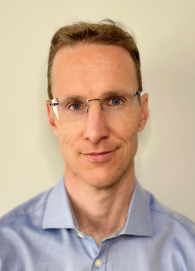 Dan Jones合作论文数The William Harvey Research Institute, Faculty of Medicine and Dentistry, Queen Mary University of London138
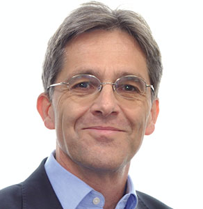 Andreas Baumbach合作论文数The William Harvey Research Institute, Faculty of Medicine and Dentistry, Queen Mary University of London108
 Rathod Krishnaraj S合作论文数Barts & London NHS Trust106
 Andrew Wragg合作论文数Department of Cardiology, Barts Health National Health Service (NHS) Trust96
 C. Bourantas合作论文数73
 Ajay Jain合作论文数Department of Chemistry, University of Roorkee72
 Charles Knight合作论文数71
 Ryo Torii合作论文数Department of Mechanical Engineering, University College London48
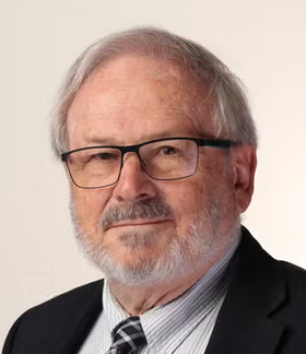 Patrick Serruys合作论文数CORRIB Research Centre for Advanced Imaging and Core Laboratory, University of Galway;National University of Ireland41Anantharaman Ramasamy合作论文数37
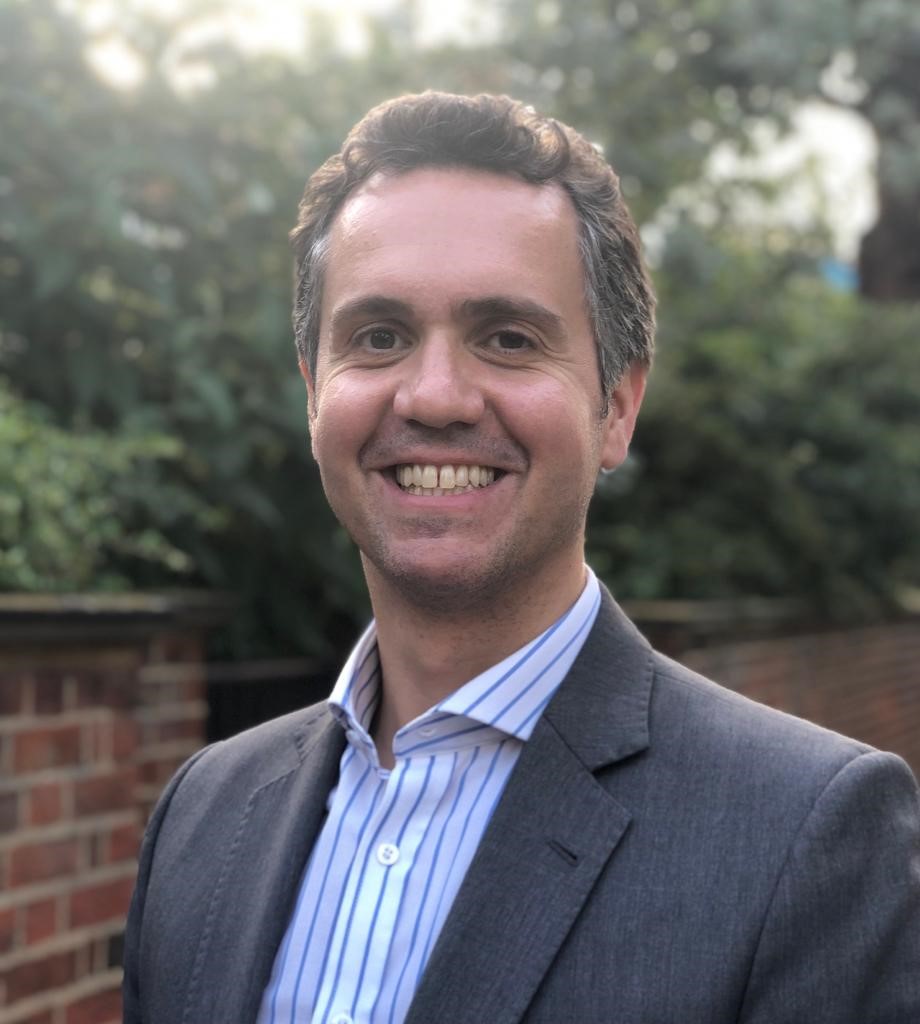 Oliver Guttmann合作论文数Barts Heart Centre, St Bartholomew's Hospital33Tom Crake合作论文数Department of Cardiology, St Bartholomew 's Hospital30Choudry Fizzah Aziz合作论文数29
 Roby Rakhit合作论文数Institute of Cardiovascular Science, University College London;CardioVascular Division, The Royal Free Hospital;The Wellington Hospital29
 James Moon合作论文数Institute of Cardiovascular Science, University College London;Mycardium AI Ltd;Chenies Mews Imaging Centre29Mick Ozkor合作论文数28
 Francesca Pugliese合作论文数28Mark Westwood合作论文数London Chest Hospital28
 John Martin合作论文数Division of Medicine, University College London;Yale School of Medicine, Yale University27R. Andrew Archbold合作论文数26
 Lewis Ceri Davies合作论文数26
 Gallagher Sean合作论文数25Simon Redwood合作论文数School of Cardiovascular Medicine & Sciences, Faculty of Life Sciences & Medicine, King’s College London25
 Jouke Dijkstra合作论文数Leiden University Medical Center24
 Amrita Ahluwalia合作论文数The William Harvey Research Institute, Faculty of Medicine and Dentistry, Queen Mary University of London22
 Elliot J Smith合作论文数Department of Cardiology, London Chest Hospital22
 Retesh Bajaj合作论文数Department of Cardiology, Barts Heart Centre21Onuma Yoshinobu合作论文数21Pitt O. Lim合作论文数21Roshan Weerackody合作论文数21
 Philip A MacCarthy合作论文数21Iqbal S Malik合作论文数20Hamshere Stephen合作论文数20Andiapen Mervyn合作论文数19
 Miles Dalby合作论文数Imperial College London;Guy's and St Thomas' NHS Foundation Trust19Simon Kennon合作论文数19Sundeep Kalra合作论文数18Kush Patel合作论文数Barts Heart Ctr18Sam Firoozi合作论文数18
 Saidi (Sam) Mohiddin合作论文数Barts Heart Centre18
 Pieter Kitslaar合作论文数Division of Image Processing, Department of Radiology, Leiden University Medical Center18Mark Whitbread合作论文数18
 Matthew Kelham合作论文数17
 Lorenz Raber合作论文数Catheterization Laboratory, Bern University Hospital;Swiss Cardiovascular Center17Didier Locca合作论文数17Rajiv Amersey合作论文数17Anne-Marie Beirne合作论文数16
 Adam Timmis合作论文数The William Harvey Research Institute, Faculty of Medicine and Dentistry, Queen Mary University of London;Bart's Heart Centre, Barts Health NHS Trust16Martin T. Rothman合作论文数16




