In the automotive industry, many car parts are being made out of plastic, synthetic, or fiber-reinforced materials. A systematic replacement of conventional metallic components is underway, mostly to save weight or reduce costs. The durability of these new constructions depends highly on the manufacturing process; hence the requirements are increasing on quality control in production lines. A typical example is car bumpers. Modern variations of car bumpers are much equipped with many sensors and connections, where a high precision in their mounting tolerances is essential. In production lines, visual inspection combined with functional testing is the widespread method for examination. Errors due to human factors are usually high. In addition, visual inspection is time consuming, which reflects on the production cost. An efficient replacement for the visual inspection hasn’t been yet available. In this paper the application of millimeter-wave imaging to the examination of car bumpers and automobile parts is presented. The mm-wave signals can penetrate the outer surface and deliver an image of the inside structure of the bumper body. Wiring, sensors, screws, tubes, and other components can be separated in the 3D image to examine their location and orientation. Material damages can be identified and localized. The mm-waves are safe to use, which is of a great importance for utilization in open environments, like production lines. Additionally, the systems do not need regular maintenance, certified professionals or specific regulatory permissions as with conventional CT systems. Moreover, modern image processing and computer vision techniques can greatly assist the analysis of the 3D image data by means of image registration, segmentation, features extraction and machine learning techniques in order to automatically search for flaws, parts and components in mm-wave imaging of automotive parts, e.g. bumpers. Thus the human effort to interpret the 3D information is largely avoided. This contribution demonstrates the capabilities of such imaging solutions with measurements of actual car bumpers with a real-time mm-wave imaging system operating at 75 GHz delivering a 2 millimeter of spatial resolution. Various image results are hence presented and discussed. More info about this article: http://ndt.net/?id=19394
The transfer of preoperative CT data into the tracking system coordinates within an operating room is of high interest for computer-aided orthopedic surgery. In this work, we introduce a solution for intra-operative ultrasound–CT registration of bones.
We propose a novel, physics-based method for detecting multi-scale tubular features in ultrasound images. The detector is based on a Hessian-matrix eigenvalue method, but unlike previous work, our detector is guided by an optimal model of vessel-like structures with respect to the ultrasound-image formation process. Our method provides a voxel-wise probability map, along with estimates of the radii and orientations of the detected tubes. These results can then be used for further processing, including segmentation and enhanced volume visualization. Most Hessian-based algorithms, including the well-known Frangi filter, were developed for CTA or MRA; they implicitly assume symmetry about the vessel centerline. This is not consistent with ultrasound data. We overcome this limitation by introducing a novel filter that allows multi-scale estimation both with respect to the vessel's centerline and with respect to the vessel's border. We use manually-segmented ultrasound imagery from 35 patients to show that our method is superior to standard Hessian-based methods. We evaluate the performance of the proposed methods based on the sensitivity and specificity like measures, and finally demonstrate further applicability of our method to vascular ultrasound images of the carotid artery, as well as ultrasound data for abdominal aortic aneurysms.
We present a new system to acquire and reconstruct 3D freehand ultrasound volumes from arbitrary 2D image acquisitions over time. Motion artifacts are significantly reduced with a novel gating approach which correlates pulse oximetry data with Doppler ultrasound. The reconstruction problem is split into a ray-based sample selection on a per-scanline basis and a backward algorithm which is based on the concept of normalized convolution. We introduce an adaptive derivation of time-domain interpolation from the correlated pulse-oximetry and Doppler signals as well as an ellipsoid kernel size for spatial interpolation based on the physical resolution of the ultrasound data. We compare pulse-oximetry to classical ECG gating and further show the suitability of our normalized pulse signal for 3D+T reconstructions. The ease of use of the setup without the need of uncomfortable triggering via ECG provides the ability to use 3D+T ultrasound in every day clinical practice.
Intravascular Ultrasound (IVUS) is a predominant imaging modality in interventional cardiology. It provides real-time cross-sectional images of arteries and assists clinicians to infer about atherosclerotic plaques composition. These plaques are heterogeneous in nature and constitute fibrous tissue, lipid deposits and calcifications. Each of these tissues backscatter ultrasonic pulses and are associated with a characteristic intensity in B-mode IVUS image. However, clinicians are challenged when colocated heterogeneous tissue backscatter mixed signals appearing as non-unique intensity patterns in B-mode IVUS image. Tissue characterization algorithms have been developed to assist clinicians to identify such heterogeneous tissues and assess plaque vulnerability. In this paper, we propose a novel technique coined as Stochastic Driven Histology (SDH) that is able to provide information about co-located heterogeneous tissues. It employs learning of tissue specific ultrasonic backscattering statistical physics and signal confidence primal from labeled data for predicting heterogeneous tissue composition in plaques. We employ a random forest for the purpose of learning such a primal using sparsely labeled and noisy samples. In clinical deployment, the posterior prediction of different lesions constituting the plaque is estimated. Folded cross-validation experiments have been performed with 53 plaques indicating high concurrence with traditional tissue histology. On the wider horizon, this framework enables learning of tissue-energy interaction statistical physics and can be leveraged for promising clinical applications requiring tissue characterization beyond the application demonstrated in this paper.
Breast cancer is the most common form of cancer in women. Early diagnosis can significantly improve lifeexpectancy and allow different treatment options. Clinicians favor 2D ultrasonography for breast tissue abnormality screening due to high sensitivity and specificity compared to competing technologies. However, inter- and intra-observer variability in visual assessment and reporting of lesions often handicaps its performance. Existing Computer Assisted Diagnosis (CAD) systems though being able to detect solid lesions are often restricted in performance. These restrictions are inability to (1) detect lesion of multiple sizes and shapes, and (2) differentiate between hypo-echoic lesions from their posterior acoustic shadowing. In this work we present a completely automatic system for detection and segmentation of breast lesions in 2D ultrasound images. We employ random forests for learning of tissue specific primal to discriminate breast lesions from surrounding normal tissues. This enables it to detect lesions of multiple shapes and sizes, as well as discriminate between hypo-echoic lesion from associated posterior acoustic shadowing. The primal comprises of (i) multiscale estimated ultrasonic statistical physics and (ii) scale-space characteristics. The random forest learns lesion vs. background primal from a database of 2D ultrasound images with labeled lesions. For segmentation, the posterior probabilities of lesion pixels estimated by the learnt random forest are hard thresholded to provide a random walks segmentation stage with starting seeds. Our method achieves detection with 99.19% accuracy and segmentation with mean contour-to-contour error < 3 pixels on a set of 40 images with 49 lesions.
Medical ultrasonic grayscale images are formed from acoustic waves following their interactions with distributed scatterers within tissues media. For accurate simulation of acoustic wave propagation, a reliable model describing unknown parameters associated with tissues scatterers such as distribution, size and acoustic properties is essential. In this work, we introduce a novel approach defining ultrasonic scatterers by incorporating a distribution of cellular nuclei patterns in biological tissues to simulate ultrasonic response of atherosclerotic tissues in intravascular ultrasound (IVUS). For this reason, a virtual phantom is generated through manual labeling of different tissue types (fibrotic, lipidic and calcified) on histology sections. Acoustic properties of each tissue type are defined by assuming that the ultrasound signal is primarily backscattered by the nuclei of the organic cells within the intima and media of the vessel wall. This resulting virtual phantom is subsequently used to simulate ultrasonic wave propagation through the tissue medium computed using finite difference estimation. Subsequently B-mode images for a specific histological section are processed from the simulated radiofrequency (RF) data and compared with the original IVUS of the same tissue section. Real IVUS RF signals for these histological sections were obtained using a single-element mechanically rotating 40MHz transducer. Evaluation is performed by trained reviewers subjectively assessing both simulated and real B-mode IVUS images. Our simulation platform provides a high image quality with a very promising correlation to the original IVUS images. This will facilitate to better understand progression of such a chronic disease from micro-level and its integration into cardiovascular disease-specific models.
Coronary artery disease leads to failure of coronary circulation secondary to accumulation of atherosclerotic plaques. In adjunction to primary imaging of such vascular plaques using coronary angiography or alternatively magnetic resonance imaging, intravascular ultrasound (IVUS) is used predominantly for diagnosis and reporting of their vulnerability. In addition to plaque burden estimation, necrosis detection is an important aspect in reporting of IVUS. Since necrotic regions generally appear as hypoechic, with speckle appearance in these regions resembling true shadows or severe signal dropout regions, it contributes to variability in diagnosis. This dilemma in clinical assessment of necrosis imaged with IVUS is addressed in this work. In our approach, fidelity of the backscattered ultrasonic signal received by the imaging transducer is initially estimated. This is followed by identification of true necrosis using statistical physics of ultrasonic backscattering. A random forest machine learning framework is used for the purpose of learning the parameter space defining ultrasonic backscattering distributions related to necrotic regions and discriminating it from non-necrotic shadows. Evidence of hunting down true necrosis in shadows of intravascular ultrasound is presented with ex vivo experiments along with cross-validation using ground truth obtained from histology. Nevertheless, in some rare cases necrosis is marginally over-estimated, primarily on account of non-reliable statistics estimation. This limitation is due to sparse spatial sampling between neighboring scan-lines at location far from the transducer. We suggest considering the geometrical location of detected necrosis together with estimated signal confidence during clinical decision making in view of such limitation.
Advances in ultrasound system development have led to a substantial improvement of image quality. Nevertheless, ultrasound attenuation and shadowing artifacts cannot be entirely avoided and continue to challenge medical image computing algorithms. In this thesis a confidence measure is introduced that emphasizes uncertainty in attenuated and/or shadow regions in ultrasound images. The measure was introduced into various ultrasound image processing applications including: 3D freehand reconstruction, shadow detection, mono- and multimodal registration, tissue classification, and bone detection.
The introduction of 3D + t medical datasets is posing new challenges to visualization. Volumetric and flow visualization are established fields offering a wide spectrum of techniques for visualizing 3D + t datasets. In this work we address the problem of visualizing the motion of organs during breathing. As opposed to flow visualization we are not directly interested in the underlying flow, but in the deformation caused by the flow. Therefore, visualization of breathing motion focuses on emphasizing the organ motion while preserving the anatomical context provided by the volumetric visualization. In this work we will discuss methods from flow and volume visualization, their applications, and introduce alternative visualization approaches for enhancing the perception of organ motion due to breathing.
Advances in ultrasound system development have led to a substantial improvement of image quality and to an increased use of ultrasound in clinical practice. Nevertheless, ultrasound attenuation and shadowing artifacts cannot be entirely avoided and continue to challenge medical image computing algorithms. We introduce a method for estimating a per-pixel confidence in the information depicted by ultrasound images, referred to as an ultrasound confidence map, which emphasizes uncertainty in attenuated and/or shadow regions. Our main novelty is the modeling of the confidence estimation problem within a random walks framework by taking into account ultrasound specific constraints. The solution to the random walks equilibrium problem is global and takes the entire image content into account. As a result, our method is applicable to a variety of ultrasound image acquisition setups. We demonstrate the applicability of our confidence maps for ultrasound shadow detection, 3D freehand ultrasound reconstruction, and multi-modal image registration.
Registration of ultrasound images is often complicated due to inherent noise. Robust similarity metrics and optimization procedures are required to facilitate medical applicability. In this paper a novel hybrid procedure, incorporating global statistics and local textural features, is proposed for the registration of envelope detected radio frequency ultrasound data. On the global scale this is achieved by Hellinger distance between distribution in images, and on the local scale by a statistics-based extension of Fuzzy Local Binary Patterns (FLBP). The proposed procedure is shown to outperform standard measures such as SSD and NCC, as well as Hellinger distance and histogram matching of standard FLBPs, in rigid registration experiments of envelope detected radio frequency data samples of the human neck.
Intravascular ultrasound (IVUS) is the most favorable imaging modality that often used in coronary artery catheterization procedures and provides cross-sectional images of arterial wall structures and extend of atherosclerosis. Although several techniques have been developed to classify atherosclerotic tissues, deploying IVUS radiofrequency (RF) backscattered signals and/or grayscale images their clinical applications have seen limited success. In this paper, we propose a unified methodological framework from data collection, histology preparation, registration, feature extraction, and classification to achieve a reliable in vitro trained tissue characterization classifier for in vivo applications. Finally, the results from proposed algorithm is compared with state of the art virtual histology (VH) technique.
Registration of pre-operative CT datasets to intra-operative 3D freehand ultrasound has been of high interest for computer assisted orthopedic surgery. Feature-based registration relies on an accurate detection of the bone surface in the B-mode ultrasound images. In this work we present a fully automatic bone detection approach for US. The pre-operative CT is utilized to create a patient-specific bone model for our joint detection-registration framework. The model provides a geometric constraint for accurate and robust detection. Simultaneously to the detection, our method yields a close estimate of the rigid transformation from US to CT, which can be used as an initialization for further refinement through sophisticated intensity-/feature-based registration methods. We evaluated our approach on datasets of the human femur acquired in a cadaver study and demonstrate a mean bone detection error of below 0.4mm.
In this paper, for the first time, we present a systematic framework to register intravascular ultrasound (IVUS) images with histology correspondences. We deployed intermediate representations of images, generating segmentation masks corresponding to lumen and media-adventitia borders for both histology and IVUS images, incorporated into a non-rigid registration framework using discrete multi-labeling and approximate curvature penalty for smoothness regularization. The resulting deformation field was then applied to the original histology image to transfer it to IVUS coordinate system. Finally, the results were quantified on 14 cross sections of interest. The main contribution of this work is that the registered results could be used for systematic labeling of tissues, which ultimately will lead to reliable construction of training dataset for feature extraction and supervised classification of atherosclerotic tissues.
The shadow regions in ultrasound (US) B-mode images are due to severe reflection of backscattered signals from dense tissue/medium interface. The loss of ultrasound energy as well as lack of textural or spectral features in these regions raise uncertainty, resulting confusion among experts decisions and developed computer-aided diagnosis (CAD) algorithms outcomes. In this paper, we present a novel uncertainty (confidence) estimation method, modeling the problem through random walk under particular constrains motivated by underlying physics of ultrasound. We demonstrate that constructed confidence maps can then be employed in different ultrasound based CAD algorithms, which ultimately improve experts qualitative and quantitative assessments. We evaluate our method on intravascular ultrasound (IVUS) radiofrequency (RF) data and quantify the results through non-linearly registered histology image as ground-truth.
Intravascular ultrasound (IVUS) is the predominant imaging modality in the field of interventional cardiology that provides real-time cross-sectional images of coronary arteries and the extent of atherosclerosis. Due to heterogeneity of lesions and stringent spatial/spectral behavior of tissues, atherosclerotic plaque characterization has always been a challenge and still is an open problem. In this paper, we present a systematic framework from in vitro data collection, histology preparation, IVUS-histology registration along with matching procedure, and finally a robust texture-derived unsupervised atherosclerotic plaque labeling. We have performed our algorithm on in vitro and in vivo images acquired with single-element 40 MHz and 64-elements phased array 20 MHz transducers, respectively. In former case, we have quantified results by local contrasting of constructed tissue colormaps with corresponding histology images employing an independent expert and in the latter case, virtual histology images have been utilized for comparison. We tackle one of the main challenges in the field that is the reliability of tissues behind arc of calcified plaques and validate the results through a novel random walks framework by incorporating underlying physics of ultrasound imaging. We conclude that proposed framework is a formidable approach for retrieving imperative information regarding tissues and building a reliable training dataset for supervised classification and its extension for in vivo applications.
In this paper, a new segmentation framework with prior knowledge is proposed and applied to the left ventricles in cardiac Cine MRI sequences. We introduce a new formulation of the random walks method, coined as guided random walks, in which prior knowledge is integrated seamlessly. In comparison with existing approaches that incorporate statistical shape models, our method does not extract any principal model of the shape or appearance of the left ventricle. Instead, segmentation is accompanied by retrieving the closest subject in the database that guides the segmentation the best. Using this techniques, rare cases can also effectively exploit prior knowledge from few samples in training set. These cases are usually disregarded in statistical shape models as they are outnumbered by frequent cases (effect of class population). In the worst-case scenario, if there is no matching case in the database to guide the segmentation, performance of the proposed method reaches to the conventional random walks, which is shown to be accurate if sufficient number of seeds is provided. There is a fast solution to the proposed guided random walks by using sparse linear matrix operations and the whole framework can be seamlessly implemented in a parallel architecture. The method has been validated on a comprehensive clinical dataset of 3D+t short axis MR images of 104 subjects from 5 categories (normal, dilated left ventricle, ventricular hypertrophy, recent myocardial infarction, and heart failure). The average segmentation errors were found to be 1.54mm for the endocardium and 1.48mm for the epicardium. The method was validated by measuring different algorithmic and physiologic indices and quantified with manual segmentation ground truths, provided by a cardiologist.
 Nassir Navab合作论文数School of Computation, Information and Technology, Technische Universitat Munchen;Department of Computer Science, Technical University of Munich22
 Amin Katouzian合作论文数Vayo Inc.;GuideAI Health9
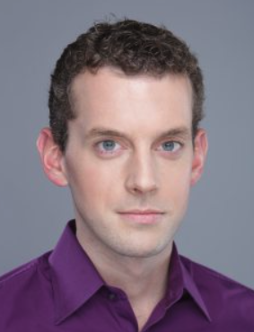 Wolfgang Wein合作论文数School of Computation, Information and Technology, Technische Universität München;ImFusion GmbH7
 Debdoot Sheet合作论文数Indian Institute of Technology Kharagpur5
 Abouzar Eslami合作论文数ZEISS Medical Technology5
 Stéphane G. Carlier合作论文数Cardiovascular Research Foundation, Columbia University Medical Center5
 Peter B. Noel合作论文数Department of Radiology, Perelman School of Medicine, University of Pennsylvania4
 Ajoy Kumar Ray合作论文数National Metallurgical Laboratory3
 Jyotir Moy Chatterjee合作论文数Medical College and Hospital Radiology Department 88 College Street 700 012 Calcutta India3
 Andrew Laine合作论文数The Fu Foundation School of Engineering and Applied Science, Columbia University;Center for Health Analytics, Data Science Institute, Columbia University3
 Seyed-Ahmad Ahmadi合作论文数NVIDIA3
 Tassilo Klein合作论文数SAP AI Research;MESASIGHT GmbH3A Katouzian合作论文数2Silvan Kraft合作论文数2
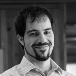 Christoph Hennersperger合作论文数Computer Aided Medical Procedures, Technical University of Munich2Michael Gkatzogias合作论文数2Konstantinos Pyrinis合作论文数2
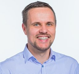 Oliver Kutter合作论文数Siemens Corporate Research, Princeton, NJ2
 Maximilian Baust合作论文数Computer Aided Medical Procedures, Technische Universitat Munchen2
 Dionysios Politis合作论文数2Koeppel, T.合作论文数1Beitzel Julian合作论文数1Drecoll Enken合作论文数1Andreas Koenig合作论文数1
 Renu Virmani合作论文数CVPath Institute Inc.1
 Mattias Hansson合作论文数School of Technology, Malmö University1Jennifer Lisauskas合作论文数1
 Elisa E. Konofagou合作论文数Department of Biomedical Engineering, School of Engineering and Applied Science, Columbia University1Masataka Nakano合作论文数CVPath Institute, Inc.1Babak Baseri合作论文数1
 Kai Boetzel合作论文数1
 Sherif Sayed Ahmed合作论文数1
 Tibor Vag合作论文数1Ernst J. Rummeny合作论文数1Annika Plate合作论文数1Paulo Waelkens合作论文数1
 Anup Sadhu合作论文数Department of Radiology, Medical College1Andreas Konig合作论文数1




