Unopposed denosumab (Dmab) discontinuation is followed by overshoot in bone turnover markers (BTM), rapid bone mineral density (BMD) loss, and elevated risk of multiple vertebral fractures. To preserve BMD gains and mitigate fracture risk it is currently recommended to administer bisphosphonates following Dmab discontinuation. We prospectively evaluate the efficacy of alendronate (ALN) in preventing BTM overshoot and BMD loss in women with postmenopausal osteoporosis (PMO) discontinuing Dmab. This is a 12-month prospective, observational study among patients treated with Dmab 60 mg every 6 months and achieved osteopenia at the lumbar spine (LS) and femoral neck (FN). Six months after the last Dmab injection participants received oral ALN 70 mg weekly for 6 months followed by 6 months without therapy (Group 1) or for 12 months (Group 2). The primary endpoint was the percent change in LS-BMD from baseline to month 12. Eighty women (39 Group 1, 41 Group 2) who had received 2-22 Dmab injections (mean 9.8) completed the study. Median age was 67 years; 27.5% had prevalent fractures and 59% had prior anti-osteoporotic therapy. At month 12, LS-BMD declined significantly in the overall cohort [-5.9% (8.2%), p < 0.001], with no difference between groups (Group 1: -6.8% vs Group 2: -5.0%, p = 0.50). FN-BMD decreased (-3.5%, p < 0.001), and similarly in both groups. Serum procollagen type 1 N-terminal propeptide (P1NP), C-terminal telopeptide of type 1 collagen (CTX), and tartrate-resistant acid phosphatase isoform 5b increased significantly; P1NP and CTX were significantly higher in Group 1 at month 12. One patient sustained multiple vertebral fractures (1.25%). In conclusion, alendronate did not consistently attenuate BMD loss and BTM rebound after denosumab discontinuation in women with PMO, with 6 months being as effective as 12 months of treatment. Importantly, both regimens were associated with low incidence of multiple vertebral fractures.
OBJECTIVE:To prospectively evaluate the association between vitamin D optimization and quality of life (QoL) and bone metabolism in adult patients with Langerhans cell histiocytosis (LCH). DESIGN:and Methods: In this prospective, uncontrolled, observational study, 45 adult LCH patients received individualized cholecalciferol supplementation for 3 months to achieve serum 25-hydroxyvitamin D (25(OH)D) levels ≥30 ng/mL. Skeletal-related and general QoL were assessed using the QUALEFFO-41 and SF-36 questionnaires, respectively. Biochemical markers of calcium metabolism and bone turnover were measured at baseline and follow-up. Correlation and exploratory linear regression analyses evaluated associations between changes in parameters. RESULTS:25(OH)D levels increased significantly (22.8 ± 10.3 to 34.3 ± 8.2 ng/mL, p < 0.0005), followed by a reduction in parathyroid hormone (PTH) (55.7 ± 21.1 to 49.0 ± 15.8 pg/mL, p = 0.024). Improvements were observed in QUALEFFO-41 subdomains (household activities, mental condition) and SF-36 domains (bodily pain, emotional role limitations). C-terminal telopeptide (CTX) levels decreased (p = 0.006), consistent with reduced bone resorption. Changes in 25(OH)D were inversely correlated with changes in PTH (r = -0.35, p = 0.024), and changes in PTH were correlated with changes in CTX (r = -0.41, p = 0.007). No correlation was found between changes in 25(OH)D and CTX. CONCLUSIONS:In this uncontrolled cohort, vitamin D optimization was associated with improvements in selected QoL domains and favourable changes in calcium metabolism and bone resorption markers, supporting routine assessment and correction of vitamin D deficiency in this population.
Denosumab (Dmab) discontinuation in osteoporosis leads to overshoot (rebound) of bone turnover, but its cause remains largely unclear. In a prospective trial, Dmab-treated Langerhans cell histiocytosis (LCH) patients showed no overshoot of bone turnover markers despite high short-term dosing. Findings suggest that total Dmab dose does not drive the overshoot of bone turnover markers. In patients with osteoporosis, the length of treatment with denosumab (Dmab) is an important risk factor for the overshoot (rebound) of bone turnover markers following its discontinuation. Whether this is due to the higher total Dmab dose given and/or the severity of the disease is unknown. To address this question, long-term follow-up of changes in bone metabolism after stopping Dmab with doses higher than those used in osteoporosis is essential. In a prospective, single-arm, open label, phase 2b clinical trial, ten adult patients with Langerhans cell histiocytosis (LCH), eight with bone lesions (four single, four multiple) and two without, were treated with Dmab sc injections 120 mg/2 months for 6 months (total 480 mg) and were followed for 24 months after the last injection. Treatment reduced bone turnover markers to about 10
High-dose intravenous steroid treatment (HDIST) represents the first choice of treatment for multiple sclerosis (MS) relapses. Chronic oral glucocorticoid (GC) administration correlates with bone loss whereas data regarding HDIST in MS are still conflicting. Twenty-five newly diagnosed MS patients (NDMSP) (median age: 37 years) were prospectively studied for the effects of HDIST on bone mineral density (BMD) and bone metabolism. Patients received 1000 mg methylprednisolone intravenously every day for 5 days followed by oral prednisolone tapering over 21 days. Bone metabolism indices were determined prior to GC, on days 2, 4, 6, and 90, and at months 6, 12, 18, and 24 post GC therapy. Femoral, lumbar-spine BMD, and whole-body measurement of adipose/lean tissue were assessed prior to GC-administration and then every six months. Ten patients completed the study. N-terminal-propeptide-procollagen-type-1 and bone-specific alkaline phosphatase showed a significant increase at day-90 (p < 0.05). A transient non-significant fall of BMD was observed at 6 months after GC-administration, which subsequently appeared to be restored. We conclude that HDIST seems not to have long-term negative effects on BMD, while the observed transient increase of bone formation markers probably indicates a high bone turnover phase to GC-administration. Additional prospective studies with larger sample size are needed.
To test the hypothesis that during treatment with denosumab osteomorphs and precursors recycle to higher number of osteoclasts with time, we measured TRAcP5b in serum taken 6 months after the last injection in postmenopausal women treated for 1–10 years. Serum TRAcP5b values were not related to time of exposure to denosumab. In women with postmenopausal osteoporosis the aetiology of the observed inverse relationship between duration of denosumab (Dmab) therapy and bone loss after its discontinuation is currently unknown. In studies in mice inhibition of RANKL is associated with an increase in osteomorphs and osteoclast precursors that recycle into osteoclasts and may accumulate with time. We hypothesized that longer inhibition of RANKL by Dmab will be followed by the synchronous formation of a larger number of osteoclasts after stopping treatment. To test this hypothesis, we measured serum TRAcP5b, a marker of osteoclast numbers, in postmenopausal women treated with Dmab for different periods of time up to 10 years. TRAcP5b, C-terminal telopeptide of type 1 collagen (CTX) and procollagen type 1 N-terminal propeptide (P1NP) were measured at 6.0 months ± 15 days after last Dmab injection in 59 women who had received Dmab for 4.0 ± 2.3 years (range 1–10 years). Of these, 38 were treatment naïve (group 1) and 21 had received other treatments prior Dmab (group 2). Duration of Dmab treatment was not related to serum TRAcP5b values or to TRAcP5b/CTX ratio either in the whole cohort or in each of the two groups separately. In contrast, serum TRAcP5b values were significantly correlated with serum CTX values (rs = 0.619; p < 0.001), but not with serum P1NP values or BMD at all skeletal sites. Our observations indicate that serum TRAcP5b, measured at 6 months after a Dmab injection, is not a useful early marker for time-dependent increased accumulation of osteoclasts in humans and for identification of patients at risk for a higher rebound increase in bone resorption.
Introduction: GnRH-analogs induce bone loss. We aimed to investigate the effects of goserelin-induced menstrual cessation (MC) and subsequent menstrual restoration (MR) on bone metabolism (BM). Methods: In this prospective cohort study, premenopausal women (PMW) with histologically verified endometriosis (n = 21) received goserelin monthly for 6 months (6 m) resulting in MC and were followed up for another 6 m after MR (12 m). Age- and BMI-matched healthy PMW (n = 20) served as controls for bone mineral density (BMD) measurements. The primary endpoint was changes in lumbar spine (LS)-BMD at 6 m and 12 m; Secondary endpoints were changes in femoral neck (FN)-BMD, bone turnover markers (P1NP and CTx), sclerostin, and expression of bone-related circulating microRNAs (miRNAs) at 6 m and 12 m. Results: Goserelin-induced MC reduced LS- and FN-BMD at 6 m (both p < 0.001). From 6 m to 12 m, LS-BMD increased (p < 0.001) but remained below baseline values (p = 0.012), whereas FN-BMD remained stable (p = 1.000). CTx and P1NP levels increased at 6 m (both p < 0.001) and decreased at 12 m (p < 0.001 and p = 0.013, respectively), while CTx (p = 1.000) alone and not P1NP (p = 0.020) returned to baseline. Sclerostin levels did not change. Relative expression of miRNAs targeting RUNX 2 and beta-catenin was significantly downregulated at 6 m compared to baseline (p < 0.001), while the expression of miRNAs targeting osteoblast and osteoclast function at both directions demonstrated a robust increase (up to 400fold) at 12 m (p < 0.001). Conclusions: Six months of goserelin-induced MC lead to significant bone loss associated with increased bone turnover and changes in the expression of bone-related miRNAs, changes that are only partially reversed at 6 m after MR.
Searchable abstracts of presentations at key conferences in endocrinology ISSN 1470-3947 (print) | ISSN 1479-6848 (online)
Discontinuation of denosumab treatment is associated with rapid bone loss that could be prevented in many patients by zoledronate (ZOL) infusion given 6 months after the last denosumab injection. The effects, however, of zoledronate administration at a later time point are unknown. We aimed to compare the 1-year effect of ZOL infusion given 6 versus 18 months following the last Dmab injection. In this extension of a previously reported 2-year randomized clinical trial, we included initially treatment-naive postmenopausal women, who became osteopenic after approximately 2.5 years of denosumab therapy, and were subjected to a single ZOL infusion at 6 months (early-ZOL, n = 27) versus 18 months (late-ZOL, n = 15) after the last Dmab injection. Annual changes in lumbar spine (LS) and femoral neck (FN) bone mineral density (BMD), and markers of bone turnover (P1NP, CTx) at 6 and 12 months following ZOL infusion were assessed. LS BMD was maintained in both early-ZOL (+ 1.7%) and late-ZOL (+ 1.8%) infusion with no difference between groups (p = 0.949). FN BMD was maintained in early-ZOL (+ 0.1%) and increased in late-ZOL (+ 3.4%) infusion with no difference between groups (p = 0.182). Compared to 6 months after last Dmab injection, the overall LS BMD change of the late-ZOL group (− 3.5%) was significantly different (p = 0.007) from that of the early-ZOL group (+ 1.7%). P1NP and CTx gradually increased in the early-ZOL group, while profoundly decreased and remained suppressed in the late-ZOL infusion. A ZOL infusion 18 months following the last Dmab injection is still useful in terms of BMD maintenance and BTM suppression. However, there is no clear clinical benefit compared to the early infusion, while any theoretical advantage is counterbalanced from the expected bone loss, especially at the LS, and the risk of rebound-associated fractures. Trial Registration: NCT02499237; July 16, 2015
Primary extranodal non-Hodgkin lymphomas that involve skeletal muscles (PSML) are infrequent with non- specific features or symptoms. Therefore, their diagnosis can be immensely convoluted since they mimic other soft tissue tumors and diseases (34). In this study, the case of a 61-year-old male patient, who presented with a history of a 6-week left thigh oedema and concomitant pain in our Emergency Department, is discussed. The patient was initially reviewed in another institution; the results of imaging studies (ultrasound scan) were consistent with deep vein thrombosis (DVT).Despite treatment, the patient's pain and swelling was exacerbating, which forced him to visit our hospital. Magnetic resonance imaging (MRI) revealed a diffused mass in his right thigh, while fine needle aspiration cytology (FNAC) yielded a diagnosis of B-cell lymphoid hyperplasia. The patient was then referred to a tertiary cancer treatment center for further management.
Carfilzomib with dexamethasone (Kd) is a well-established regimen for the treatment of relapsed/refractory multiple myeloma (RRMM). There is limited information for the effects of Kd on myeloma-related bone disease. This non-interventional study aimed to assess skeletal-related events (SREs) and bone metabolism in patients with RRMM receiving Kd, in the absence of any bone-targeted agent. Twenty-five patients were enrolled with a median of three prior lines of therapy; 72% of them had evidence of osteolytic bone disease at study entry. During Kd treatment, the rate of new SREs was 28%. Kd produced a clinically relevant (≥30%) decrease in C-telopeptide of collagen type-1 (p = 0.048) and of tartrate-resistant acid phosphatase-5b (p = 0.002) at 2 months. This reduction was at least partially due to the reduction in the osteoclast regulator RANKL/osteoprotegerin ratio, at 2 months (p = 0.026). Regarding bone formation, there was a clinically relevant increase in osteocalcin at 6 months (p = 0.03) and in procollagen type I N-propeptide at 8 months post-Kd initiation. Importantly, these bone metabolism changes were independent of myeloma response to treatment. In conclusion, Kd resulted in a low rate of SREs among RRMM patients, along with an early, sustained and clinically relevant decrease in bone resorption, which was accompanied by an increase in bone formation, independently of myeloma response and in the absence of any bone-targeted agent use.
Context: Serum expression of microRNAs (miRs) related to bone metabolism is affected by antiosteoporotic treatment. Objective: To investigate the effect of sequential treatments on miR expression in postmenopausal women with osteoporosis. Design: Observational, open label, nonrandomized clinical trial. Setting: A single-center outpatient clinic. Patients and Interventions: Denosumab (Dmab) was administered for 12 months in 37 women who were treatment-naive (naive group) (n = 11) or previously treated with teriparatide (TPTD group) (n = 20) or zoledronate (ZOL group) (n = 6). Main Outcome Measures: Relative serum expression of miRs linked to bone metabolism at 3 and 6 months of Dmab treatment. Results: Baseline relative expression of miR-21a-5p, miR-23a-3p, miR-29a-3p, and miR-338-3p was higher in the TPTD group, while the relative expression of miR-21a-5p was lower in the ZOL group compared to the naive group. Dmab decreased the relative expression of miR-21a-5p at 3 months (fold change [FC] 0.43, P < 0.001) and 6 months (FC 0.34, P < 0.001), and miR-338-3p and miR-2861 at 6 months (FC 0.31, P = 0.041; FC 0.52, P = 0.016, respectively) in the whole cohort. In subgroup analyses, Dmab decreased the relative expression of miR-21a-5p, miR29a-3p, miR-338-3p, and miR-2861 at 3 months (FC 0.13, P < 0.001; FC 0.68, P = 0.044; FC 0.46, P = 0.012; and FC 0.16, P < 0.001, respectively) and 6 months (FC 0.1, P <0.001; FC 0.52, P < 0.001; FC 0.04, P = 0.006; and FC 0.2, P < 0.001, respectively) only within the TPTD group. Conclusions: TPTD treatment potentially affects the expression of the pro-osteoclastogenic miR21a-5p and miRs related to the expression of osteoblastic genes RUNX2 (miR-23a-3p), COL1 (miR29a-3p), and HDAC5 (miR-2861), while sequential treatment with Dmab acts in the opposite direction.
Abstract Depending on osteoporosis severity a long-term treatment, often in the form of a sequential regimen, may be required. The expression of microRNAs (miRs) related to bone metabolism in the serum is potentially affected by anti-osteoporotic treatment. Here, we investigated the effect of sequential treatments on microRNA expression profile in the serum. Methods: This is an observational, open label, non-randomized clinical trial that included 37 postmenopausal women with osteoporosis who were treated with denosumab (Dmab) for 1 year. Patients had been previously treated for 2 years with either teriparatide (n=20), or zolendronate (n=6), or were treatment-naïve (n=11). We evaluated changes in the relative serum expression of selected miRs linked to bone metabolism at 3 and 12 months of Dmab treatment at each group separately. Results: In the group of patients who were previously treated with teriparatide, the relative expressions of miR-21a-5p, miR-29a, and miR-2861 were significantly decreased at both 3 months (fold change 0.13, p<0.001 for miR-21a-5p; fold change 0.68, p=0.044 for miR-29a; fold change 0.16, p<.0001 for miR-2861), and 12 months (fold change 0.09, p<0.001 for miR-21a-5p; fold change 0.65, p=0.044 for miR-29a; fold change 0.19, p<0.001 for miR-2861) of Dmab treatment. The relative expression of miR-23a-3p was also significantly decreased (fold change 0.65, p<0.001) at 12 months of Dmab treatment. The relative expression of miRs was not changed among patients previously treated with zolendronate. No change was also observed during Dmab treatment in previously treatment-naïve osteoporotic women. Conclusions: The expression of circulatings miRs linked to bone metabolism during Dmab treatment is depended on previous treatment status. Patients previously on teriparatide treatment present alterations of the relative expression of miRs related to the expression of key osteoblastic genes such as RUNX-2 (miR-23), collagen type 1 (miR-29a) and HDAC5 gene (miR-2861) during subsequent treatment with Dmab Our data suggest that teriparatide may influence the subsequent anti-resorptive effect of Dmab on bone metabolism at post-transcriptional level.
Background Independent patient-related and procedure-related factors increase the risk of pancreatitis after endoscopic retrograde cholangiopancreatography (post-ERCP pancreatitis [PEP]). Non-steroidal anti-inflammatory drugs (NSAIDs) have demonstrated efficacy in reducing the incidence of PEP. This study investigated the difference in the incidence of PEP between intramuscular and rectal prophylactic administration of diclofenac before ERCP. Methods We performed a retrospective analysis of data from 516 patients who underwent ERCP during the period 2014-2017. The route of diclofenac administration (rectal or intramuscular), patient-related and procedure-related risk factors, as well as serum amylase levels 18 h after the endoscopic procedure and immediate bleeding during ERCP were recorded and evaluated. Results The overall incidence of PEP was 4.5%, without significant differences between the rectal (5.2%) and intramuscular (3.9%) routes of administration. The factor that appeared to be of significance was pre-cut sphincterotomy, since patients who underwent that procedure showed a higher probability of PEP (P=0.05; odds ratio 2.67, 95% confidence interval). Intraprocedural bleeding was almost twice as frequent in the rectal compared to the intramuscular group. Pancreatic stent placement did not appear to be statistically significant in the prevention of PEP, either alone or in combination with diclofenac administration. Conclusions The results of our study did not reveal any statistically significant difference between the rectal or intramuscular administration of diclofenac in the prevention of PEP, contradicting the results of the majority of studies and meta-analyses published so far. One of the known risk factors associated with increased risk of PEP was also confirmed.
Introduction: Chemotherapy associated osteoporosis is a severe problem in patients with malignant diseases as it increases the risk for fractures and deteriorates quality of life. There are very limited data in the literature for the effect of chemotherapy on bone metabolism of adult patients with Non-Hodgkin Lymphoma (NHL). Thus, there is lack of formal recommendation regarding bone investigations at baseline or prophylactic bone management during treatment administration. The aim of this study was to perform a thorough assessment of bone remodeling in newly diagnosed patients with NHL, prior and post chemotherapy administration, to provide insight on the mechanisms of bone loss in these patients. Methods : Patients with NHL who received frontline treatment were eligible for participation in this study. Exclusion criteria included patients with lymphoma bone-involvement or with known osteoporosis under medication, previous bone fractures, BMD T-scores <-2.0, creatinine clearance <60 mL/min, prior bisphosphonate or significant steroid use for other medical reasons, dental and endocrine problems, metabolic bone diseases and previous radiotherapy to lumbar spine. Bone Mineral Density (BMD) of the lumbar spine (L1-L4, antero-posterior view), and femoral neck (FN) was measured on day 1 of cycle 1 (baseline) and on day 30 post the last cycle of chemotherapy. The following serum markers of bone remodeling were measured on samples collected on the days of DXA in NHL patients and in 44 healthy controls of similar age and gender, using ELISA methodology: (i) osteoclast regulators [sRANKL, osteoprotegerin (OPG)]; (ii) osteoblast regulators [dickkopf-1 (Dkk-1), parathyroid hormone (PTH) and vitamin-D]; (iii) bone resorption markers: NTX, CTX, and TRACP-5b; and (iii) bone formation markers [bone-specific alkaline phosphatase (bALP) and osteocalcin (OC)]. Results: Sixty-one newly diagnosed patients with NHL were prospectively enrolled: 42 (68.9%) patients had diffuse large B-cell lymphoma, 6 (9.8 %) follicular lymphoma (grade III), 4 (6.6%) mantle-cell lymphoma, 7 (11.5%) marginal-zone lymphoma and 2 (3.3%) T-cell NHL. Fifty-four patients (88.5%) received R-CHOP (47 every 21 days and 7 every 14 days), 4 (6.6%) received R-CVP and 3 (4.9%) CHOP as first-line therapy. At baseline, NHL patients had a median T-score of L1-L4 BMD of -0.71 (range -4.27 to +4.36) and of FN BMD of -0.79 (-4.01 to +2.49). The administration of chemotherapy resulted in a dramatic reduction of BMD in L1-L4 (median T-score: -1.12; range -4.49 to +4.34; p=0.001 and median T-score of the lumbar vertebra with the major loss: median T score -1.45; range: -4.84 to +29; p=0.001) and in FN BMD (median T-score: -0.95; range: -3.68 to +2.12; p=0.0001) compared to baseline values. The reduction of L1-L4 BMD post-chemotherapy and of vertebrae BMD with major loss was more profound in males than in females (p=0.001) and in patients of >55 years compared to all others (p=0.0001). Patients who received 8 cycles of chemotherapy had a greater reduction of L1-L4 (p=0.0001), of vertebra with major loss (p=0.003) and of FN (p=0.0001) BMD compared to patients who received 6 cycles of chemotherapy. This reduction was irrespective of the NHL stage (I/II vs. III/IV). At baseline, patients had decreased levels of OC (0.6 vs. 10.4 ng/ml in controls; p=0.001) and increased levels of TRACP-5b (1.82 vs. 1.52 U/L in controls; p=0.005), with no other alterations in bone markers studied. The administration of chemotherapy resulted in a dramatic increase of markers of bone resorption, CTX (6.93 vs. 0.6 ng/ml, p=0.008) and TRACP-5b (2.78 vs. 1.82 U/L; p=0.0001). Markers of bone formation and Dkk-1 were also increased: bALP (26.2 vs. 19.0 U/L; p=0.0001), OC (18.6 vs. 0.6 ng/mL; p=0.0001) and Dkk-1 (192.2 vs. 166.5 pg/mL, p=0.005 and), respectively. There was a greater increase of CTX (p=0.04), sRANKL/OPG (p=0.015), TRACP-5b (p=0-.03), bALP (p=0.003) and OC (p<0.0001) in patients who received 8 cycles of chemotherapy compared to all others. During study period, one patient had a pathological fracture in his right FN. Conclusions : Our study suggests that first-line chemotherapy (immuno-chemotherapy for the vast majority of patients) results in high bone turnover, which leads to increased bone loss and reduced BMD of L1-L4 and FN in NHL patients. These patients should benefit from the prophylactic use of bone-targeted agents, i.e. bisphosphonates, denosumab or romosozumab. Terpos: Amgen: Honoraria, Other: Travel expenses, Research Funding; Celgene: Honoraria; Genesis: Honoraria, Research Funding; Janssen: Honoraria, Other: Travel expenses, Research Funding; Takeda: Honoraria, Other: Travel expenses, Research Funding; Medison: Honoraria. Vassilakopoulos:Roche: Honoraria, Membership on an entity's Board of Directors or advisory committees; Celgene / GenesisPharma: Honoraria, Membership on an entity's Board of Directors or advisory committees; Amgen: Honoraria, Membership on an entity's Board of Directors or advisory committees; Abbvie: Honoraria, Membership on an entity's Board of Directors or advisory committees; Novartis: Honoraria, Membership on an entity's Board of Directors or advisory committees; Takeda: Honoraria, Membership on an entity's Board of Directors or advisory committees; WinMedica: Honoraria, Membership on an entity's Board of Directors or advisory committees. Makras:Amgen: Honoraria, Research Funding; Glaxo: Honoraria; Eli-Lilly: Honoraria; Pfizer: Honoraria; Leo: Honoraria; Genesis: Honoraria; UCB: Honoraria. Angelopoulou:Janssen: Honoraria, Membership on an entity's Board of Directors or advisory committees, Research Funding; Gilead: Honoraria, Membership on an entity's Board of Directors or advisory committees, Research Funding; Novartis: Honoraria, Membership on an entity's Board of Directors or advisory committees, Research Funding; Takeda: Honoraria, Membership on an entity's Board of Directors or advisory committees, Research Funding; Celgene / GenesiaPharma: Honoraria, Membership on an entity's Board of Directors or advisory committees, Research Funding; BMS: Research Funding; MSD: Honoraria, Membership on an entity's Board of Directors or advisory committees, Research Funding; Amgen: Membership on an entity's Board of Directors or advisory committees, Research Funding; Abbvie: Honoraria, Membership on an entity's Board of Directors or advisory committees, Research Funding. Panayiotidis:Bayer: Other: Support of clinical trial. Dimopoulos:Sanofi Oncology: Research Funding.
 Evangelos Terpos合作论文数Department of Clinical Therapeutics, Faculty of Medicine, National and Kapodistrian University of Athens77
 Athanasios D Anastasilakis合作论文数35
 Stergios A Polyzos合作论文数First Laboratory of Pharmacology, School of Medicine, Aristotle University of Thessaloniki34Dimitrios Christoulas合作论文数31
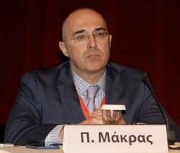 Polyzois Makras合作论文数Departments of Endocrinology;Leiden University;Medical Center;Medical Center, Leiden University26
 Meletios A. Dimopoulos合作论文数Department of Clinical Therapeutics, School of Medicine, National and Kapodistrian University of Athens25
 Efstathios Kastritis合作论文数Department of Clinical Therapeutics, National and Kapodistrian University of Athens23Gkotzamanidou Maria合作论文数18Evangelos Eleutherakis-Papaiakovou合作论文数Dept Clin Therapeut, Univ Athens16Maria Gavriatopoulou合作论文数School of Medicine, University of Athens15Ersi Voskaridou合作论文数14Kanellias Nikolaos合作论文数12
 Maria P. Yavropoulou合作论文数Aristotle University of Thessaloniki, AHEPA University Hospital11Magdalini Migkou合作论文数Department of Clinical Therapeutics, University of Athens School of Medicine11D. Christoulas合作论文数10Kokkoris Panagiotis合作论文数9Ntanasis-Stathopoulos Ioannis合作论文数Dept Clin Therapeut, Natl & Kapodistrian Univ Athens8Efstratios Moralidis合作论文数Ippokration General Hospital, Aristotle University8
 Arsos G合作论文数Department of Nuclear Medicine III, School of Medicine, Aristotle University of Thessaloniki8Marina D. Kita合作论文数7Repa Konstantina合作论文数7Eirini Katodritou合作论文数7
 Theodoros P Vassilakopoulos合作论文数Department of Haematology, National and Kapodistrian University of Athens;Laiko Hospital7
 Zoe Efstathiadou合作论文数Department of Internal Medicine, University of Ioannina School of Medicine7
 Maria Angelopoulou合作论文数Department of Hematology, National and Kapodistrian University of Athens7
 Jannis Kountouras合作论文数7Kokkoris P合作论文数6Veroniki Komninaka合作论文数6Panayiotidis Panayiotis合作论文数1st Department of Propaedeutic Internal Medicine, National and Kapodistrian University of Athens Medical School, General Hospital “LAIKO”6Georgios Boutsikas合作论文数6Konstantinos Anargyrou合作论文数6
 Maria Roussou合作论文数Dept Clin Therapeut, Natl & Kapodistrian Univ Athens5M. C. Kyrtsonis合作论文数School of Medicine, National and Kapodistrian University of Athens5Zafeiriadou Efthimia合作论文数5Vryonidou Andromachi合作论文数'Amalia Fleming' General Hospital, 'Attikon' University Hospital5
 Maria Tzetis合作论文数First Department of Pediatrics, Athens University5
 Gregory Kaltsas合作论文数First Department of Internal Medicine, National & Kapodistrian University of Athens;University Hospital Coventry and Warwickshire5
 Sophocles Lanitis合作论文数2nd Surgical Department and Surgical Oncology Unit of “Korgialenio-Benakio”,, Red Cross Hospital5
 John Meletis合作论文数5A. Gkiomisi合作论文数4Cornelia Bratengeier合作论文数4Tsionos Konstantinos合作论文数4
 Alexandros Apostolopoulos合作论文数4
 Periklis Makrythanasis合作论文数Department of Genetic Medicine and Development, University of Geneva;National and Kapodistrian University of Athens4
 Tina Bagratuni合作论文数School of Medicine, University of Athens4
 Chris Zavos合作论文数Peptiko.Gr4Socrates E. Papapoulos合作论文数4Kalliopi Patsiaoura合作论文数Department of Pathology, Ippokration General Hospital4Ioannis Litsas合作论文数4




