Normal routine electroencephalograms (EEGs) can cause delays in the diagnosis and treatment of epilepsy, especially in drug-resistant patients and those without structural abnormalities. There is a need for alternative quantitative approaches that can inform clinical decisions when traditional visual EEG review is inconclusive. We leverage a large population EEG database (N = 13,652 recordings, 12,134 unique patients) and an independent cohort of patients with focal epilepsy (N = 121) to investigate whether normal EEG segments could support the diagnosis of focal epilepsy. We decomposed expertly graded normal EEGs (N = 6,242) using unsupervised tensor decomposition to extract the dominant spatio-spectral patterns present in a clinical population. We then, using the independent cohort of patients with focal epilepsy, evaluated whether pattern loadings of normal interictal EEG segments could classify focal epilepsy, the epileptogenic lobe, presence of lesions, and drug response. We obtained six physiological patterns of EEG spectral power and connectivity with distinct spatio-spectral signatures. Both pattern types together effectively differentiated patients with focal epilepsy from non-epileptic controls (mean AUC 0.78) but failed to classify the epileptogenic lobe. Spectral power-based patterns best classified drug-resistant epilepsy (mean AUC 0.73) and lesional epilepsy (mean AUC 0.67), albeit with high variability across patients. Our findings support that visibly normal patient EEGs contain subtle quantitative differences of clinical relevance. Further development may yield normal EEG-based computational biomarkers that can augment traditional EEG review and epilepsy care.
Introduction:Scalp electroencephalography (EEG) is a cornerstone in the diagnosis and treatment of epilepsy, but routine EEG is often interpreted as normal without identification of epileptiform activity during expert visual review. The absence of interictal epileptiform activity on routine scalp EEGs can cause delays in receiving clinical treatment. These delays can be particularly problematic in the diagnosis and treatment of people with drug-resistant epilepsy (DRE) and those without structural abnormalities on MRI (i.e., MRI negative). Thus, there is a clinical need for alternative quantitative approaches that can inform diagnostic and treatment decisions when visual EEG review is inconclusive. In this study, we leverage a large population-level routine EEG database of people with and without focal epilepsy to investigate whether normal interictal EEG segments contain subtle deviations that could support the diagnosis of focal epilepsy. Data & Methods:We identified multiple epochs representing eyes-closed wakefulness from 19-channel routine EEGs of a large and diverse neurological patient population (N=13,652 recordings, 12,134 unique patients). We then extracted the average spectral power and phase-lag-index-based connectivity within 1-45Hz of each EEG recording using these identified epochs. We decomposed the power spectral density and phase-based connectivity information of all the visually reviewed normal EEGs (N=6,242) using unsupervised tensor decompositions to extract dominant patterns of spectral power and scalp connectivity. We also identified an independent set of routine EEGs of a cohort of patients with focal epilepsy (N= 121) with various diagnostic classifications, including focal epilepsy origin (temporal, frontal), MRI (lesional, non-lesional), and response to anti-seizure medications (responsive vs. drug-resistant epilepsy). We analyzed visually normal interictal epochs from the EEGs using the power-spectral and phase-based connectivity patterns identified above and evaluated their potential in clinically relevant binary classifications. Results:We obtained six patterns with distinct interpretable spatio-spectral signatures corresponding to putative aperiodic, oscillatory, and artifactual activity recorded on the EEG. The loadings for these patterns showed associations with patient age and expert-assigned grades of EEG abnormality. Further analysis using a physiologically relevant subset of these loadings differentiated patients with focal epilepsy from controls without history of focal epilepsy (mean AUC 0.78) but were unable to differentiate between frontal or temporal lobe epilepsy. In temporal lobe epilepsy, loadings of the power spectral patterns best differentiated drug-resistant epilepsy from drug-responsive epilepsy (mean AUC 0.73), as well as lesional epilepsy from non-lesional epilepsy (mean AUC 0.67), albeit with high variability across patients. Significance:Our findings from a large population sample of EEGs suggest that normal interictal EEGs of patients with epilepsy contain subtle differences of predictive value that may improve the overall diagnostic yield of routine and prolonged EEGs. The presented approach for analyzing normal EEGs has the capacity to differentiate several diagnostic classifications of epilepsy, and can quantitatively characterize EEG activity in a scalable, expert-interpretable, and patient-specific fashion. Further technical development and clinical validation may yield normal EEG-derived computational biomarkers that could augment epilepsy diagnosis and assist clinical decision-making in the future.
OBJECTIVE:Cognitive deficits are one of the most debilitating comorbidities in epilepsy and other neurodegenerative, neuropsychiatric, and neurodevelopmental brain disorders. Current diagnostic and therapeutic options are limited and lack objective measures of the underlying neural activities. In this study, electrophysiological biomarkers that reflect cognitive functions in clinically validated batteries were determined to aid diagnosis and treatment in specific brain regions. METHODS:We employed the Cambridge Neuropsychological Test Automated Battery (CANTAB) tasks to probe memory and executive functions in 86 patients with epilepsy undergoing clinical electroencephalography (EEG) monitoring. EEG electrode signals during performance of particular battery tasks were decomposed to identify specific frequency bands and cortical areas that differentiated patients with impaired, normal, and good standardized performance according to their age and gender. RESULTS:The anterior prefrontal cortical EEG power in the theta frequency band was consistently lower in patients with impaired memory and executive function performance (z-score < -1). This effect was evident in all four behavioral measures of executive, visual, spatial, and working memory functions and was confined to the cortical area of all four frontal pole electrodes (Nz, Fpz, Fp1, and Fp2). SIGNIFICANCE:Theta EEG power in the anterior prefrontal cortex provides simple, accessible, and objective electrophysiological measure of memory and executive functions in epilepsy. Our results suggest a feasible clinical biomarker for diagnosis, monitoring, and treatment of cognitive deficits with emerging targeted neuromodulation approaches.
Eidetic memory has been reported in children and in patients with synesthesia but is otherwise thought to be a rare phenomenon. Presented herein is a patient with right-sided language dominance, as proven via multiple functional imaging and neuropsychometric methods, who has a seizure onset zone in the right temporo-parietal-occipital cortex. This patient's medically refractory epilepsy and thus hyperactive cortex could possibly contribute to near eidetic ability with paired-associates learning tasks (in both short-term and long-term retention). There are reports of epilepsy negatively affecting memory, but as far as the authors are aware to date, there is limited evidence of any lesion enhancing cognitive functions (whether through direct lesion or via compensatory mechanism) that would be localized to a seizure onset zone in the dominant temporo-parietal-occipital junction.
Low frequency brain rhythms facilitate communication across large spatial regions in the brain and high frequency rhythms are thought to signify local processing among nearby assemblies. A heavily investigated mode by which these low frequency and high frequency phenomenon interact is phase-amplitude coupling (PAC). This phenomenon has recently shown promise as a novel electrophysiologic biomarker, in a number of neurologic diseases including human epilepsy. In 17 medically refractory epilepsy patients undergoing phase-2 monitoring for the evaluation of surgical resection and in whom temporal depth electrodes were implanted, we investigated the electrophysiologic relationships of PAC in epileptogenic (seizure onset zone or SOZ) and non-epileptogenic tissue (non-SOZ). That this biomarker can differentiate seizure onset zone from non-seizure onset zone has been established with ictal and pre-ictal data, but less so with interictal data. Here we show that this biomarker can differentiate SOZ from non-SOZ interictally and is also a function of interictal epileptiform discharges. We also show a differential level of PAC in slow-wave-sleep relative to NREM1-2 and awake states. Lastly, we show AUROC evaluation of the localization of SOZ is optimal when utilizing beta or alpha phase onto high-gamma or ripple band. The results suggest an elevated PAC may reflect an electrophysiology-based biomarker for abnormal/epileptogenic brain regions.
Identifying abnormal patterns in electroencephalography (EEG) remains the cornerstone of diagnosing several neurological diseases. The current clinical EEG review process relies heavily on expert visual review, which is unscalable and error-prone. In an effort to augment the expert review process, there is a significant interest in mining population-level EEG patterns using unsupervised approaches. Current approaches rely either on two-dimensional decompositions (e.g., principal and independent component analyses) or deep representation learning (e.g., auto-encoders, self-supervision). However, most approaches do not leverage the natural multi-dimensional structure of EEGs and lack interpretability. In this study, we propose a tensor decomposition approach using the canonical polyadic decomposition to discover a parsimonious set of population-level EEG patterns, retaining the natural multi-dimensional structure of EEG recordings $(\mathbf{time}\times \mathbf{space} \times \mathbf{frequency})$ . We then validate their clinical value using a cohort of patients with varying stages of cognitive impairment. Our results show that the discovered patterns reflect physiologically meaningful features and accurately classify the stages of cognitive impairment (healthy vs mild cognitive impairment vs Alzheimer's dementia) with substantially fewer features compared to classical and deep learning-based baselines. We conclude that the decomposition of population-level EEG tensors recovers expert-interpretable EEG patterns that can aid in studying smaller specialized clinical cohorts.
Background: The Coronavirus Disease 2019 (COVID-19) is a global pandemic that has killed over 1.5 million people worldwide. A constellation of multisystem involvement with SARS-CoV-2 has been reported. COVID-19 has been shown to affect the human nervous system, however, both the extent and severity of involvement have yet to be fully elucidated. In this manuscript, we aimed to better understand the effect of COVID-19 on neuro-respiratory status by studying COVID-19 patients who presented with central apnea. Methodology: We analyzed patient characteristics, clinical outcomes, laboratory results, and imaging results of three patients with symptomatic, PCR-proven COVID-19 and episodes of central apnea. Results: Of the three patients included in this study, two patients developed new central apnea, and one patient developed an exacerbation of underlying central apnea despite COVID-19 treatments with systemic steroids and remdesivir. All occurred, on average, 15 days after the onset of COVID-19 symptoms. At 1-year follow-up, all patients experienced complete resolution of apneic breathing. Conclusions: Physicians should be vigilant for the presentation of COVID-19 with central apnea. Central apnea may be a complication in patients with severe COVID-19 infection. More research is warranted to further understand this association.
Background Posterior Reversible Encephalopathy Syndrome (PRES) is a neurotoxic state characterized by seizures, headache, vision change, paresis, and altered mental status. PRES has an important place in medicine due to the wide variety of causative diseases, infections, and medications that precipitate its mysterious onset. Although exposure to medications, particularly immunosuppressants, cancer chemotherapy, and biologic drugs, is a common occurrence in patients who develop PRES, Mepolizumab has never before been associated. Case presentation This report of a 67-year-old male patient outlines the first reported case of Mepolizumab-induced PRES in the literature. Conclusions Treatment of severe asthma, asthma-exacerbations, and diseases such as eosinophilic granulomatosis with polyangiitis (formerly Churg-Strauss) with Mepolizumab is rapidly gaining popularity ever since the drug’s recent FDA-approval. This report aims to raise awareness of this potentially life-threatening and previously unreported side effect of Mepolizumab since early identification of the causative agent is the key to preventing the severe neurologic disability and possible death that may occur from the delayed treatment of PRES.
The recent availability of large datasets in bio-medicine has inspired the development of representation learning methods for multiple healthcare applications. Despite advances in predictive performance, the clinical utility of such methods is limited when exposed to real-world data. This study develops model diagnostic measures to detect potential pitfalls before deployment without assuming access to external data. Specifically, we focus on modeling realistic data shifts in electrophysiological signals (EEGs) via data transforms and extend the conventional task-based evaluations with analyses of a) the model's latent space and b) predictive uncertainty under these transforms. We conduct experiments on multiple EEG feature encoders and two clinically relevant downstream tasks using publicly available large-scale clinical EEGs. Within this experimental setting, our results suggest that measures of latent space integrity and model uncertainty under the proposed data shifts may help anticipate performance degradation during deployment.
Abstract Introduction Low Frequency brain rhythms facilitate communication across large spatial regions in the brain and high frequency rhythms are thought to signify local processing among nearby assemblies. A heavily investigated mode by which these low frequency and high frequency phenomenon interact is Phase-Amplitude Coupling )PAC). This phenomenon has recently shown promise as a novel electrophysiologic biomarker, in a number of neurologic diseases. In 10 patients undergoing phase-2 monitoring for the evaluation of surgical resection and in whom temporal depth electrodes were implanted, we investigated electrophysiologic relationships of PAC in epileptogenic (seizure onset zone or SOZ) and non-epileptogenic tissue (non-SOZ). That this biomarker can differentiate pathologic from non-pathologic brain and has been established with ictal and pre-ictal data, but less so with interictal data. Here we show that this biomarker can differentiate interictally. We also show PAC activity is related to interictal epileptiform discharges and high frequency activity. Importantly, we also show a differential level of PAC in slow-wave-sleep from NREM1-2 and awake. And finally we show that localization of pathologic tissue sensitivity and specificity is optimal when utilizing beta or alpha phase onto high-gamma or Ripple with knowledge of the sleep stage. Illustrating some of the physiologic nature of this biomarker in human epilepsy will provide a basis for understanding the mechanism of neurologic disease and normal physiology of brain communication, details which are at this point ready to be utilized in neurotechnological therapies to treat and understand both. Methods Per Institutional Review Board protocol, 10 patients who were under evaluation for resective surgery for MRE at Mayo Clinic in Rochester MN were included in this study. IRB approved the study and necessary consenting procedures were followed prior to any data acquisition. All subjects had bilaterally placed intracranial depth electrodes with usually 8 contacts. In some cases not all contacts could be used for data acquisition (hardware or recording problems). 6 Subjects of this cohort had scalp and EMG recordings concurrently placed for the purposes of sleep scoring. Subject recordings were ignored for POD-1 as anesthetics were dissipating. Subjects then stayed in the ICU ranging from 3-12 days before explanation.Pathological tissue identified as seizure onset zone (SOZ) was determined from phase II monitoring and determined by a trained neurologist. Sleep staging was done with expert-in-the-loop semi automated methods described elsewhere but overseen by a trained neurologist. Behavioral state was determined with scalp EEG signals and verified by a neurologist board certified in sleep medicine. All EEG recordings were bandpass filtered 0.3-75Hz and 60Hz notch filtered for scoring. Visual sleep scoring was in accordance with standard methods with modification for replacing the electrooculogram (EOG) recording with FP1, FP2, FPZ scalp electrodes. Wakefulness was determined by the presence of eye blinks visualized in fronto-parietal scalp leads, accompanied by posteriorly dominant alpha rhythms (8 - 12 Hz) comprising >50% of the epoch. Slow-wave sleep (N3) was scored when high-voltage (>75 uV) delta (0.5 - 3 Hz) frequency scalp EEG activity was present in at least 20% of the epoch (i.e., at least 6 s within a 30 s epoch) in the frontal derivations using conventional International 10-20 System electrode placements (FP1, FP2, FZ, F3, F4, CZ, C3, C4, O1, O2, and Oz). Phase Amplitude Coupling with Coherency Angle CFC. A Hanning taper n points is the length of the sliding time window. Next, the coherency CFC(fmodulating,fmodulated) was estimated between signal {Xt} and the estimate of the time-course of power{Pt(fmodulated)} for a given frequency fmodulating. The coherence was the absolute value of the CFC . The phase difference between the signal at fmodualting and the power at fmodulated is given by the angle of the coherency arg(CFC). In this case γ refer to a 1024 points Hanning window and * to the complex conjugate. This allowed us to characterize the phase-to-power cross-frequency interaction with respect to f and fmodulated sensor by sensor. The spike detection algorithm was utlized to evaluated successive 1 minute blocks of iEEG and removes artifact channels. Individual channels were defined as average slope greater than 10 SD outside of mean slopes of all channels. Second, iEEG was bandpass filtered 20-50Hz to identify possible spikes, where a sharp discharge must last between 20-70ms. Absolute amplitudes of peaks greater than 4SD of channel mean amplitude were noted as potential spike locations for further consideration. Third, raw iEEG was bandpass filtered (2nd order Butterworth) 1-35Hz. A scaling factor is determined by finding a value that will bring the median of all channel amplitudes to 70uV. All channels are multiplied by this scaling factor. Once the data have been scaled, the amplitude and slope of each half-wave of the potential spikes identified previously in step 2 are calculated and the values are compared to static thresholds (Total amplitude of both half-waves > 600μV, slope of each half-wave > 7μV/ms, duration of each half-wave > 10ms). Potential spikes with half-waves that exceed these thresholds are marked as interictal spikes.HFOs were detected using a Hilbert transform-based method, as previously reported. Here, the discrete time series is transformed into an analytic signal, where the real part is the original signal, and the imaginary part is the Hilbert transform of the original, x(t). Results Holding constant the frequency for amplitude to include all high activity (30-175Hz), beta is the best localizing (not statistically significant from alpha) band. Holding the frequency for phase, constant, there is no statistical difference between LG, HG, and ripple in terms of pathological bain tissue localization. Examining different frequencies for phase in varied behavioral/sleep states using 12 minute awake segments were used in all 10 patients, localization is best via AUROC when delta is the frequency band used to calculate PAC in Slow Wave Sleep / NREM3.Interictal Epileptiform spiking is seen with much higher regularity, although correlated and seen in the SOZ. A great deal of the associated elevations in wide-spectrum (0.5-30 modulating 65-175Hz) PAC (here defined as >2 SD across the entire 2 hour period and assessed across all channels) occurred with IEDs. Since 3 second segments were used to calculate PAC values, sometimes 2 IEDs were within a single epoch although in a small minority of observations. PAC elevations are seen with IEDs and with HFO, although to a greater extend with spikes. However, some IEDs are not associated with elevations in PAC. In fact most detected IEDs were not associated with elevated PAC values or with HFOs for that matter. A minority of IEDs are associated with elevated PAC values and HFOs. At the group and individual levels the average spike count per patient, grouping all SOZ and non-SOZ electrodes together, there is not a statistically significant effect, but if the average count is taken per electrode within a single patient, a significant difference was noted at p = .0031. Subjects on average had 15 (SEM 0.15) electrodes and SOZ electrode counts of 3 (SEM 0.71). When analyzing IED + elevated PAC, grouped SOZ electrodes among patients and within individuals show a strongly significant effect (.0004). When taking into account electrode numbers within each group this significance only increases (.0001). At the individual level, significance of p<0.05 is seen for all but one patient in this cohort. Conclusion Sleep Stage is critical in the analysis of pathological brain from non-pathological brain and electrophysiologic biomarkers behave differently in the different behavioral states. Here we found evidence to support his proposition in the following ways: low frequency delta phase modulates a broad high frequency amplitude in N3 and has relevance for brain pathology. Phase-Amplitude Coupling is increased in pathologic tissue. Peaks in PAC occur sporadically and infrequently in these patients. PAC is correlated with Interictal Epileptiform Discharges and High Frequency Oscillations however most Interictal Epileptiform Discharges are unrelated to peaks in Phase-Amplitude Coupling. Low Frequency Activity in Beta-band modulates broad high frequency power across low gamma, high gamma, and ripple bands. Support (If Any) Neurophysiological mapping and stimulation of the human brain for memory enhancement. Funded by EUO - PIS via BioTechMed Center, Department of Multimedia Systems, Faculty of Electronics, Telecommunication and Informatics, Gdansk University of Technology
Hyperammonemia is a condition that may result after solid organ transplantation, particularly lung transplantation. However, it is very uncommon for this presentation to occur more than 30 days post-transplantation. Hyperammonemia and the resulting encephalopathy typically manifest with altered sensorium, a clinical situation which is not often included in the differential diagnosis of presumed nonconvulsive status epilepticus (NCSE). Seizures are common among this subset of patients with hyperammonemia and may be refractory to traditional treatments. Evidence of elevated intracranial pressure by invasive monitoring and neuroimaging findings of diffuse cerebral edema are commonly reported. Here we examine the therapeutic importance of identifying the specific cause of hyperammonemic encephalopathy, a condition which may result in status epilepticus and ultimately cerebral edema or even brain death.
Clinical trials are conducted to evaluate the efficacy of new treatments. Clinical trials involving multiple treatments utilize the randomization of treatment assignments to enable the evaluation of treatment efficacies in an unbiased manner. Such evaluation is performed in post hoc studies that usually use supervised-learning methods that rely on large amounts of data collected in a randomized fashion. That approach often proves to be suboptimal in that some participants may suffer and even die as a result of having not received the most appropriate treatments during the trial. Reinforcement-learning methods improve the situation by making it possible to learn the treatment efficacies dynamically during the course of the trial, and to adapt treatment assignments accordingly. Recent efforts using multi-arm bandits, a type of reinforcement-learning method, have focused on maximizing clinical outcomes for a population that was assumed to be homogeneous. However, those approaches have failed to account for the variability among participants that is becoming increasingly evident as a result of recent clinical-trial-based studies. We present a contextual-bandit-based online treatment optimization algorithm that, in choosing treatments for new participants in the study, takes into account not only the maximization of the clinical outcomes as well as the patient characteristics. We evaluated our algorithm using a real clinical trial dataset from the International Stroke Trial. We simulated the online setting by sequentially going through the data of each participant admitted to the trial. Two bandits (one for each context) were created, with four choices of treatments. For a new participant in the trial, depending on the context, one of the bandits was selected. Then, we took three different approaches to choose a treatment: (a) a random choice (i.e., the strategy currently used in clinical trial settings), (b) a Thompson sampling-based approach, and (c) a UCB-based approach. Success probabilities of each context were calculated separately by considering the participants with the same context. Those estimated outcomes were used to update the prior distributions within the bandit corresponding to the context of each participant. We repeated that process through the end of the trial and recorded the outcomes and the chosen treatments for each approach. We also evaluated a context-free multi-arm-bandit-based approach, using the same dataset, to showcase the benefits of our approach. In the context-free case, we calculated the success probabilities for the Bernoulli sampler using the whole clinical trial dataset in a context-independent manner. The results of our retrospective analysis indicate that the proposed approach performs significantly better than either a random assignment of treatments (the current gold standard) or a multi-arm-bandit-based approach, providing substantial gains in the percentage of participants who are assigned the most suitable treatments. The contextual-bandit and multi-arm bandit approaches provide 72.63% and 64.34% gains, respectively, compared to a random assignment.
The recent availability of large datasets in bio-medicine has inspired the development of representation learning methods for multiple healthcare applications. Despite advances in predictive performance, the clinical utility of such methods is limited when exposed to real-world data. Here we develop model diagnostic measures to detect potential pitfalls during deployment without assuming access to external data. Specifically, we focus on modeling realistic data shifts in electrophysiological signals (EEGs) via data transforms, and extend the conventional task-based evaluations with analyses of a) model’s latent space and b) predictive uncertainty, under these transforms. We conduct experiments on multiple EEG feature encoders and two clinically relevant downstream tasks using publicly available large-scale clinical EEGs. Within this experimental setting, our results suggest that measures of latent space integrity and model uncertainty under the proposed data shifts may help anticipate performance degradation during deployment.
Routine scalp EEG is essential in the clinical diagnosis and management of epilepsy. However, a normal scalp EEG (based on expert visual review) recorded from a patient with epilepsy can cause delays in diagnosis and clinical care delivery. Here, we investigated whether normal EEGs might contain subtle electrophysiological dues of epilepsy. Specifically, we investigated (i) whether there arc indicators of abnormal brain electrophysiology in normal EEGs of epilepsy patients, and (ii) whether such abnormalities are modulated by the side of the brain generating seizures in focal epilepsy. We analysed awake scalp EEG recordings of age-matched groups of 144 healthy individuals and 48 individuals with drug-resistant focal epilepsy who had normal scalp EEGs. After preprocessing, using a bipolar montage of eight channels, we extracted the fraction of spectral power in the alpha band (8-13 Hz) relative to a wide band of 0.5-40 Hz within 10-s windows. We analysed the extracted features for (i) the extent to which people with drug-resistant focal epilepsy differed from healthy subjects, and (ii) whether differences within the drug-resistant focal epilepsy patients were related to the hemisphere generating seizures. We then used those differences to classify whether an EEG is likely to have been recorded from a person with drug-resistant focal epilepsy, and if so, the epileptogenic hemisphere. Furthermore, we tested the significance of these differences while controlling for confounders, such as acquisition system, age and medications. We found that the fraction of alpha power is generally reduced (i) in drug-resistant focal epilepsy compared to healthy controls, and (ii) in right-handed drug-resistant focal epilepsy subjects with left hemispheric seizures compared to those with right hemispheric seizures, and that the differences are most prominent in the frontal and temporal regions. The fraction of alpha power yielded area under curve values of 0.83 in distinguishing drug-resistant focal epilepsy from healthy and 0.77 in identifying the epileptic hemisphere in drug-resistant focal epilepsy patients. Furthermore, our results suggest that the differences in alpha power are greater when compared with differences attributable to acquisition system differences, age and medications. Our findings support that EEG-based measures of normal brain function, such as the normalized spectral power of alpha activity, may help identify patients with epilepsy even when an EEG does not contain any epileptiform activity, recorded seizures or other abnormalities. Although alpha abnormalities are unlikely to be disease-specific, we propose that such abnormalities may provide a higher pre-test probability for epilepsy when an individual being screened for epilepsy has a normal EEG on visual assessment.
Many autoimmune diseases can affect the central nervous system, and their varying clinical presentations often confound a straightforward diagnosis. In this report, we describe a unique presentation of CLIPPERS syndrome. To our knowledge, this is the first case to demonstrate significant supratentorial involvement with symmetric and non-confluent lesions in the medial orbitofrontal cortex; additionally, this is the second case to describe an association between diagnoses of hypothyroidism and CLIPPERS.
INTRODUCTION:Creutzfeldt-Jakob disease (CJD) is a prion protein disorder of significant consequence and currently incurable. Diagnosis can be challenging early in the disease course. CJD can present in many ways but often fits a pattern of cognitive problems, cerebellar disturbance, behavioral/psychological changes, and perhaps myoclonus.CASE REPORT:We herein present the case of a 69-year-old White male with subacute progressive bulbar and limb weakness over ten weeks period. Early on, he was diagnosed with amyotrophic lateral sclerosis versus autoimmune-related bulbar neuropathy and treated as such. However, he continued to deteriorate clinically that prompted another admission, upon readmission, his cerebrospinal fluid RTQuick and 14-3-3 from the National Prion Disease Pathology Surveillance Center (NPDPSC) did eventually return positive. Hence he was diagnosed with CJD.CONCLUSIONS:CJD may present with progressive bulbar symptoms similar to acute inflammatory demyelinating polyradiculoneuropathy (MF variant), motor neuron disease, or autoimmune brainstem encephalitis. It becomes even higher on the differentials especially with no response to immunotherapy.
This paper presents a domain-guided approach for learning representations of scalp-electroencephalograms (EEGs) without relying on expert annotations. Expert labeling of EEGs has proven to be an unscalable process with low inter-reviewer agreement because of the complex and lengthy nature of EEG recordings. Hence, there is a need for machine learning (ML) approaches that can leverage expert domain knowledge without incurring the cost of labor-intensive annotations. Self-supervised learning (SSL) has shown promise in such settings, although existing SSL efforts on EEG data do not fully exploit EEG domain knowledge. Furthermore, it is unclear to what extent SSL models generalize to unseen tasks and datasets. Here we explore whether SSL tasks derived in a domain-guided fashion can learn generalizable EEG representations. Our contributions are three-fold: 1) we propose novel SSL tasks for EEG based on the spatial similarity of brain activity, underlying behavioral states, and age-related differences; 2) we present evidence that an encoder pretrained using the proposed SSL tasks shows strong predictive performance on multiple downstream classifications; and 3) using two large EEG datasets, we show that our encoder generalizes well to multiple EEG datasets during downstream evaluations.
Objective: Our goal was to analyze the electrophysiological response to direct electrical stimulation (DES) systematically applied at a wide range of parameters and anatomical sites, with particular focus on neural activities associated with memory and cognition. Methods: We used a large set of intracranial EEG (iEEG) recordings with DES from 45 subjects with electrodes implanted both subdurally on the cortical surface and subcortically into the brain parenchyma. Subjects were stimulated in blocks of alternating frequency and amplitude parameters during quiet wakefulness. Results: Stimulating at different frequencies and amplitudes of electric current revealed a persistent pattern of response in the slow and the fast neural activities. In particular, amplification of the theta (4-7 Hz) and attenuation of the gamma (29-52 Hz) power-in-band was observed with increasing the stimulation parameters. This opposite effect on the low and high frequency bands was found across a network of selected local and distal sites proportionally to the proximity and magnitude of the electric current. Power increase in the theta and decrease in the gamma band was driven by the total electric charge delivered with either increasing the frequency or amplitude of the stimulation current. This inverse effect on the theta and gamma activities was consistently observed in response to different stimulation frequencies and amplitudes. Conclusion: Our results suggest a uniform DES effect of amplifying theta and suppressing gamma neural activities in the human brain. Significance: These findings reveal the utility of simple power-in-band features for understanding and optimizing the effects of electrical stimulation on brain functions.
Engineering technology plays a pivotal role in the delivery of health care in under-resourced countries by providing an infrastructure to improve patient outcomes. However, sustainability of these technologies is difficult in these settings oftentimes due to limited resources or training. The framework presented in this editorial focuses on establishing medical and laboratory equipment sustainability in developing countries and is comprised of four steps: 1) establishing reliable in-country relationships with stakeholders, 2) identifying needs for sustainable solutions locally, 3) exploring potential solutions and assessing their effort-to-impact ratios, and 4) working with strategic partners to implement solutions with clear performance metrics. By focusing on the sustainability of donated equipment instead of the equipment itself, this method presented distinguishes itself from other philanthropic endeavors in the field by seeking to establish preventive maintenance habits that can impact clinical outcomes of a community long term. Application of this methodology is reported in the Original Research Article "A Low-Cost Humidity Control System to Protect Microscopes in a Tropical Climate" by Asp et. al.
 Gregory A. Worrell合作论文数Department of Neurology, Mayo Clinic;Department of Physiology & Biomedical Engineering, Mayo Clinic38
 Michael R. Sperling合作论文数Department of Neurology, Sidney Kimmel Medical College, Thomas Jefferson University;Jefferson Comprehensive Epilepsy Center, Thomas Jefferson University Hospital;Clinical Neurophysiology Laboratory, Thomas Jefferson University Hospital22
 Michal Kucewicz合作论文数Brain and Mind Electrophysiology Laboratory, BioTechMed Center, Multimedia Systems Department, Faculty of Electronics Telecommunications and Informatics, Gdansk University of Technology19
 Benjamin H. Brinkmann合作论文数Section of Nuclear Medicine, Mayo Clinic and Mayo Foundation19
 Kathryn A. Davis合作论文数Department of Neurology, Perelman School of Medicine, University of Pennsylvania18
 Vaclav Kremen合作论文数Faculty of Electrical Engineering, Czech Technical University in Prague18
 Barbara C. Jobst合作论文数Department of Medicine, Dartmouth Medical School18
 Malik Ghannam合作论文数Dept Neurol, Univ Iowa Hosp & Clin17
 Yogatheesan Varatharajah合作论文数Department of Electrical and Computer Engineering, The Grainger College of Engineering, University of Illinois at Urbana-Champaign17
 Bradley Lega合作论文数Department of Neurological Surgery, Medical School, UT Southwestern Medical Center;Department of Neurology, Medical School, UT Southwestern Medical Center;Department of Psychiatry, Medical School, UT Southwestern Medical Center16
 Robert E. Gross合作论文数Department of Neurosurgery, School of Medicine, Emory University15Richard J. T. Gorniak合作论文数15
 Daniel S. Rizzuto合作论文数Volen Center for Complex Systems, Brandeis University14
 Michael J. Kahana合作论文数Department of Psychology University of Pennsylvania11
 Ashwini Sharan合作论文数Neurosurgery Department, Thomas Jefferson University10
 Joel Stein合作论文数Department of Rehabilitation Medicine, Weill Cornell Medicine, Cornell University9
 Sandhitsu Das合作论文数Perelman School of Medicine, University of Pennsylvania9
 Ezzyat Youssef合作论文数Department of Psychology and Center for Neural Science, New York University8
 Ravishankar K Iyer合作论文数Departments of Electrical & Computer Engineering, The Grainger College of Engineering, University of Illinois Urbana-Champaign;Armored Computing, Inc.;Center for Reliable and High-Performance Computing, University of Illinois at Urbana-Champaign8
 Sameer Anil Sheth合作论文数Baylor College of Medicine8
 Matt Stead合作论文数8
 Dana Ghazaleh合作论文数7Laura R Miller合作论文数7
 Paul A Wanda合作论文数Dept Psychol, Univ Penn7
 Neeraj Wagh合作论文数University of Illinois at Urbana-Champaign6
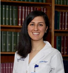 Fatemeh (Mehraneh) Khadjevand合作论文数Interdicsiplinary Neuroscience Research Program (INRP),, Tehran University of Medical Sciences6
 Cory S. Inman合作论文数Dept Psychol, Emory Univ6
 Kareem A. Zaghloul合作论文数Department of Neurosurgery, University of Pennsylvania5
 Jamie J. Van Gompel合作论文数Mayo Clinic5Samarth Rawal合作论文数4Zachary J Waldman合作论文数4
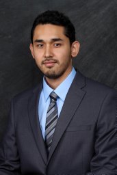 Azizullah Beran合作论文数Departments of Internal Medicine.4Boney Joseph合作论文数Mayo Clin, Dept Psychiat & Psychol, 200 First St SW, Rochester, MN 55905 USA4Krishnakant Saboo合作论文数Dept Elect Comp Engn Urban, Univ Illinois4
 Joshua Jacobs合作论文数Department of Neurology, University of Chicago;Neuroscience Institute, University of Chicago4
 Shennan Aibel Weiss合作论文数Department of Neurology, Columbia University4
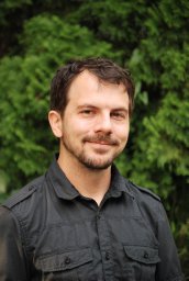 Andrew J. Watrous合作论文数4
 Sang Ah Lee合作论文数Department of Psychology, Harvard University4Jonathan Miller合作论文数3Tanner Ferderer合作论文数3Jionghao Wei合作论文数3
 Hari Guragain合作论文数Georgia State Univ3Bahareh Elahian合作论文数Department of Neuroscience Thomas Jefferson University Philadelphia PA3Tom Coffey合作论文数3
 Jan Cimbalnik合作论文数International Center for Research Consortium, and St. Anne's Hospital3Shoichi Shimamoto合作论文数2Matthew Stead合作论文数2Irena Balzekas合作论文数2
 Jetter Robertson合作论文数2




