Defects in pancreatic beta cell turnover are implicated in the pathogenesis of type 2 diabetes by genetic markers for diabetes. Decreased beta cell neogenesis could contribute to diabetes. The longevity and turnover of human beta cells is unknown; in rodents <1 year old, a half-life of 30 days is estimated. Intracellular lipofuscin body (LB) accumulation is a hallmark of ageing in neurons. To estimate the lifespan of human beta cells, we measured beta cell LB accumulation in individuals aged 1–81 years.
Diabetic neuropathy may be a result of disturbed vasomotion, the spontaneous rhythmic oscillations of vessel diameter, flow, or pressure in the microcirculation. To examine the differences in vasomotion prior to overt diabetes and with diagnosed diabetes, metabolic and blood flow measurements were obtained from three metabolically distinct groups of nonhuman primates: normoglycemic, metabolic syndrome/prediabetic, and overtly diabetic. Three of these indices characterize spectral properties of microcirculatory vasomotion: (1) the fraction of spectral power in a band around 2.0 Hz, (2) the ratio of spectral power in a band around 1.0 Hz to that around 2.0 Hz, (3) the Hurst exponent. The rest were more traditional measures including (4) fasting plasma Glucose (FPG), (5) glycosylated hemoglobin level (HbA1c), (6) fasting insulin levels (IRI), (7) average systolic blood pressure, and (8) average diastolic blood pressure. We found that the mean fraction of the spectral power of the vasomotion in the 2 Hz band is significantly greater in the nondiabetic than in the diabetic group. Similarly, the ratio of the spectral power in the 1.0 Hz band to that in the 2.0 Hz band was significantly higher among the diabetic subjects than in the nondiabetic and prediabetic subjects. Principal component analysis (PCA) of the ensemble of eight measures revealed that values of the first principal component in the diabetic group were highly and significantly different from those of the normal and prediabetic groups, which are also significantly different from each other. Furthermore, the spectral measures of vasomotion offered a distinct advantage over the traditional physiological variables in distinguishing the normal subjects from the prediabetic group, and are likely to reflect very early autonomic dysfunction.
The PPAR‐α agonist, K‐111 (Kowa), mitigates dyslipidemia and insulin resistance in obese, hypertriglyceridemic rhesus monkeys. Some of the cellular mechanisms of action of this agent to improve characteristics of the metabolic syndrome include in adipose tissue, a decrease in basal LPL activity, and in skeletal muscle during a euglycemic hyperinsulinemic clamp, an increase in glycogen synthase and LPL activities and carnitine palmitoyltransferase 1 mRNA and a decrease in triglyceride (TG) content. To determine whether changes in liver lipid metabolism were involved in the effect of K‐111 to alleviate dyslipidemia, total extractable LPL activity and protein, TG content and TNF‐α protein in liver samples from 6 obese monkeys before and after 7 weeks of K‐111 (3 mg/kg/day) were measured. LPL activity was reduced (p<0.05), whereas LPL protein was not affected by K‐111, suggesting posttranslational modification of LPL by K‐111. TG content and TNF‐α protein were reduced to the greatest extent by K‐111 in those monkeys with the highest baseline TG content and TNF‐α mRNA (baseline TG vs. K‐111 effect on TG, r=−0.80, p<0.05 and on TNF‐α, r=−0.97, p<0.001). TNF‐α protein was lower after K‐111 compared to baseline (p=0.08). We conclude that the beneficial effects of K‐111 include changes in liver lipid metabolism and that K‐111 may be useful in treating hepatic steatosis and inflammation.
Diabetes mellitus develops spontaneously in middle-aged, obese rhesus monkeys, thus making them a good model for examining the effects of co-morbid factors on the development of end-organ damage. Changes in structure and function in the eyes of one monkey who spontaneously developed type 2 diabetes are reported here. This animal had concomitant hypertension, high levels of triglycerides and serum cholesterol, and a low fraction of high-density lipoprotein. The eyes showed intraretinal hemorrhages and large areas of retinal capillary nonperfusion. Indo-cyanin green (ICG) angiography revealed a large area of non- or poorly perfused choriocapillaris in one eye, and immunohistochemistry showed loss of viable choriocapillaries in this region. Both basal laminar deposits and hard drusen were present on areas of Bruch's membrane adjacent to nonviable choriocapillaris. Blood flow via the nasal posterior ciliary arteries to this section of choroid was not detectable by color duplex Doppler ultrasound, indicating contribution of extraocular vascular disease to ischemia in this eye. There was a severe decline in number of photoreceptor inner and outer segments, and corresponding reductions in the multifocal electroretinogram (ERG), in the areas of choriocapillaris loss. The ganzfeld ERG indicated loss in both inner and outer retinal function. Much of the ganglion cell layer was absent throughout the retina, possibly reflecting the effect of diabetes as well as chronic open angle glaucoma; the latter diagnosis supported by elevated intraocular pressures and excavated optic disks. In summary, high resolution, enzyme histochemical and histopathological analyses of a diabetic hypertensive monkey retina and choroid after serial functional in vivo analyses have demonstrated the relationship between vascular dysfunction and visual function loss. Choroidal vascular dysfunction in both large and small vessels was associated with age-related macular degeneration-like changes in Bruch's membrane and photoreceptor degeneration.
Some physiological measures change with age, but the existence of age-related disorders such as type 2 diabetes raises questions about which patterns reflect progressive pathology and which are manifestations of aging. Here we report a retrospective investigation of age-related physiological changes in rhesus monkeys that developed diabetes (D group, n = 65) or exhibited healthy aging (N group, n = 88). Data were available on clinical chemistries, hematology, glucose tolerance, and insulin sensitivity based on oral and intravenous glucose tolerance tests and euglycemic, hyperinsulinemic clamp assessments. Individuals contributed data for an average of 7.6 years, when they were between 5 and 30 years of age. Only glucose disappearance rate, insulin sensitivity rate, and high density lipoprotein levels changed significantly with age in the nondiabetic group. In the diabetic group, significant decreases in glucose tolerance were evident by middle age (age 14 y), and fasting insulin first increased before diabetes was diagnosed, and then declined with advancing age.
 Nl Bodkin合作论文数58
 Heidi Ortmeyer合作论文数44Klc Jen合作论文数18Tigno Xenia T合作论文数14BL METZGER合作论文数13samuel a clark合作论文数11Ga Lutty合作论文数9
 D. Scott Mcleod合作论文数Wilmer Eye Institute, Johns Hopkins Hospital9Kunimoto Hotta合作论文数7Sy Ding合作论文数7
 Mark Johnson合作论文数Johnson Lab, Department of Chemistry, Yale University7Gustafson T A合作论文数7t alexander合作论文数6N L Bodkin合作论文数6John F. Morris合作论文数6s m steidl合作论文数5Sy Kim合作论文数5
 Robert Wolfe合作论文数Department of Biostatistics, School of Public Health, University of Michigan5Gp Schielke合作论文数4Ejp Dekoning合作论文数4S Yoshioka合作论文数4
 Alan R. Shuldiner合作论文数University of Maryland School of Medicine;University of Maryland Clinical and Translational Research Institute;Division of Endocrinology, Diabetes and Nutrition, University of Maryland;Regeneron Pharmaceuticals, Inc.4SBP Charge合作论文数3robert w flower合作论文数3L. van de Laar合作论文数3Js Hannah合作论文数3Z Huang合作论文数3
 Joseph Larner合作论文数Department of Pharmacology, University of Virginia3E. J. P. de Koning合作论文数2R. Shilling合作论文数2Ejp De Koning合作论文数2Jl Morton合作论文数2Sb Pek合作论文数2CJ GOODNER合作论文数2LM SPEEGLE合作论文数2T Funahashi合作论文数2M Harrell合作论文数2T Otsuji合作论文数2s mcevoy合作论文数2
 Barbara Howard合作论文数Center for the Study of Sex Differences in Health, Aging & Disease, Georgetown University Medical Center2
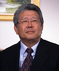 Yuji Matsuzawa (松澤佑次)合作论文数Division of Metabolic Medicine, Osaka University;Sumitomo Hospital2
 Joanna Poulton (Jo Poulton)合作论文数Nuffield Department of Women's & Reproductive Health, Medical Sciences Division, University of Oxford2Kikuko Hotta合作论文数2DJ KOERKER合作论文数2
 Chittaranjan Yajnik合作论文数Diabetes Unit, KEM Hospital Research Centre;Peninsula College of Medicine and Dentistry, University of Exeter2MA Lane合作论文数1Rice F L合作论文数1E. de Koning合作论文数1H Kuriyama合作论文数1




