PS-OCT imaging of retinal blood vessels reveals early indicators of coronary artery disease, offering a non-invasive and cost-effective screening method. This technique detects subtle vascular changes before clinical manifestation of cardiovascular disease. (c) 2025 The Author(s).
Preliminary data obtained with PS-OCT through the eyes of hypertensives suggests a mechanistic connection between arterial health and hypertension. In this study we want to determine whether arterial health may be more predictive of negative clinical outcomes than hypertension and blood glucose. Patients with various stages of hypertension, diabetes and coronary artery disease were recruited from Fiona Stanley Hospital (Perth). They were subsequently imaged with PS-OCT. The data were analyzed for retinal vessel wall thickness and vessel wall birefringence. We demonstrated that the combination of blood vessel wall tissue structure and wall thickness, a recognized clinical biomarker (Afsharan et al, BOE, 2021), could diagnose hypertension and diabetes with high sensitivity and specificity. PS-OCT measurements can detect the smallest changes related to cardiovascular disease in the retina before the disease manifests itself clinically. The method is cheap, noninvasive and easy to apply, which makes it highly suitable for screening, especially in underserved communities.
Terahertz (THz) imaging and optical coherence tomography (OCT) provide complementary information with similar length scales. In addition to OCT's extensive use in ophthalmology, both methods have shown some promise for other medical applications and non-destructive testing. In this paper, we present an iterative algorithm that combines the information from OCT and THz imaging at two different measurement locations within an object to determine both the depth of the reflecting layers at the two locations and the unknown refractive index of the medium for both the OCT wavelengths and THz frequencies. We validate this algorithm using a silicone test object with embedded layers and show that the depths and refractive index values obtained from the algorithm agreed with the measured values to within 3.3%. We further demonstrate for the first time that OCT and THz images can be co-registered and aligned using unsupervised image registration. Hence we show that a combined OCT/THz system can provide unique information beyond the capability of the separate modalities alone, with possible applications in the medical, industrial and pharmaceutical sectors.
We present the use of sub-micron resolution optical coherence tomography (OCT) in quality inspection for printed electronics. The device used in the study is based on a supercontinuum light source, Michelson interferometer and high-speed spectrometer. The spectrometer in the presented spectral-domain optical coherence tomography setup (SD-OCT) is centered at 600 nm and covers a 400 nm wide spectral region ranging from 400 nm to 800 nm. Spectra were acquired at a continuous rate of 140,000 per second. The full width at half maximum of the point spread function obtained from a Parylene C sample was 0.98 mu. In addition to Parylene C layers, the applicability of sub-micron SD-OCT in printed electronics was studied using PET and epoxy covered solar cell, a printed RFID antenna and a screen-printed battery electrode. A commercial SD-OCT system was used for reference measurements.
Retinal dystrophies (RD) are blinding diseases affecting visual acuity mostly at young age. Intrinsic optical signals (IOS) on optical coherence tomography (OCT) may give topographical information on injure of retinal function in these patients. We demonstrate light response of the healthy and diseased human retina by IOS on a commercially available spectral-domain OCT. Significant IOS could be measured in the healthy retina and in unchanged retinal sectors of the RD patients. Main responses were located in the outer retina (photoreceptors) and the nerve fiber layer. In affected areas of RD eyes IOS were significantly reduced or even absent. Functional OCT imaging was able to give information about retinal function in RD patients on a micrometer scale. These results could be of value for refined disease analysis and control of upcoming gene therapy studies.
Cone phototransduction is a cornerstone of visual process and defects in phototransduction underlie many ocular diseases. We describe an optical method for direct in vivo observation of physiological processes accompanying phototransduction. The method takes advantage of interference among multiple reflections that straddle the cone outer segment, where phototransduction is known to begin. This self-interference is highly sensitive to phase changes within the outer segment such as those caused by changes in its refractive index, scattering properties, and size. A high-speed flood-illumination retina camera equipped with adaptive optics was used to observe changes in reflectance of individual cones following delivery of varying levels of visible stimulation. This pattern of changing reflectance, termed “scintillation”, was analyzed in terms of its amplitude, onset, and frequency, and their varying dependence upon stimulus energy. Measurements were taken on four subjects. Amplitude of scintillation was determined to be uncorrelated with stimulus energy (p[[gt]]0.1), onset of scintillation occurred within 10 ms after stimulus, and scintillation frequency was found to depend upon stimulus energy. Repeatability tests and quantitative comparison between subjects were conducted. Possible physiological origins for the phase changes underlying scintillation are discussed and a simple mathematical model is presented.
We present ultra-high resolution optical coherence tomography (OCT) structural intensity and optical Doppler tomography (ODT) flow velocity images of the human retina in vivo. The ultra-high speed OCT system is based on Spectral Domain or Fourier Domain technology, which provides a sensitivity advantage over conventional OCT of more than 2 orders of magnitude. This sensitivity improvement allows video rate OCT and ODT cross sectional imaging of retinal structures. Images will be presented with axial resolutions of 6 and 3.5 microns. We observed small features in the inner and outer plexiform layers, which are believed to be small blood vessels. Flow velocity images will be presented showing pulsatile flow in retinal arteries and veins.
We present ultra-high resolution optical coherence tomography (OCT) structural intensity and optical Doppler tomography (ODT) flow velocity images of the human retina in vivo. The ultra-high speed OCT system is based on Spectral Domain or Fourier Domain technology, which provides a sensitivity advantage over conventional OCT of more than 2 orders of magnitude. This sensitivity improvement allows video rate OCT and ODT cross sectional imaging of retinal structures. Images will be presented with axial resolutions of 6 and 3.5 microns. We observed small features in the inner and outer plexiform layers, which are believed to be small blood vessels. Flow velocity images will be presented showing pulsatile flow in retinal arteries and veins.
Thinning of the retinal nerve fiber layer and changes in retinal nerve fiber layer birefringence may both precede clinically detectable glaucomatous vision loss. We present in vivo thickness and depth-resolved birefringence measurements of the human retinal nerve fiber layer (RNFL) by use of polarization-sensitive optical coherence tomography (PS-OCT). Using a fiber-based PS-OCT setup real-time images of the human retina in vivo were recorded, co-registered with retinal video images of the location of PS-OCT scans. PS-OCT scans around the optic nerve head (ONH) of two healthy young volunteers were made using 10 concentric circles of increasing radius. Both the mean retinal nerve fiber layer thickness and mean retinal nerve fiber birefringence for each of 48 sectors on a circle were determined. The retinal nerve fiber layer thickness and birefringence varied as a function of sector around the ONH. Measured double pass phase retardation per unit depth values around the ONH range between 0.10 and 0.35 degrees/microm. The retinal nerve fiber layer becomes thinner with increasing distance from the ONH. In contrast, the birefringence does not vary significantly with increasing distance from the ONH.
We demonstrate a high- speed multi- functional spectral- domain optical coherence tomography system, using a broadband light source centered at 1.3 mu m and two InGaAs line scan cameras capable of acquiring individual axial scans in 24.4 mu s, at a rate of 18,500 axial scans per second. Fundamental limitations on the accuracy of phase determination as functions of signal- to- noise ratio and lateral scan speed are presented and their relative contributions are compared. The consequences of phase accuracy are discussed for both Doppler and polarization- sensitive OCT measurements. A birefringence artifact and a calibration procedure to remove this artifact are explained. Images of a chicken breast tissue sample acquired with the system were compared to those taken with a time- domain OCT system for birefringence measurement verification. The ability of the system to image pulsatile flow in the dermis and to perform functional imaging of large volumes demonstrates the clinical potential of multifunctional spectral- domain OCT. (C) 2005 Optical Society of America.
We introduce a method to determine the retinal nerve fiber layer (RNFL) thickness in OCT images based on anisotropic noise suppression and deformable splines. Spectral-Domain Optical Coherence Tomography (SDOCT) data was acquired at 29 kHz A-line rate with a depth resolution of 2.6 mum and a depth range of 1.6 mm. Areas of 9.6x6.4 mm2 and 6.4x6.4 mm2 were acquired in approximately 6 seconds. The deformable spline algorithm determined the vitreous-RNFL and RNFL-ganglion cell/inner plexiform layer boundary, respectively, based on changes in the reflectivity, resulting in a quantitative estimation of the RNFL thickness. The thickness map was combined with an integrated reflectance map of the retina and a typical OCT movie to facilitate clinical interpretation of the OCT data. Large area maps of RNFL thickness will permit better longitudinal evaluation of RNFL thinning in glaucoma.
Spectral-Domain Polarization-Sensitive Optical Coherence Tomography (SD-PS-OCT) is a technique developed to measure the thickness and birefringence of the nerve fiber layer in vivo as a tool for the early diagnosis of glaucoma. A clinical SD-PS-OCT system was developed and scans were made around the optic nerve head (ONH) using ten concentric circles of increasing diameter. One healthy volunteer was imaged. Retinal nerve fiber layer thickness and birefringence information was extracted from the data. Polarization-sensitive OCT images were acquired at video rate (29 frames per second (fps), 1000 A-lines / frame) and at 7 fps (1000 A-lines / frame). The last setting improved the signal to noise ratio by approximately 6 dB. Birefringence measurements on the healthy volunteer gave similar results as earlier reported values that were obtained with a time-domain setup. The measurement time was reduced from more than a minute to less than a second.
OBJECTIVE To introduce a new ophthalmic optical coherence tomography technology that allows unprecedented simultaneous ultra-high speed and ultra-high resolution. METHODS Using a superluminescent diode source, a clinically viable ultra-high speed, ultra-high resolution spectral domain optical coherence tomography system was developed. RESULTS In vivo images of the retina, the optic nerve head, and retinal blood flow were obtained at an ultra-high speed of 34.1 microseconds (ms) per A-scan, which is 73 times faster than commercially available optical coherence tomography instruments. Single images (B-scans) consisting of 1000 A-scans were acquired in 34.1 ms, allowing video rate imaging at 29 frames per second with an axial resolution of 6 mum. Using a different source in a slightly slower configuration, single images consisting of 500 A-scans were acquired in 34 ms, allowing imaging at 29 frames per second at an axial resolution of 3.5 microm, which is 3 times better than commercially available optical coherence tomography instruments. The amount of energy directed into the eye in both cases, 600 microW, is less than that of the Stratus OCT3 and is safe for intrabeam viewing for up to 8 hours at the same retinal location. CONCLUSION Spectral domain optical coherence tomography technology enables ophthalmic imaging with unprecedented simultaneous ultra-high speed and ultra-high resolution.
 B. Hyle Park合作论文数Harvard Medical School, Massachusetts General Hospital31
 Johannes F. De Boer合作论文数Department of Physics and Astronomy, Vrije Universiteit Amsterdam29
 Mark C. Pierce合作论文数Laser Photonics Research Group, University of Manchester25
 Brett Bouma合作论文数Institute for Medical Engineering & Science, Massachusetts Institute of Technology;Dermatology, Massachusetts General Hospital, Harvard University;Center for Biomedical OCT Research and Translation14
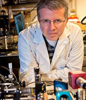 Guillermo J. Tearney合作论文数Harvard Medical School, Harvard University;School of Dental Medicine, Harvard University13TC Chen合作论文数7N Nassif合作论文数5Mircea Mujat合作论文数5
 Joo Chulmin合作论文数5
 Nader A Nassif合作论文数5
 Michael Hackmann合作论文数School of Human Sciences, The University of Western Australia4
 Seok-Hyun Andy Yun合作论文数Mass General Research Institute, Massachusetts General Hospital;Wellman Center for Photomedicine, Massachusetts General Hospital;Harvard Medical School4
 Taner Akkin合作论文数Department of Biomedical Engineering, University of Minnesota4david sampson合作论文数3A. Cairncross合作论文数3Li Qingyun合作论文数3
 Karol Karnowski合作论文数3
 Donald T. Miller合作论文数School of Optometry, Indiana University3
 Girish Dwivedi合作论文数3Peter B. Noble合作论文数Division of Clinical Sciences, Telethon Institute for Child Health Research3J. Elliot合作论文数2
 Hadi Afsharan合作论文数Opt Biomed Engn Lab, Univ Western Australia2
 Qiang Wang合作论文数School of Optometry, Indiana University2Wanlin Gao合作论文数2
 Ravi S. Jonnal合作论文数School of Optometry, Indiana University2Alan L. James合作论文数2Omer P. Kocaoglu合作论文数2
 Teresa C. Chen合作论文数Department of Ophthalmology, Harvard Medical School, Harvard University2N. Lan合作论文数1B. Thompson合作论文数1R L Sheridan合作论文数1eli weinberg合作论文数1kay wang合作论文数1AJ Fitzgerald合作论文数1andy yun合作论文数1Eungwang Seo合作论文数Dept Phys, Chinese Univ Hong Kong1pierce合作论文数1
 Xin Tie合作论文数University of Wisconsin–Madison1Kris Nilsen合作论文数Swinburne Univ1
 Adam Gibson合作论文数Institute for Sustainable Heritage, The Bartlett Faculty of the Built Environment, University College London1
 Dilusha Silva, K.K.M.B.合作论文数1Thomas Rajesh合作论文数1
 Weihua Gao合作论文数Meta1
 Abdul Ihdayhid合作论文数Harry Perkins Institute of Medical Research;CoraMetix;Fiona Stanley Hospital1
 Lauri Janne合作论文数1
 C.B. Hoyng (Carel)合作论文数Donders Institute for Brain, Cognition and Behaviour, Radboud University;Faculty of Medical Sciences, Radboud University1
 Milen Shishkov合作论文数Wellman Center for Photomedicine, Massachusetts General Hospital1
 Tapio Fabritius合作论文数Computational Optics Group;University of Tsukuba;Computational Optics Group, University of Tsukuba1
 Rafal Sliz合作论文数University of Oulu, University of Oulu1




