The ability of adult epithelial cells to rewire their cell fate programme in response to injury has emerged as a new paradigm in stem cell biology. This plasticity supersedes the concept of strict stem cell hierarchies, granting cells access to a wider repertoire of fate choices. Yet, in order to prevent a disordered cellular response, this process must be finely regulated. Here we investigate the little-known regulatory processes that restrict fate permissibility in adult cells, and keep plasticity in check. Using a 3D regenerative culture system, that enables co-culturing epithelium and stroma of different origins, we demonstrate that oesophageal cells exposed to the ectopic signals of the dermis are capable of switching their identity towards skin. Lineage tracing experiments and histological analysis, however, reveal that the oesophageal-to-skin lineage conversion process is highly inefficient, pointing to the existence of barriers limiting cell fate re-specification. Single-cell RNA sequencing capturing the temporality of this process shows that cells transitioning towards skin identity resist the natural progression towards tissue maturation by remaining in a persistent regenerative state marked by a particularly strong hypoxic signature. Gain and loss of function experiments demonstrate that the HIF1a-SOX9 axis acts as a key modulator of epithelial cell fate plasticity, restricting changes in identity during tissue regeneration. Taken together, our results reveal the existence of lineage conversion barriers that must be resolved for cells to respond to signals instructing alternative fate choices, shedding light on the principles underlying the full regenerative capacity of adult epithelial cells.
Oncogenic PIK3CA mutations generate large clones in aging human esophagus. Here we investigate the behavior of Pik3ca mutant clones in the normal esophageal epithelium of transgenic mice. Expression of a heterozygous Pik3caH1047R mutation drives clonal expansion by tilting cell fate toward proliferation. CRISPR screening and inhibitor treatment of primary esophageal keratinocytes confirmed the PI3K-mTOR pathway increased mutant cell competitive fitness. The antidiabetic drug metformin reduced mutant cell advantage in vivo and in vitro. Conversely, metabolic conditions such as type 1 diabetes or diet-induced obesity enhanced the competitive fitness of Pik3caH1047R cells. Consistently, we found a higher density of PIK3CA gain-of-function mutations in the esophagus of individuals with high body mass index compared with those with normal weight. We conclude that the metabolic environment selectively influences the evolution of the normal epithelial mutational landscape. Clinically feasible interventions to even out signaling imbalances between wild-type and mutant cells may limit the expansion of oncogenic mutants in normal tissues.
Aging epithelia are colonized by somatic mutations, which are subjected to selection influenced by intrinsic and extrinsic factors. The lack of suitable culture systems has slowed the study of this and other long-term biological processes. Here, we describe epithelioids, a facile, cost-effective method of culturing multiple mouse and human epithelia. Esophageal epithelioids self-maintain without passaging for at least 1 year, maintaining a three-dimensional structure with proliferative basal cells that differentiate into suprabasal cells, which eventually shed and retain genomic stability. Live imaging over 5 months showed that epithelioids replicate in vivo cell dynamics. Epithelioids support genetic manipulation and enable the study of mutant cell competition and selection in three-dimensional epithelia, and show how anti-cancer treatments modulate competition between transformed and wild-type cells. Finally, a targeted CRISPR-Cas9 screen shows that epithelioids recapitulate mutant gene selection in aging human esophagus and identifies additional drivers of clonal expansion, resolving the genetic networks underpinning competitive fitness. Epithelioids are genetically stable, self-sustaining three-dimensional cultures. They may be used to investigate various aspects of epithelial biology over several months without need for passaging. In this paper, mouse epithelioids are used to identify drivers of clonal expansion in the esophagus.
Interactions between mutant cells and their environment play a key role in determining cancer susceptibility. However, our understanding of how the pre-cancer microenvironment contributes to early tumorigenesis remains limited. Here, we show that newly emerging tumours at their most incipient stages shape their microenvironment in a critical process that determines their survival. Analysis of nascent squamous tumours in the upper gastrointestinal tract of the mouse reveals that the stress response of early tumour cells instructs the underlying mesenchyme to form a supportive “pre-cancer niche”, which dictates the long-term outcome of epithelial lesions. Stimulated fibroblasts beneath emerging tumours activate a wound healing response that triggers a dramatic remodelling of the underlying extracellular matrix, resulting in the formation of a fibronectin-rich stromal scaffold that promotes tumour growth. Functional heterotypic 3D culture assays and in vivo grafting experiments, combining carcinogen-free healthy epithelium and tumour-derived stroma, demonstrate that the pre-cancerous niche alone is sufficient to confer tumour properties to healthy epithelial cells. We propose a model where both mutations and the stromal response to genetic stress defines the likelihood of early tumours to survive and progress towards more advanced disease stages. ### Competing Interest Statement The authors have declared no competing interest.
ABSTRACT Studying long-term biological processes such as the colonization of aging epithelia by somatic mutant clones has been slowed by the lack of suitable culture systems. Here we describe epithelioids, a facile, cost-effective method of culturing multiple mouse and human epithelia. Esophageal epithelioids self-maintain without passaging for at least a year, recapitulating the 3D structure, cell dynamics, transcriptome, and genomic stability of the esophagus. Live imaging over 5 months showed epithelioids replicate in vivo cell dynamics. Epithelioids enable the study of cell competition and mutant selection in 3D epithelia, and how anti-cancer treatments modulate the competition between transformed and wild type cells. Epithelioids are a novel method with a wide range of applications in epithelial tissues, particularly the study of long term processes, that cannot be accessed using other culture models.
Tylosis with oesophageal cancer (TOC) is a rare syndrome associated with focal non-epidermolytic palmoplantar keratoderma (NEPPK), a high, lifetime risk of developing oesophageal squamous cell carcinoma (OSCC), and gain-of-function mutations in RHBDF2 (encoding iRhom2). To date, there is a lack of a useful and representative model to study the development of NEPPK and OSCC in vivo, which might aid our understanding of hyperproliferative skin disease and cancer predisposition. CRISPR-Cas9 was used to generate a novel mouse model of TOC that contains a heterozygous Rhbdf2I156T/+ mutation which mirrors the UK TOC family mutation, RHBDF2I186T/+. Mice were comprehensively phenotyped and bulk and spatial RNA-sequencing was performed on forepaw skin. First observations found that TOC mice developed thickened, reddened footpads around ∼8 months of age, and TOC paw epidermis was significantly thicker than wildtype comparisons. This matched the human condition, in which NEPPK presents late and is restricted to weight-bearing areas. Bulk RNA-Sequencing identified an upregulation of keratin genes, Krt14, Krt16, and Krt17; alarmins, S100a8 and S100a9; and the AP-1 transcription factor subunit, Fos. Pathway analysis revealed that TNF-α signalling was enriched in TOC forepaw skin, which matched human TOC skin, and is directly linked to iRhom2, which regulates the TNF-α sheddase, ADAM17. Spatial RNA-Sequencing was performed to reveal the exact location of these transcriptomic events, and gene expression was found to vary considerably between footpad and sole regions. Moreover, Fos was upregulated in TOC sole, which mirrored the bulk data, and highlights a possible role for AP-1 in NEPPK development. We introduce a new, clinically representative mouse model of the NEPPK and OSCC syndrome, TOC. Mice present with focal paw lesions, and transcriptomic analyses have revealed several genes which might be key drivers of focal NEPPK. Further, this model may enhance our understanding of hyperproliferative skin biology.
The ability of epithelial cells to rewire their cell fate program beyond their physiological repertoire has become a new paradigm in stem cell biology. This plasticity leaves behind the concept of strict stem cell hierarchies, opening up new exciting questions about its limits and underlying regulation. Here we developed a heterotypic 3D culture system to study the mechanisms modulating changes in the identity of adult esophageal epithelial cells. We demonstrate that, when exposed to the foreign stroma of adult skin, esophageal cells transition towards hair follicle identity and architecture. Heterotypic transplantation experiments recapitulated this cell fate conversion process in vivo . Single-cell RNA sequencing and histological analysis, capturing the temporality of this process, reveal that most esophageal cells switching towards skin identity remain in an intermediate state marked by a transient regenerative profile and a particularly strong hypoxic signature. Inhibition of HIF1a establishes the central role of this pathway in regulating epithelial cell plasticity, driving cells away from their transition state in favor of cell fate conversion. ### Competing Interest Statement The authors have declared no competing interest.
Summary Human epithelial tissues accumulate cancer-driver mutations with age 1–7 , yet tumor formation remains rare. The positive selection of these mutations argues they alter the behavior and fitness of proliferating cells 8–10 . Hence, normal adult tissues become a patchwork of mutant clones competing for space and survival, with the fittest clones expanding by eliminating their less-competitive neighbors 9–12 . However, little is known about how such dynamic competition in normal epithelia impacts early tumorigenesis. Here we show that the majority of newly formed esophageal tumors are eliminated through competition with mutant clones in the surrounding normal epithelium. We followed the fate of microscopic tumors in a mouse model of esophageal carcinogenesis. Most neoplasms are rapidly lost despite no indication of tumor cell death, decreased proliferation, or an anti-tumor immune response. Deep-sequencing of 10-day and 1-year-old tumors shows evidence of genetic selection on the surviving neoplasms. Induction of highly competitive clones in transgenic mice increased tumor removal, while pharmacologically inhibiting clonal competition reduced tumor loss. The results are consistent with a model where survival of early neoplasms depends on their competitive fitness relative to that of mutant clones in the adjacent normal tissue. We have identified an unexpected anti-tumorigenic role for mutant clones in normal epithelium by purging early neoplasms through cell competition, thereby preserving tissue integrity.
Human epithelial tissues accumulate cancer-driver mutations with age1–9, yet tumour formation remains rare. The positive selection of these mutations suggests that they alter the behaviour and fitness of proliferating cells10–12. Thus, normal adult tissues become a patchwork of mutant clones competing for space and survival, with the fittest clones expanding by eliminating their less competitive neighbours11–14. However, little is known about how such dynamic competition in normal epithelia influences early tumorigenesis. Here we show that the majority of newly formed oesophageal tumours are eliminated through competition with mutant clones in the adjacent normal epithelium. We followed the fate of nascent, microscopic, pre-malignant tumours in a mouse model of oesophageal carcinogenesis and found that most were rapidly lost with no indication of tumour cell death, decreased proliferation or an anti-tumour immune response. However, deep sequencing of ten-day-old and one-year-old tumours showed evidence of selection on the surviving neoplasms. Induction of highly competitive clones in transgenic mice increased early tumour removal, whereas pharmacological inhibition of clonal competition reduced tumour loss. These results support a model in which survival of early neoplasms depends on their competitive fitness relative to that of mutant clones in the surrounding normal tissue. Mutant clones in normal epithelium have an unexpected anti-tumorigenic role in purging early tumours through cell competition, thereby preserving tissue integrity. The rarity of tumour formation despite the high proportion of cancer-driver mutations in epithelia is explained by the competitive fitness of tumour cells relative to that of surrounding mutant epithelial cells.
The ability of epithelial cells to rewire their cell fate program beyond their physiological repertoire has become a new paradigm in stem cell biology. This plasticity leaves behind the concept of strict stem cell hierarchies, opening up new exciting questions about its limits and underlying regulation. Here we developed a heterotypic 3D culture system to study the mechanisms modulating changes in the identity of adult esophageal epithelial cells. We demonstrate that, when exposed to the foreign stroma of adult skin, esophageal cells transition towards hair follicle identity and architecture. Heterotypic transplantation experiments recapitulated this cell fate conversion process in vivo. Single-cell RNA sequencing and histological analysis, capturing the temporality of this process, reveal that most esophageal cells switching towards skin identity remain in an intermediate state marked by a transient regenerative profile and a particularly strong hypoxic signature. Inhibition of HIF1a establishes the central role of this pathway in regulating epithelial cell plasticity, driving cells away from their transition state in favor of cell fate conversion.
Summary Normal human tissues progressively accumulate cells carrying mutations. Activating mutations in PIK3CA generate large clones in the aging human esophagus, but the underlying cellular mechanisms are unclear. Here, we tracked mutant PIK3CA esophageal progenitor cells in transgenic mice by lineage tracing. Expression of an activating heterozygous Pik3ca H1047R mutation in single progenitor cells tilts cell fate towards proliferation, generating mutant clones that outcompete their wild type neighbors. The mutation leads to increased aerobic glycolysis through the activation of Hif1α transcriptional targets compared with wild type cells. We found that interventions that level out the difference in activation of the PI3K/HIF1α/aerobic glycolysis axis between wild type and mutant cells attenuate the competitive advantage of Pik3ca H1047R mutant cells in vitro and in vivo . Our results suggest that clinically feasible interventions that even out signaling imbalances between wild type and mutant cells may limit the expansion of oncogenic mutants in normal epithelia.
During aging, progenitor cells acquire mutations, which may generate clones that colonize the surrounding tissue. By middle age, normal human tissues, including the esophageal epithelium (EE), become a patchwork of mutant clones. Despite their relevance for understanding aging and cancer, the processes that underpin mutational selection in normal tissues remain poorly understood. Here, we investigated this issue in the esophageal epithelium of mutagen-treated mice. Deep sequencing identified numerous mutant clones with multiple genes under positive selection, including Notch1, Notch2 and Trp53, which are also selected in human esophageal epithelium. Transgenic lineage tracing revealed strong clonal competition that evolved over time. Clone dynamics were consistent with a simple model in which the proliferative advantage conferred by positively selected mutations depends on the nature of the neighboring cells. When clones with similar competitive fitness collide, mutant cell fate reverts towards homeostasis, a constraint that explains how selection operates in normal-appearing epithelium. Deep sequencing and lineage tracing analysis of esophageal epithelium of mutagen-treated aging mice leads to a model in which the proliferative advantage of positively selected mutations depends on the competitive fitness of neighboring cells.
Adult tissues such as the epidermis of the skin and the epithelium lining the esophagus are continuously turned over throughout life. Cells are shed from the tissue surface and replaced by cell division. Yet, the cellular mechanisms that underpin these tissues homeostasis remain poorly established, having important implications for wound healing and carcinogenesis. Lineage tracing, in which of a cohort of proliferating cells and their descendants are genetically labelled in transgenic mice, has been used to study the fate behavior of the proliferating cells that maintain these tissues. However, based on this technique, distinct mutually irreconcilable models, differing in the implored number and hierarchy of proliferating cell types, have been proposed to explain homeostasis. To elucidate which of these conflicting scenarios should prevail, here we performed cell proliferation assays across multiple body sites in transgenic H2BGFP mouse epidermis and esophagus. Cell-cycle properties were then extracted from the H2BGFP dilution kinetics and adopted in a common analytic approach for a refined analysis of a new lineage-tracing experiment and eight published clonal data sets from esophagus and different skin territories. Our results show H2BGFP dilution profiles remained unimodal over time, indicating the absence of slow-cycling stem cells across all tissues analyzed. We find that despite using diverse genetic labelling approaches, all lineage-tracing data sets are consistent with tissues maintenance by a single population of proliferating cells. The outcome of a given division is unpredictable but, on average the likelihood of producing proliferating and differentiating cells is balanced, ensuring tissue homeostasis. The fate outcomes of sister cells are anticorrelated. We conclude a single cell population maintains squamous epithelial homeostasis.
Endothelial cells line the lumen of all blood vessels and play a critical role in maintaining the barrier function of the vasculature. Sealing of the vessel wall between adjacent endothelial cells is facilitated by interactions involving junctionally expressed transmembrane proteins, including tight junctional molecules, such as members of the junctional adhesion molecule family, components of adherence junctions, such as VE-Cadherin, and other molecules, such as platelet endothelial cell adhesion molecule. Of importance, a growing body of evidence indicates that the expression of these molecules is regulated in a spatiotemporal manner during inflammation: responses that have significant implications for the barrier function of blood vessels against blood-borne macromolecules and transmigrating leukocytes. This review summarizes key aspects of our current understanding of the dynamics and mechanisms that regulate the expression of endothelial cells junctional molecules during inflammation and discusses the associated functional implications of such events in acute and chronic scenarios.
Leukocyte‐endothelial cell interaction regulates immune cell trafficking and is a key feature of inflammatory responses as mediated by complex series of molecular and cellular events. In recent years numerous metabolic, catabolic and redox sensitive cellular pathways have also been implicated in the regulation of leukocyte trafficking. In particular, autophagy, a house keeping process aimed at the orderly degradation and recycling of defunct cellular components, has been linked to the development of numerous inflammatory conditions. As much of these works have focussed on the occurrence of autophagy in immune cells, the aim of the present study was to investigate the potential role of endothelial cell (EC) autophagy in neutrophil transendothelial cell migration (TEM). Autophagy functions at a low basal level in many cell types through the formation of autophagosomes (AP). These organelles can be identified by their association with the membrane‐bound lipid modified form of autophagy protein microtubule‐associated protein 1A/1B‐light chain 3 (LC3) through development of characteristic LC3‐punctae. To investigate autophagy in venular ECs in vivo , we established a high resolution confocal microscopy method to observe formation of LC3‐punctae in cremasteric postcapillary venules using GFP‐LC3 transgenic mice. Briefly, GFP‐LC3 mice were injected locally with fluorescently‐labelled non‐blocking anti‐PECAM‐1 antibody, in order to label ECs, prior to analysis of tissues by confocal microscopy. GFP‐LC3 punctae were quantified in 3D using Imaris Bitplane Software. With this approach, venular ECs were found to exhibit a low level of basal autophagy that was significantly enhanced (~75%) in response to the conventional autophagy stimulus of starvation. To investigate the potential involvement of this reaction in neutrophil trafficking, the effect of numerous inflammatory stimuli were tested in the GFP‐LC3 mice. These studies identified local LPS (300ng; 4 h reaction) as an effective inducer of LC3 punctae formation in venular ECs in vivo. Local application of the early autophagy inhibitor 3‐methyladenine (3‐MA) suppressed formation of LC3 punctae in response to both starvation and LPS, indicating its efficacy in inhibiting stimulated EC autophagy. Local 3‐MA however had no impact on LPS‐induced neutrophil extravasation, suggesting that inhibition of canonical autophagy in ECs does not affect acute and local neutrophil migration. On‐going studies are aimed at investigating the role of EC autophagy in regulation of neutrophil trafficking in other inflammatory reactions and through the use of EC‐specific autophagy deficient mice. Collectively the findings provide the first direct evidence for the occurrence of autophagy in microvascular endothelial cells in vivo and provide a platform for further exploration of this phenomenon in inflammatory conditions. Support or Funding Information This work was supported by generous funds from The Wellcome Trust.
Tracking the fate of individual cells and their progeny by clonal analysis has redefined the concept of stem cells and their role in health and disease. The maintenance of cell turnover in adult tissues is achieved by the collective action of populations of stem cells with an equal likelihood of self-renewal or differentiation. Following injury stem cells exhibit striking plasticity, switching from homeostatic behavior in order to repair damaged tissues. The effects of disease states on stem cells are also being uncovered, with new insights into how somatic mutations trigger clonal expansion in early neoplasia.
Endothelial cells line the lumen of all blood vessels and play a critical role in maintaining the barrier function of the vasculature. Sealing of the vessel wall between adjacent endothelial cells is facilitated by interactions involving junctionally expressed transmembrane proteins, including tight junctional molecules, such as members of the junctional adhesion molecule family, components of adherence junctions, such as VE-Cadherin, and other molecules, such as platelet endothelial cell adhesion molecule. Of importance, a growing body of evidence indicates that the expression of these molecules is regulated in a spatiotemporal manner during inflammation: responses that have significant implications for the barrier function of blood vessels against blood-borne macromolecules and transmigrating leukocytes. This review summarizes key aspects of our current understanding of the dynamics and mechanisms that regulate the expression of endothelial cells junctional molecules during inflammation and discusses the associated functional implications of such events in acute and chronic scenarios.
Breaching endothelial cells (ECs) is a decisive step in the migration of leukocytes from the vascular lumen to the extravascular tissue, but fundamental aspects of this response remain largely unknown. We have previously shown that neutrophils can exhibit abluminal-to-luminal migration through EC junctions within mouse cremasteric venules and that this response is elicited following reduced expression and/or functionality of the EC junctional adhesion molecule-C (JAM-C). Here we demonstrate that the lipid chemoattractant leukotriene B4 (LTB4) was efficacious at causing loss of venular JAM-C and promoting neutrophil reverse transendothelial cell migration (rTEM) in vivo. Local proteolytic cleavage of EC JAM-C by neutrophil elastase (NE) drove this cascade of events as supported by presentation of NE to JAM-C via the neutrophil adhesion molecule Mac-1. The results identify local LTB4-NE axis as a promoter of neutrophil rTEM and provide evidence that this pathway can propagate a local sterile inflammatory response to become systemic.
The incidence of cardiac disease is age and sex dependent, but the mechanisms governing these associations remain poorly understood. Mitochondria are the organelles in charge of producing energy for the cells, and their malfunction has been linked to cardiovascular disease and heart failure. Interestingly, heart mitochondrial content and functionality are also age and sex dependent. Here we investigated the combinatory effects of age and sex in mitochondrial bioenergetics that could help to understand their role on cardiac disease. Cardiac mitochondria from 6- and 24-month-old male and female Wistar rats were isolated, and the enzymatic activities of the oxidative-phosphorylative complexes I, III, and IV and ATPase, as well as the protein levels of complex IV, β-ATPase, and mitochondrial transcription factor A (TFAM), were measured. Furthermore, heart DNA content, citrate synthase activity, mitochondrial protein content, oxygen consumption, and H2O2 generation were also determined. Results showed a reduction in heart mitochondrial mass and functionality with age that correlated with increased H2O2 generation. Moreover, sex-dependent differences were found in several of these parameters. In particular, old females exhibited a significant loss of mitochondrial function and increased relative H2O2 production compared with their male counterparts. The results demonstrate a sex dimorphism in the age-associated defects on cardiac mitochondrial function.
Junctional adhesion molecule C (JAM‐C) is a transmembrane protein with significant roles in regulation of endothelial cell (EC) functions, including immune cell recruitment and angiogenesis. As these responses are important in promoting tumor growth, the role of EC JAM‐C in tumor development was investigated using the ID8 syngeneic model of ovarian cancer. Within 10–15 wk, intraperitoneally injected ID8 cells form multiple tumor deposits and ascites that resemble human high‐grade serous ovarian cancer. Compared to wild‐type mice, survival in this model was increased in EC JAM‐C knockouts (KOs; 88 vs. 96 d, P=0.04) and reduced in EC JAM‐C transgenics (88 vs. 78.5 d, P=0.03), mice deficient in or overexpressing EC JAM‐C, respectively. While tumor growth was significantly reduced in EC JAM‐C KOs (87% inhibition at 10 wk, P<0.0005), this was not associated with alterations in tumor vessel density or immune cell infiltration. However, tumor microvessels from EC JAM‐C‐deficient mice exhibited reduced pericyte coverage and increased vascular leakage, suggesting a role for EC JAM‐C in the development of functional tumor vessels. These findings provide evidence for a role for EC JAM‐C in tumor growth and aggressiveness as well as recruitment of pericytes to newly formed blood vessels in a model of ovarian cancer.—Leinster, D. A., Colom, B., Whiteford, J. R., Ennis, D. P., Lockley, M., McNeish, I. A., Aurrand‐Lions, M., Chavakis, T., Imhof, B. A., Balkwill, F. R., Nourshargh, S., Endothelial cell junctional adhesion molecule C plays a key role in the development of tumors in a murine model of ovarian cancer. FASEB J. 27, 4244–4253 (2013). www.fasebj.org
 Sussan Nourshargh合作论文数16
 Maria Alcolea合作论文数Wellcome - MRC Cambridge Stem Cell Institute, University of Cambridge;Oncology Department, University of Cambridge14
 Beat Imhof合作论文数Department of Pathology and Immunolog, Faculty of Medicine, University of Geneva10
 Phil Jones合作论文数Wellcome - MRC Cambridge Stem Cell Institute, University of Cambridge/MRC Cancer Unit, University of Cambridge/Department of Ocology, University of Cambridge Department of Ocology, University of Cambridge9Francisco García-Palmer合作论文数9
 Albert Herms合作论文数Equip de Proliferació i Senyalització Cel·lular, Institut d'Investigacions Biomèdiques August Pi i Sunyer (IDIBAPS), 08036 Barcelona, Spain8
 Gabriel Piedrafita合作论文数Dept. of Biochemistry and Molecular Biology, University Complutense of Madrid8Abigail Woodfin合作论文数8
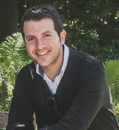 David Fernandez-Antoran合作论文数Wellcome Sanger Institute7
 Ong Swee Hoe合作论文数Genome Institute of Singapore7Triantafyllos Chavakis合作论文数7
 C. King合作论文数Wellcome Sanger Institute6Mathieu-Benoit Voisin合作论文数6Joanna C. Fowler合作论文数5Murai Kasumi合作论文数5
 Gerstung Moritz合作论文数Dept Biosyst Sci & Engn, Swiss Fed Inst Technol5Dentro Stefan合作论文数5Magdalena Gianotti合作论文数5Roshan Sood合作论文数Wellcome Sanger Institute4Michael W J Hall合作论文数University of Cambridge4
 Benjamin Hall合作论文数MRC Cancer Unit, University of Cambridge4Meda Paolo合作论文数4
 Jordi Oliver合作论文数Institut Universitari d'Investigació en Ciències de la Salut, Universitat de les Illes Balears4Michel Aurrand-Lions合作论文数4
 Krishnaa Mahbubani合作论文数Lucy Cavendish College, University of Cambridge;Department of Haematology, University of Cambridge4
 Emilie Abby合作论文数3
 Ilias Moutsopoulos合作论文数3Argyro Kalogeropoulou合作论文数Univ Patras, Fac Med, Dept Physiol, Stem Cell Biol Lab, Rion 26504, Achaia, Greece3Beyrau Martina合作论文数3Bodkin Jennifer Victoria合作论文数3Isabel Lladó合作论文数3
 Pilar Roca合作论文数Institut Universitari d’Investigació en Ciències de la Salut (IUNICS),, Universitat de les Illes Balears3david a leinster合作论文数2
 Dorothee Caille合作论文数2Ines Ferreira合作论文数2
 Adamo Valle合作论文数IUNICS, Univ Balearic Isl2
 Steven M. Albelda合作论文数Department of Medicine, Perelman School of Medicine, University of Pennsylvania;Thoracic Oncology Research Laboratory, University of Pennsylvania;Albelda Lab, Center for Cellular Immunotherapies, University of Pennsylvania2
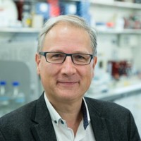 Bart Vanhaesebroeck合作论文数Research Department of Oncology, University College London;Cancer Institute, University College London2
 Wabik Agnieszka合作论文数Hutchison-MRC Research Centre, University of Cambridge2
 Costantino Pitzalis合作论文数William Harvey Research Institute, Barts and The London School of Medicine and Dentistry, Queen Mary University of London2
 Ed Rainger合作论文数Institute of Cardiovascular Sciences, College of Medical and Dental Sciences, University of Birmingham2
 Mauro Perretti合作论文数The William Harvey Research Institute, Faculty of Medicine and Dentistry, Queen Mary University of London2
 Christopher Bryant合作论文数Department of Biology and Biochemistry, University of Bath;and.;Department of Biology and Biochemistry, University of Bath2Diapouli Frantzeska-Maria合作论文数2
 Natalia Reglero-Real合作论文数Centro de Biología Molecular Severo Ochoa, Universidad Autónoma de Madrid2
 Gerard Nash合作论文数Institute of Cardiovascular Sciences, College of Medical and Dental Sciences, University of Birmingham2John V. Priestley合作论文数2
 Irina Mohorianu合作论文数School of Computing Sciences, University of East Anglia2
 Sharon Averill合作论文数2




