We have cloned the zebrafish homolog of the receptor tyrosine kinase flk-1 to provide us with a tool to study normal vascular pattern formation in the developing zebrafish embryo and to compare it to mutants in which vascular pattern is perturbed. We find that during normal development the first angioblasts arise laterally in the mesoderm and then migrate medially to form the primordia of the large axial vessels, the dorsal aorta (axial artery) and the axial vein. Lumen formation occurs shortly before onset of circulation at 24 hr postfertilization. We examined the specification of vascular progenitors in the mutant cloche, which fails to form both vessels and blood. cloche lacks all flk-expressing cells and therefore appears to lack angioblasts. The axial vessels of the trunk form in close proximity to notochord and endoderm, which may provide cues for their formation. The dorsal aorta is normally just ventral to the notochord; the axial vein is just below the dorsal aorta and above the endoderm. floating head (flh) and no tail (ntl) mutants both have defects in the formation of notochord. Both are cell-autonomous lesions, flh abolishing notochord and ntl preventing its differentiation. In both mutants the dorsal aorta fails to form, while formation of the axial vein is less affected. Mosaic analysis of mutant embryos shows that transplanted wild-type cells can become notochord in mutant flh embryos. In these mosaic embryos flh cells expressing flk assemble at the midline, beneath the wild-type notochord, and form an aortic primordium. This suggests that signals from the notochord may guide angioblasts in the fashioning of the dorsal aorta. The notochord seems to be less important for the formation of the vein.
As part of a large-scale mutagenesis screen of the zebrafish genome, we have identified 58 mutations that affect the formation and function of the cardiovascular system. The cardiovascular system is particularly amenable for screening in the transparent zebrafish embryo because the heart and blood vessels are prominent and their function easily examined. We have classified the mutations affecting the heart into those that affect primarily either morphogenesis or function. Nine mutations clearly disrupt the formation of the heart. cloche deletes the endocardium. In cloche mutants, the myocardial layer forms in the absence of the endocardium but is dysmorphic and exhibits a weak contractility. Two loci, miles apart and bonnie and clyde, play a critical role in the fusion of the bilateral tubular primordia. Three mutations lead to an abnormally large heart and one to the formation of a diminutive, dysmorphic heart. We have found no mutation that deletes the myocardial cells altogether, but one, pandora, appears to eliminate the ventricle selectively. Seven mutations interfere with vascular integrity, as indicated by hemorrhage at particular sites. In terms of cardiac function, one large group exhibits a weak beat. In this group, five loci affect both chambers and seven a specific chamber (the atrium or ventricle). For example, the weak atrium mutation exhibits an atrium that becomes silent but has a normally beating ventricle. Seven mutations affect the rhythm of the heart causing, for example, a slow rate, a fibrillating pattern or an apparent block to conduction. In several other mutants, regurgitation of blood flow from ventricle to atrium is the most prominent abnormality, due either to the absence of valves or to poor coordination between the chambers with regard to the timing of contraction. The mutations identified in this screen point to discrete and critical steps in the formation and function of the heart and vasculature.
 Mark C. Fishman合作论文数Fishman Lab, Department of Stem Cell & Regenerative Biology, Harvard University4
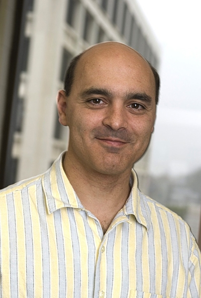 Brant M. Weinstein合作论文数National Institute of Child Health and Human Development, National Institutes of Health4
 Fabrizio C. Serluca合作论文数2Bl Roman合作论文数2Vn Pham合作论文数2Tae-Lin Huh合作论文数2F Zwartkruis合作论文数1L Solnica-Krezel合作论文数1AM VOGEL合作论文数1Pn Bennett合作论文数1
 Jau-Nian Chen合作论文数Department of Molecular Cell and Developmental Biology, College of Life Sciences, University of California, Los Angeles1
 Steffen E Meiler合作论文数Department of Anesthesiology and Perioperative Medicine, Medical College of Georgia1
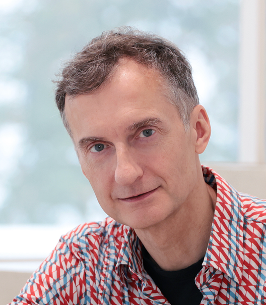 Alexander F. Schier合作论文数The Center for Molecular Life Sciences, University of Basel1Kerri S Warren合作论文数1Lawson Nathan D合作论文数1
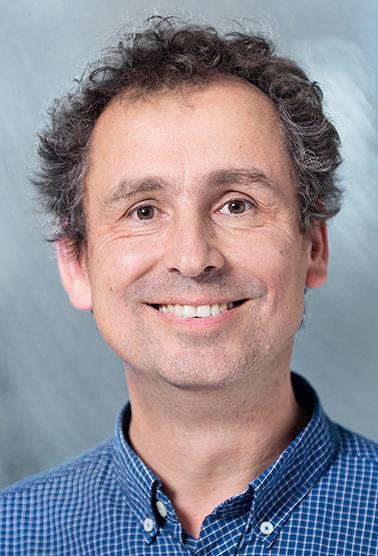 Stephan Neuhauss合作论文数Department of Molecular Life Sciences, University of Zurich1
 Wolfgang Driever合作论文数Institute of Biology I, University of Freiburg1Jarema Malicki合作论文数1Derek L. Stemple合作论文数1
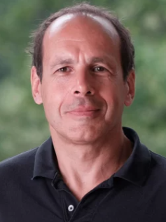 Didier Stainier合作论文数Department of Developmental Genetics, Max Planck Institute for Heart and Lung Research1




