Supplemental Table 1. MDS International Working Group response criteria for hematologic improvement Supplemental Table 2. Differences in Baseline Patient Characteristics Supplemental Table 3. Responses by Cytogenetics Supplemental Table 4. Responses by Mutation Supplemental Table 5. Study Representativeness Table
Supplemental Figure 1. Overall Survival Responders vs Non-Responders Supplemental Figure 2. Overall Survival Among Groups
During aging, hematopoietic stem cell (HSC) function progressively declines which can lead to reduced blood cell production and regeneration. This work uncovered that cell surface presentation of P-selectin (CD62P, encoded by Selp) increases in a large fraction of aging HSCs driven by a proinflammatory milieu in mice. Notably, expression of P-selectin molecularly and functionally dichotomized the aging HSC pool; stem cells presenting with highly abundant P-selectin were hallmarked by aging-associated gene expression programs and reduced repopulation capacity upon regenerative stress. Ectopic expression of Selp in young HSCs was sufficient to impair long-term reconstitution potential and impair erythropoiesis. Mechanistically, we uncovered that P-selectin receptor activation by its primary ligand, P-selectin glycoprotein ligand-1, suppressed aging-associated gene expression, and, reversely, lack of P-selectin signaling led to HSC premature aging. Collectively, our study uncovered a functional role of P-selectin engagement in regulating HSC regeneration and driving stem cell aging when perturbed.
During aging, hematopoietic stem cell (HSC) function progressively declines, which can lead to reduced blood cell production and regeneration. This work uncovered that cell surface presentation of P-selectin (CD62P, encoded by Selp) increases in a large fraction of aging HSCs driven by a proinflammatory milieu in mice. Notably, expression of P-selectin molecularly and functionally dichotomized the aging HSC pool; stem cells presenting with highly abundant P-selectin were hallmarked by aging-associated gene expression programs and reduced repopulation capacity upon regenerative stress. Ectopic expression of Selp in young HSCs was sufficient to impair long-term reconstitution potential and impair erythropoiesis. Mechanistically, we uncovered that P-selectin receptor activation by its primary ligand, P-selectin glycoprotein ligand-1 (PSGL-1), suppressed aging-associated gene expression and conversely, lack of P-selectin signaling led to HSC premature aging. Collectively, our study uncovered a functional role of P-selectin engagement in regulating HSC regeneration and driving stem cell aging when perturbed.
Mechanisms governing the maintenance of blood-producing hematopoietic stem and multipotent progenitor cells (HSPCs) are incompletely understood, particularly those regulating fate, ensuring long-term maintenance, and preventing aging-associated stem cell dysfunction. We uncovered a role for transitory free cytoplasmic iron as a rheostat for adult stem cell fate control. We found that HSPCs harbor comparatively small amounts of free iron and show the activation of a conserved molecular response to limited iron-particularly during mitosis. To study the functional and molecular consequences of iron restriction, we developed models allowing for transient iron bioavailability limitation and combined single-molecule RNA quantification, metabolomics, and single-cell transcriptomic analyses with functional studies. Our data reveal that the activation of the limited iron response triggers coordinated metabolic and epigenetic events, establishing stemness-conferring gene regulation. Notably, we find that aging-associated cytoplasmic iron loading reversibly attenuates iron-dependent cell fate control, explicating intervention strategies for dysfunctional aged stem cells.
Acute myeloid leukemia (AML) is initiated and sustained by a hierarchy of leukemia stem cells (LSCs), and elimination of this cell population is required for curative therapies. Here we show that transmembrane and immunoglobulin domain containing 2 (TMIGD2), a recently discovered co-stimulatory immune receptor, is aberrantly expressed by human AML cells, and can be used to identify and enrich functional LSCs. We demonstrate that TMIGD2 is required for the development and maintenance of AML and self-renewal of LSCs but is not essential for normal hematopoiesis. Mechanistically, TMIGD2 promotes proliferation, blocks myeloid differentiation and increases cell-cycle of AML cells via an ERK1/2-p90RSK-CREB signaling axis. Targeting TMIGD2 signaling with anti-TMIGD2 monoclonal antibodies attenuates LSC self-renewal and reduces leukemia burden in AML patient-derived xenograft models but has negligible effect on normal hematopoietic stem/progenitor cells. Thus, our studies reveal the function of TMIGD2 in LSCs and provide a promising therapeutic strategy for AML.
Hematopoietic stem cells (HSCs) are routinely mobilized from the bone marrow (BM) to the blood circulation for clinical transplantation. However, the precise mechanisms by which individual stem cells exit the marrow are not understood. This study identified cell-extrinsic and molecular determinants of a mobilizable pool of blood-forming stem cells. We found that a subset of HSCs displays macrophage-associated markers on their cell surface. Although fully functional, these HSCs are selectively niche-retained as opposed to stem cells lacking macrophage markers, which exit the BM upon forced mobilization. Macrophage markers on HSCs could be acquired through direct transfer by trogocytosis, regulated by receptor tyrosine-protein kinase C-Kit (CD117), from BM-resident macrophages in mouse and human settings. Our study provides proof of concept that adult stem cells utilize trogocytosis to rapidly establish and activate function-modulating molecular mechanisms.
Malignancies are reliant on glutamine as an energy source and a facilitator of aberrant DNA methylation. We demonstrate preclinical synergy of telaglenastat (CB-839), a selective glutaminase inhibitor, combined with azacytidine (AZA), followed by a single-arm, open-label, phase 1b/2 study in persons with advanced myelodysplastic syndrome (MDS). The dual primary endpoints evaluated clinical activity, safety and tolerability; secondary endpoints evaluated pharmacokinetics, pharmacodynamics, overall survival, event-free survival and duration of response. The dose-escalation study included six participants and the dose-expansion study included 24 participants. Therapy was well tolerated and led to an objective response rate of 70% with (marrow) complete remission in 53% of participants and a median overall survival of 11.6 months, with evidence of myeloid differentiation in responders determined by single-cell RNA sequencing. Glutamine transporter solute carrier family 38 member 1 in MDS stem cells was associated with clinical responses and predictive of worse prognosis in a large MDS cohort. These data demonstrate the safety and efficacy of CB-839 and AZA as a combined metabolic and epigenetic approach in MDS. ClinicalTrials.gov identifier: NCT03047993 . DiNardo et al. perform a phase 1b/2 clinical trial of telaglenastat (CB-839) in combination with azacytidine in persons with advanced myelodysplastic syndromes and report on the treatment safety and efficacy, including a definition of clinical responders.
Dysregulated apoptosis and proliferation are fundamental properties of cancer, and microRNAs (miRNA) are critical regulators of these processes. Loss of miR- 15a/16- 1 at chromosome 13q14 is the most common genomic aberration in chronic lympho-cytic leukemia (CLL). Correspondingly, the deletion of either murine miR- 15a/16- 1 or miR- 15b/16- 2 locus in mice is linked to B cell lymphoproliferative malignancies. However, unexpectedly, when both miR-15/16 clusters are eliminated, most double knockout (DKO) mice develop acute myeloid leukemia (AML). Moreover, in patients with CLL, significantly reduced expression of miR-15a, miR-15b, and miR-16 associates with progression of myelodysplastic syndrome to AML, as well as blast crisis in chronic myeloid leukemia. Thus, the miR-15/16 clusters have a biological relevance for myeloid neoplasms. Here, we demonstrate that the myeloproliferative phenotype in DKO mice correlates with an increase of hematopoietic stem and progenitor cells (HSPC) early in life. Using single -cell transcriptomic analyses, we presented the molecular underpinning of increased myeloid output in the HSPC of DKO mice with gene signatures suggestive of dysregulated hematopoiesis, metabolic activities, and cell cycle stages. Functionally, we found that multipotent progenitors (MPP) of DKO mice have increased self- renewing capacities and give rise to significantly more progeny in the granulocytic compartment. Moreover, a unique transcriptomic signature of DKO MPP correlates with poor out-come in patients with AML. Together, these data point to a unique regulatory role for miR-15/16 during the early stages of hematopoiesis and to a potentially useful biomarker for the pathogenesis of myeloid neoplasms.
PDF file - 646K, Supp Fig 1: Pathways involved by hypermethylated genes in PV / ET. Supp Fig 2: Validation of HELP assay findings by MassArray bisulfite analysis Supp Fig 3: Pathways involved by genes that are overexpressed and hypomethylated at promoters by the Jak2V617F mutant. Supp Fig 4: Jak2V617F mutation does not have significant effects on global DNA methylation profiles
Background: Cancer patients show increased morbidity with COVID-19 and need effective immunization strategies. Many healthcare regulatory agencies recommend administering ‘booster’ doses of COVID-19 vaccines beyond the standard two-dose series, for this group of patients. Therefore, studying the efficacy of these additional vaccine doses against SARS-CoV-2 and variants of concern is of utmost importance in this immunocompromised patient population Methods: We conducted a prospective single arm clinical trial enrolling patients with cancer that had received two doses of mRNA or one dose of AD26.CoV2.S vaccine and administered a third dose of mRNA vaccine. We further enrolled patients that had no or low responses to three mRNA COVID vaccines and assessed the efficacy of a fourth dose of mRNA vaccine. Efficacy was assessed by changes in anti-spike antibody, T-cell activity, and neutralization activity, which were again assessed at baseline and 4 weeks. Results: We demonstrate that a third dose of COVID-19 vaccine leads to seroconversion in 57% of patients that were seronegative after primary vaccination series. The immune response is durable as assessed by anti-SARS-CoV-2 (anti-S) antibody titers, T-cell activity, and neutralization activity against wild-type (WT) SARS-CoV2 and BA1.1.529 at 6 months of follow-up. A subset of severely immunocompromised hematologic malignancy patients that were unable to mount an adequate immune response (titer <1000 AU/mL) after the third dose and were treated with a fourth dose in a prospective clinical trial which led to adequate immune boost in 67% of patients. Low baseline IgM levels and CD19 counts were associated with inadequate seroconversion. Booster doses induced limited neutralization activity against the Omicron variant. Conclusions: These results indicate that third dose of COVID vaccine induces durable immunity in cancer patients and an additional dose can further stimulate immunity in a subset of patients with inadequate response. Funding: Leukemia Lymphoma Society, National Cancer Institute. Clinical trial number: NCT05016622 .
Aberrant overexpression of Interleukin-8 (IL8) has been reported in Myelodysplastic Syndromes (MDS), Acute Myeloid Leukemia (AML), Myeloproliferative Neoplasms (MPNs) and other myeloid malignancies. IL8 (CXCL8) is a CXC chemokine that is secreted by aberrant hematopoietic stem and progenitors as well as other cells in the tumor microenvironment. IL8 can bind to CXCR1/CXCR2 receptors and activate oncogenic signaling pathways, and also increase the recruitment of myeloid derived suppressor cells to the tumor microenvironment. IL8/CXCR1/2 overexpression has been associated with poorer prognosis in MDS and AML and increased bone marrow fibrosis in Myelofibrosis. Preclinical studies have demonstrated benefit of inhibiting the IL8/CXCR1/2 pathways via restricting the growth of leukemic stem cells as well as normalizing the immunosuppressive microenvironment in tumors. Targeting the IL8-CXCR1/2 pathway is a potential therapeutic strategy in myeloid neoplasms and is being evaluated with small molecule inhibitors as well as monoclonal antibodies in ongoing clinical trials. We review the role of IL8 signaling pathway in myeloid cancers and discuss future directions on therapeutic targeting of IL8 in these diseases.
Supp Fig 1 : SFRP1 methylation with coculture Supp Fig 2 : Validation of methylation changes with stroma and leukemic cell coculture Supp Fig 3: FRZB upregulation in MDS stroma after 5-Azacytidine treatment SuppFig 4: FRZB knockdown leads to partial inhibition of erythroid differentiation induced by treatment of MDS stromawith 5-Aza SuppFig 5: The expression of WNT signature genes in MDS and control marrow derived CD34+ cells
Culture conditions in which hematopoietic stem cells (HSCs) can be expanded for clinical benefit are highly sought after. To elucidate regulatory mechanisms governing the maintenance and propagation of human HSCs ex vivo, we screened libraries of annotated small molecules in human cord blood cells using an optimized assay for detection of functional HSCs during culture. We found that the antifungal agent ciclopirox ethanolamine (CPX) selectively supported immature CD34+CD90+ cells during culture and enhanced their long-term in vivo repopulation capacity. Purified HSCs treated with CPX showed a reduced cell division rate and an enrichment of HSC-specific gene expression patterns. Mechanistically, we found that the HSC stimulating effect of CPX was directly mediated by chelation of the intracellular iron pool, which in turn affected iron-dependent proteins and enzymes mediating cellular metabolism and respiration. Our findings unveil a significant impact of iron homeostasis in regulation of human HSCs, with important implications for both basic HSC biology and clinical hematology.
PDF file - 846K, Supplementary Table 1: Genes hypermethylated in PV / ET Supplementary Table 2: Genes hypermethylated in PMF Supplementary Table 3: Genes hypomethylated in PMF Supplementary Table 4: Genes hypermethylated in PMF cases with ASXL1 mutations / deletions Supp Table 5: Biological pathways affected by hypermethylated genes in ASXL1 mutated/deleted cases of PMF Supplementary Table 6: Genes hypermethylated in MPN cases with TET2 mutations Supp Table 7: Biological pathways affected by hypermethylated genes in TET2 mutated cases of MPNs
Curtailed protein translation ensures stemness and multipotency in embryonic and adult tissue-specific stem cells. In this issue of Cell, a study led by Zhao and colleagues uncovered increased susceptibility of hematopoietic stem cells (HSC) to iron-dependent programmed necrotic cell death (ferroptosis) as a consequence of low protein synthesis.
Acute myeloid leukemia (AML) is a clinically and molecularly heterogeneous group of mostly incurable hematologic malignancies driven by transformed stem cells (leukemic stem cells, LSC) endowed with enhanced self-renewal capacity and blocked myeloid differentiation. Insights into pathways of LSC maintenance will provide key targets for curative therapies. We have previously shown that chaperone-mediated autophagy (CMA) is essential for metabolic adaptation in HSC to regenerative stress (Dong et al., Nature 2021). To test the relevance of CMA in LSC, we developed new mouse models of AML with concomitant CMA deficiency through conditionally deleting (Vav-iCre driven) Lysosome-associated membrane protein 2 (Lamp2a, L2A) which is specific and rate-limiting for this autophagy pathway. We characterized self-renewal and myeloid differentiation potential of leukemia cells upon L2A deletion by gold-standard serial ex vivo colony assays and in vivo bone marrow transplantation assays. Our data thus far have revealed that CMA blockade causes compromised ex vivo self-renewal of LSC and significantly prolongs survival of recipients upon adoptive cell transfer compared to L2AWT controls, consistent with compromised LSC maintenance. Moreover, CMA deficient LSC presented with significantly increased myeloid differentiation compared to CMA competent LSC, suggesting that CMA impacts molecular pathways that favor self-renewal over differentiation commitment. We are currently building precise humanized models and employ degron-mediated ablation of L2A to test the consequences of CMA ablation in human AML. If successful, our study will provide exciting preclinical rationales for the testing of CMA inhibitors for eradication of LSC, particularly alongside current mainstay therapies, as a novel and potential curative option for patients with myeloid malignancies. Acute myeloid leukemia (AML) is a clinically and molecularly heterogeneous group of mostly incurable hematologic malignancies driven by transformed stem cells (leukemic stem cells, LSC) endowed with enhanced self-renewal capacity and blocked myeloid differentiation. Insights into pathways of LSC maintenance will provide key targets for curative therapies. We have previously shown that chaperone-mediated autophagy (CMA) is essential for metabolic adaptation in HSC to regenerative stress (Dong et al., Nature 2021). To test the relevance of CMA in LSC, we developed new mouse models of AML with concomitant CMA deficiency through conditionally deleting (Vav-iCre driven) Lysosome-associated membrane protein 2 (Lamp2a, L2A) which is specific and rate-limiting for this autophagy pathway. We characterized self-renewal and myeloid differentiation potential of leukemia cells upon L2A deletion by gold-standard serial ex vivo colony assays and in vivo bone marrow transplantation assays. Our data thus far have revealed that CMA blockade causes compromised ex vivo self-renewal of LSC and significantly prolongs survival of recipients upon adoptive cell transfer compared to L2AWT controls, consistent with compromised LSC maintenance. Moreover, CMA deficient LSC presented with significantly increased myeloid differentiation compared to CMA competent LSC, suggesting that CMA impacts molecular pathways that favor self-renewal over differentiation commitment. We are currently building precise humanized models and employ degron-mediated ablation of L2A to test the consequences of CMA ablation in human AML. If successful, our study will provide exciting preclinical rationales for the testing of CMA inhibitors for eradication of LSC, particularly alongside current mainstay therapies, as a novel and potential curative option for patients with myeloid malignancies.
Abstract Purpose: Thrombocytopenia is a serious complication of myelodysplastic syndromes (MDS) associated with an increased bleeding risk and worse prognosis. Eltrombopag (ELT), a thrombopoietin receptor agonist, can increase platelet counts and reverse anti-megakaryopoietic effects of lenalidomide (LEN) in preclinical studies. We hypothesized ELT would reduce the incidence of thrombocytopenia in MDS. Patients and Methods: We conducted a Phase II multicenter trial of ELT and LEN in adult patients with low- or intermediate-1–risk MDS with symptomatic or transfusion-dependent anemia or thrombocytopenia (NCT01772420). Thrombocytopenic patients were started on ELT and subsequently treated with LEN after platelets were increased. Patients without thrombocytopenia were started on LEN monotherapy and treated with ELT if they became thrombocytopenic. Results: Fifty-two patients were enrolled; mean age was 71 years (range 34–93). Overall response rate (ORR) in the intention-to-treat population was 35% (18/52). ELT monotherapy led to ORR of 33.3% (7/21), 29% achieving hematologic improvement (HI)-Platelets, and 24% bilineage responses. LEN monotherapy had 38% ORR (6/16) with all responders achieving HI-Erythroid. Fifteen patients received both ELT and LEN with ORR of 33.3%, 20% achieved HI-Erythroid, and 20% HI-Platelets with 13% bilineage responses. Median duration of response was 40 weeks for ELT (range 8–ongoing), 41 weeks (25–ongoing) for LEN, and 88 weeks (8.3–ongoing) for ELT/LEN. Non-hematologic grade 3–4 treatment-related adverse events were infrequent. Among patients on ELT, 2 had major bleeding events, 1 had a reversible increase in peripheral blasts, and 1 developed marrow fibrosis after 6 years on ELT. Conclusions: ELT and LEN are well tolerated and effective in achieving hematologic improvement in patients with low-/intermediate-risk MDS.
 Ulrich G. Steidl合作论文数90
 Amit K. Verma合作论文数Division of Hemato-Oncology, Department of Oncology, Albert Einstein College of Medicine, Montefiore Einstein;Comprehensive Cancer Center, Montefiore Einstein;Roshon Therapeutics;ClinStreet;Stelexis BioSciences, Inc.56Tushar V Bhagat合作论文数Prince Sattam bin Abdulaziz University28Boris Bartholdy合作论文数27Kith Pradhan合作论文数23Victor Thiruthuvanathan合作论文数Blood Canc Inst, Montefiore Med Ctr23
 Shastri Aditi合作论文数Montefiore Medical Center, Albert Einstein College of Medicine;Montefiore Medical Center, Albert Einstein College of Medicine23
 Barreyro Laura合作论文数Department of Medicine (Oncology),, Albert Einstein College of Medicine20
 Tihomira I. Todorova合作论文数19
 Ioannis Mantzaris合作论文数18
 Gaurav Choudhary合作论文数Department of Molecular Pharmacology, Physiology, and Biotechnology, Brown University18Narayanagari Swathi合作论文数16Jiahao Chen合作论文数Blood Canc Inst, Montefiore Med Ctr14
 Jacqueline Boultwood合作论文数Radcliffe Department of Medicine, University of Oxford14
 Andrea Pellagatti合作论文数Radcliffe Department of Medicine, University of Oxford13Yu Yiting合作论文数12Yun-Ruei Kao合作论文数11Okoye-Okafor Ujunwa C合作论文数11Amittha Wickrema合作论文数Section of Hematology/Oncology, Department of Medicine, The University of Chicago11Luis Carvajal合作论文数Asterisk with author names denotes non-ASH members.10Nandini Ramachandra合作论文数10
 John M. Greally合作论文数Department of Pediatrics, Albert Einstein College of Medicine, Yeshiva University;Department of Genetics, Albert Einstein College of Medicine, Yeshiva University10Kambhampati Suman合作论文数9
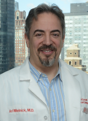 Ari M. Melnick合作论文数Departments of Medicine and Pharmacology, Weill Cornell Medical College9
 Bartenstein Matthias合作论文数Montefiore Medical Center, Albert Einstein College of Medicine;Montefiore Medical Center, Albert Einstein College of Medicine9
 Christian Steidl合作论文数Graduate Program in Bioinformatics, Faculty of Science, The University of British Columbia9Shanisha Gordon-Mitchell合作论文数8
 Kelly Mitchell合作论文数Health Sciences Center, Texas Tech University8
 Daniel G. Tenen合作论文数Division of Medical Sciences, Harvard Medical School;Beth Israel Deaconess Medical Center8
 Aluri Srinivas合作论文数Department of Microbiology and Cell Biology, Indian Institute of Science8
 Maximilian Christopeit合作论文数Center for Oncology, Universitätsklinikum Hamburg-Eppendorf;University Cancer Center Hamburg, Universitätsklinikum Hamburg-Eppendorf8
 Kira Gritsman合作论文数Albert Einstein Coll Med, Dept Cell Biol, Bronx, NY 10467 USA8
 Robert Stanley合作论文数7Madhuri Tatiparthy合作论文数7
 Maria M. Aivalioti合作论文数Dept Cell Biol, Montefiore Med Ctr7Ira Braunschweig合作论文数7
 Mario Pujato合作论文数Cincinnati Childrens Hosp Med Ctr, Ctr Autoimmune Genom & Etiol, Cincinnati, OH 45229 USA7
 Sanchari Bhattacharyya合作论文数Albert Einstein Coll Med, Bronx, NY 10461 USA7Sohal Davendra P S合作论文数7
 Roth Michael合作论文数7Li Zhou合作论文数7
 Constantine S. Mitsiades合作论文数Department of Medicine, Dana-Farber Cancer Institute;Department of Medicine, Harvard Medical School7
 Manuel Aivado合作论文数7
 Carolina Schinke合作论文数7Yuhong Ma合作论文数6
 Prafulla Bhagat合作论文数6
 Sakshi Jasra合作论文数Canc Ctr, Univ Vermont6Lumie Benard合作论文数6




