Background/Objectives: Formation of tight contacts between oral soft tissue and dental implants is a significant challenge in contemporary implantology. An essential role in this process is played by oral epithelial cells. In the present study, we investigated how titanium and zirconia surfaces with different roughness influence various parameters of oral epithelial cells in vitro. Methods: We used the human oral squamous carcinoma Ca9-22 cell line and cultured them on the following surfaces: machined smooth titanium (TiM) and zirconia (ZrM) surfaces, as well as sandblasted and acid-etched titanium moderately rough (SLA) and zirconia (ZLA) surfaces. Cell proliferation/viability was measured by CCK-8 assay, and cell morphology was analyzed by fluorescent microscopy. The gene expression of interleukin (IL)-8, intercellular adhesion molecule (ICAM)-1, E-cadherin, integrin (ITG)-α6, and ITG-β4 was measured by qPCR, and the content of IL-8 in conditioned media by ELISA. Results: At the initial culture phase, cell proliferation was promoted by rougher surfaces. Differences in cell attachment were observed between machined and moderately rough surfaces. Machined surfaces were associated with slightly higher IL-8 levels (p < 0.05). Furthermore, both ZLA and SLA surfaces promoted the expression of (ITG)-α, ITG-β4, and ICAM-1 in Ca9-22 cells (p < 0.05). Surface material had no impact on the investigated parameters. Conclusions: Under the limitations of this in vitro study, some properties of oral epithelial cells, particularly the immunological and barrier function, are moderately modified by roughness but not by material. Hence, the roughness of the implant surface might play a role in the quality of the peri-implant epithelium.
Angiogenesis is essential for skeletal development, bone healing, and regeneration. Improved non-destructive, three-dimensional (3D) imaging of the vasculature within bone tissue benefits many research areas, especially implantology and tissue engineering. X-ray microcomputed tomography (microCT) is a well-suited non-destructive 3D imaging technique for bone morphology. For microCT-based detection of vessels, it is paramount to use contrast enhancement. Limited differences in radiopacity between perfusion agents and mineralized bone make their distinct segmentation problematic and have been a major drawback of this approach. A decalcification step resolves this issue but inhibits the simultaneous assessment of bone microstructure and vascular morphology. The problem of contrasting becomes further complicated in samples with metal implants. This study describes contrast-enhanced microCT-based visualization of vasculature within bone tissue in small and large animal models, also in the vicinity of the metal implants. We present simultaneous microvascular and bone imaging in murine tibia, a murine bone metastatic model, the pulp chamber, gingiva, and periodontal ligaments. In a large animal model (minipig), we performed visualization and segmentation of different tissue types and vessels in the hemimandible containing metal implants. We further demonstrate the potential of dual-energy imaging in distinguishing bone tissue from the applied contrast agents. This work introduces a non-destructive approach for 3D imaging of vasculature within soft and hard tissues near metal implants in a large animal model.
OBJECTIVES:The aim of the present study was to compare a novel tapered, double-threaded self-tapping tissue-Level design implant (TLC) to a well-established parallel walled tissue-level (TL) implant in terms of primary and secondary stability over time. MATERIALS AND METHODS:Test TLC (n = 10/per timepoint) and control TL (n = 10/per timepoint) implants were placed in the mandible of minipigs and left for submerged healing for 3, 6, and 12 weeks. Maximum insertion torque and implant stability quotient (ISQ) were measured for each implant at placement. Osseointegration and cortical bone maintenance were histologically evaluated by measuring total bone-to-implant contact (BIC) and first bone-to-implant contact (fBIC). RESULTS:A significantly higher maximum insertion torque was measured for the test implant TLC compared to the control TL implant (57.83 ± 24.73 Ncm and 22.62 ± 23.16 Ncm, respectively; p < .001). The mean ISQ values were comparable between the two implant types (75.00 ± 6.70 for TL compared to 75.40 ± 3.20 for TLC, p = .988). BIC was comparable between both implant types at each of the evaluated time points. The fBIC was found to be significantly more coronal at 12 weeks for the TLC implant compared to the TL implant (0.31 ± 0.83 mm for TLC compared to -0.22 ± 0.85 for TL, p = .027). CONCLUSION:The novel tapered tissue level design implant showed improved primary stability and an overall improved crestal bone height maintenance compared to the parallel walled design at 12 weeks.
AIMS:To test whether titanium surface roughness disparity might be used to specifically guide the behavior of gingiva fibroblasts and keratinocytes, thereby improving the quality of soft tissue (ST) integration around abutments. METHODS:Titanium discs resembling the roughness of enamel (M) or cementum (MA) were created with normal or increased hydrophilicity and used as substrates for human fibroblasts and keratinocytes. Adhesion and proliferation assays were performed to assess cell-type specific responses upon encountering the different surfaces. Additionally, immunofluorescence and qPCR analyses were performed to study more in depth the behavior of fibroblasts and keratinocytes on MA and M surfaces, respectively. RESULTS:While enamel-like M surfaces supported adhesion, growth and a normal differentiation potential of keratinocytes, cementum-emulating MA surfaces specifically impaired the growth of keratinocytes. Vice versa, MA surfaces sustained regular adhesion and proliferation of fibroblasts. Yet, a more intimate adhesion between fibroblasts and titanium was achieved by an increased hydrophilicity of MA surfaces, which was associated with an increased expression of elastin. CONCLUSION:The optimal titanium implant abutment might be achieved by a bimodal roughness design, mimicking the roughness of enamel (M) and cementum with increased hydrophilicity (hMA), respectively. These surfaces can selectively elicit cell responses favoring proper ST barrier by impairing epithelial downgrowth and promoting firm adhesion of fibroblasts.
Surface design plays a critical role in determining the integration of dental implants with bone tissue. Femtosecond laser-texturing has emerged as a breakthrough technology offering excellent uniformity and reproducibility in implant surface features. However, when compared to state-of-the-art sandblasted and acid-etched surfaces, laser-textured surface designs typically underperform in terms of osseointegration. This study investigates the capacity of a bio-inspired femtosecond laser-textured surface design to enhance osseointegration compared to state-of-the-art sandblasted & acid-etched surfaces. Laser-texturing facilitates the production of an organized trabeculae-like microarchitecture with superimposed nano-scale laser-induced periodic surface structures on both 2D and 3D samples of titanium-zirconium-alloy. Following a boiling treatment to modify the surface chemistry, improving wettability to a contact angle of 10°, laser-textured surfaces enhance fibrin network formation when in contact with human whole blood, comparable to state-of-the-art surfaces. In vitro experiments demonstrate that laser-textured surfaces significantly outperform state-of-the-art surfaces with a 2.5-fold higher level of mineralization by bone progenitor cells after 28 days of culture. Furthermore, in vivo evaluations reveal superior biomechanical integration of laser-textured surfaces after 28 days of implantation. Notably, during abiological pull-out tests, laser-textured surfaces exhibit comparable performance, suggesting that the observed enhanced osseointegration is primarily driven by the biological response to the surface.
PURPOSE:To histometrically compare the osseointegration and crestal bone healing of a novel tapered, self-cutting tissue-level test implant with a standard tissue-level control implant in a submerged healing regimen. MATERIALS AND METHODS:In a mandibular minipig model, implants were inserted and evaluated histometrically after a healing period of 3, 6, and 12 weeks. The primary outcome was the evaluation of bone-to-implant contact (BIC) and secondary outcomes were primary stability as per insertion torque and first BIC (fBIC). Outcomes for the test and control implants were compared using Wilcoxon signed-rank tests and mixed linear regression models. RESULTS:Insertion torque values were significantly higher for the test (50.0 ± 26.4 Ncm) compared to the control implants (35.2 ± 19.7 Ncm, p = .0071). BIC values of test implants were non-inferior to those of control implants over the investigated study period. After 12 weeks, the corresponding values measured were 81.62 ± 11.12% and 90.41 ± 4.81% (p = .1763) for test and control implants, respectively. Similarly, no statistical difference was found for fBIC values, except for the 12 weeks outcome that showed statistically lower values for the test (-675.58 ± 590.88 μm) compared to control implants (-182.75 ± 197.40 μm, p = .0068). CONCLUSIONS:Novel self-cutting tissue-level implants demonstrated noninferior osseointegration and crestal bone height maintenance to the tissue-level implants. Histometric outcomes between both implants demonstrated test implants were statistically noninferior to control implants, despite substantial differences in the bone engagement mechanism and resulting differences in insertion torque and qualitative bone healing patterns.
Surgeons treating fractures with many small osteochondral fragments have often expressed the clinical need for an adhesive to join such fragments, as an adjunct to standard implants. If an adhesive would maintain alignment of the articular surfaces and subsequently heal it could result in improved clinical outcomes. However, there are no bone adhesives available for clinical indications and few pre-clinical models to assess safety and efficacy of adhesive biomaterial candidates. A bone adhesive candidate based on water, α-TCP and an amino acid phosphoserine was evaluated in-vivo in a novel murine bone core model (preliminary results presented EORS 2019) in which excised bone cores were glued back in place and harvested @ 0, 3, 7, 14, 28 and 42days. Adhesive pull-out strength was demonstrated 0–28 days, with a dip at 14 days increasing to 11.3N maximum. Histology 0–42 days showed the adhesive progressively remodelling to bone in both cancellous and cortical compartments with no signs of either undesirable inflammation or peripheral ectopic bone formation. These favourable results suggested translation to a large animal model.A porcine dental extraction socket model was subsequently developed where dental implants were affixed only with the adhesive. Biomechanical data was collected @ 1, 14, 28 and 56 days, and histology at 1,14,28 and 56 days. Adhesive strength assessed by implant pull-out force increased out to 28 days and maintained out to 56 days (282N maximum) with failure only occurring at the adhesive bone interface. Histology confirmed the adhesive's biocompatibility and osteoconductive behavior. Additionally, remodelling was demonstrated at the adhesive-bone interface with resorption by osteoclast-like cells and followed by new bone apposition and substitution by bone. Whilst the in-vivo dental implant data is encouraging, a large animal preclinical model is needed (under development) to confirm the adhesive is capable of healing, for example, loaded osteochondral bone fragments.Acknowledgements: The murine study was supported, in part, by the Swedish Foundation for Strategic Research (#RMA15-0110).
AimsTo histologically compare osseointegration and crestal bone healing between newly introduced tapered, self-cutting bone-level test implants and tapered bone-level control implants in sites with fully healed sites.MethodsSixty-six implants (33 test, 33 control) were placed 1 mm subcrestally in a minipig model and underwent qualitative histologic and quantitative histometric analyses after 3, 6 and 12 weeks of submerged healing. The primary and secondary outcomes were the bone-to-implant contact (BIC) and first bone-to-implant contact (fBIC). Outcomes between the test and control implants were statistically compared.ResultsThe BIC values of the test implants were comparable and non-inferior over the time points studied, except for the 12 weeks time point which showed statistically significantly higher BIC values of the test (88.07 +/- 5.35%) compared to the control implants (80.88 +/- 7.51%) (p = .010). Similarly comparable and non-inferior were the fBIC values, except for the 6-week outcome, which showed statistically higher values for the test (-546.5 +/- 450.80 mu m) compared to the control implants (-75.7 +/- 100.59 mu m). fBIC results for the test implants were qualitatively more stable and consistent between test time points.ConclusionNovel self-cutting bone-level test implants demonstrated superior osseointegration and similar bone levels compared to conventional bone-level implants after a healing period of 12 weeks in healed ridges.
OBJECTIVES:To investigate the impact of a Ti-Sr-O technology, applied to either a turned surface or an SLA surface, on the mechanical robustness of osseointegration, benchmarked against the SLActive surface.MATERIAL AND METHODS:Ti discs (6.25-mm-diameter and 2-mm-thick) with three different surfaces were inserted on the proximal-anterior part of the tibial plateau of adult Swedish loop rabbits: (I) turned surface modified with Ti-Sr-O (turned + Ti-Sr-O), (II) SLA surface modified with Ti-Sr-O (SLA + Ti-Sr-O), and (III) SLActive surface (SLActive). Following a healing period of 2 weeks and 4 weeks, the pull-out (PO) force needed to detach the discs from the bone was assessed, as a surrogate of osseointegration.RESULTS:The SLActive surface exhibited statistically significant higher median PO forces, compared with the SLA + Ti-Sr-O surfaces at both 2- and 4 weeks post-op (p > .05). In this study, no single turned + Ti-Sr-O surface disk was integrated.CONCLUSIONS:The tested Ti-Sr-O technology failed to enhance osseointegration; however, this finding may be related to the inappropriateness of the rabbit tibia plateau model for assessing third-generation implant surface technologies, due to the limited diffusion and clearance at the disk-bone interface.
To examine factors influencing the primary stability of dental implants when stabilized in over-sized osteotomies using a calcium phosphate-based adhesive cement was the objective.Using implant removal torque measurements as a surrogate for primary stability, we examined the influence of implant design features (diameter, surface area, and thread design), along with cement gap size and curing time, on the resulting primary implant stability.Removal torque values scaled with implant surface area and increasing implant diameters. Cement gap size did not alter the median removal torque values; however, larger gaps were associated with an increased spread of the measured values. Among the removal torque values measured, all were found to be above 32 Ncm which is an insertion torque threshold value commonly recommended for immediate loading protocols.The adhesive cement show potential for offering primary implant stability for different dental implant designs. In this study, the primary parameters influencing the measured removal torque values were the implant surface area and diameter. As the liquid cement prevents the use of insertion torque, considering the relationship between insertion and removal torque, removal torque can be considered a reliable surrogate for primary implant stability for bench and pre-clinical settings.At present, the primary stability of dental implants is linked to the quality of the host bone, the drill protocol, and the specific implant design. The adhesive cement might find applications in future clinical settings for enhancing primary stability of implants under circumstances where this cannot be achieved conventionally.
Angiogenesis is essential for skeletal development, bone healing and regeneration. Various research areas, especially implantology and tissue engineering, would benefit from improved three-dimensional (3D) imaging of the vasculature within bone tissue.X-ray microtomography (microCT) is a well-suited non-destructive 3D imaging technique for bone morphology. For the detection of vessels, a contrast-enhanced microCT-imaging must be used. Limited contrast between perfusion agents and mineralized bone has been the major drawback of this approach, making their distinct segmentation problematic. A decalcification step resolves this issue but inhibits simultaneous assessment of intracortical bone microstructure and vascular morphology. Moreover, the problem of contrasting becomes further compounded in samples with metal implants.This study describes μAngiofil-enhanced microCT-based visualization of vasculature within bone tissue in various small and large animal models, with and without decalcification. We present simultaneous microvascular and bone imaging in murine tibia, murine bone metastatic model, pulp chamber, gingiva and periodontal ligaments. In a large animal model (minipig) we perform visualization and segmentation of different tissue types and vessels in the hemimandible containing metal implants.Our manuscript introduces the first non-destructive approach for 3D imaging of the vasculature within soft and hard tissues in the vicinity of metal implants in a large animal model.### Competing Interest StatementThe authors have declared no competing interest.
To develop a new preclinical model to study early implant loss, where local infection conditions would impair the implant osseointegration. Forty-eight smooth, 2.9-mm diameter experimental implants were placed in the mandible of 8 beagle dogs (3 in each side). In half of the animals (test group, n = 24 implants), the implants received ligatures around the implant-abutment connection. In the other half, no ligatures were placed (control group, n = 24 implants). Four weeks later, implants were extracted in a flapless approach and standard 3.3-mm diameter SLActive implants were placed into the same osteotomy site without any further drilling. Eight weeks after the second implantation, animals were sacrificed and analyzed in terms of implant survival. After 8 weeks of healing, 4 implants were lost in the control group and 14 in the test group. This corresponded to a 17.4
OBJECTIVES:Immediate implant placement and loading is a practice that continues to gain traction in implant dentistry because it reduces treatment time and improves satisfaction. Novel implant designs that facilitate increased primary stability, while not compromising osseointegration and long-term survival are important to offer immediate solutions for missing teeth. Here, we hypothesize that fully tapered implants can obtain successful osseointegration with high survival rates after immediate loading in fresh extraction sockets and healed sites.MATERIALS AND METHODS:A total of 13 swine with 73 implants were evaluated. Fully tapered or apically tapered implants were placed in extraction sockets and healed sites. Insertion torque and resonance frequency analysis were determined at placement and euthanasia. Animals were evaluated at: placement, and 1-week and 12-weeks after placement. Bone to Implant Contact (BIC), Bone Area/Total Area (BA/TA), and first BIC (fBIC) analyses were conducted.RESULTS:The fully tapered implant achieved similar primary stability with lower insertion torque at placement. Apically and fully tapered implants had comparable BIC (50.1% vs 59.4%) and ISQ (82.5 vs 80.3) values by 12 weeks in healed sites. In extraction sockets, BIC and ISQ for the apically tapered implant was 35.8% and 73.2 and 37.8% and 79.2 for the fully tapered implants, respectively.CONCLUSIONS:In this short-term study, immediately loaded fully tapered implants obtained high survival with similar osseointegration ability as apically tapered implants when placed in healed sites and fresh extraction sockets. Fully tapered implants show promise for use in immediate loading and immediate placement.
AIM:To histologically evaluate the influence of (1) loading and (2) grafting on osseointegration and peri-implant soft-tissue healing at immediately placed, self-cutting progressive tissue-level implants (TLX) in a minipig model.MATERIALS AND METHODS:TLX implants (n = 56) were immediately placed following the extraction of the mandibular first and second premolars, bilaterally, in a total of n = 14 minipigs. In each animal, the implant sites were allocated to the following four groups: (1) unloaded with simultaneous grafting using a bovine bone mineral; (2) unloaded without grafting; (3) loaded with simultaneous grafting; and (4) loaded without grafting. Histomorphometric assessments at 4 and 12 weeks (n = 7 animals each) included primary (i.e., bone-to-implant contact [BIC]) and secondary outcome measures (e.g., first BIC [fBIC], junctional epithelium length [JE], connective tissue contact length [CTC], biological width [BW = JE + CTC]).RESULTS:At 4 weeks, mean BIC values ranged from 74.5 ± 11.6% in Group 2 to 83.8 ± 13.3% in Group 1, and, at 12 weeks, from 75.5% ± 7.9% in Group 2 to 79.9 ± 8.6% in Group 1. Multivariate linear mixed regression did not reveal any associations between BIC and implant loading or grafting at 4 and 12 weeks. At 12 weeks, significantly higher fBIC values were noted in Group 2 when compared with Group 1. All groups showed comparable JE, CTC, and BW values.CONCLUSIONS:Implant loading and grafting had no major effects on osseointegration and peri-implant soft tissue healing at TLX implants.
OBJECTIVES:Thermal and mechanical stresses during osteotomy preparation can impair implant osseointegration. This study investigated implant osseointegration following the measurement of temperature exposure during osteotomy drilling, varying drill design, sequence, and drill wear.MATERIALS AND METHODS:36 tapered implants were placed in a mandibular minipig model after guided drilling of implant osteotomies using 4 different groups: (1) control drills with a conservative, sequential drilling sequence, (2) control drills using a shortened drill sequence (PF), (3) novel test drill displaying an optimized drill design and surface treatment, PF, and (4) aged test drill, PF. Intraosseous temperatures during drilling were measured using a temperature probe. BIC, fBIC, and tissue reactions were histomorphometrically derived after 2 and 8 weeks of healing.RESULTS:Compared to control drills (1) or (2), test drills (3) resulted in significantly lower maximum temperatures ((35.4 (CI 30.2-40.5)°C vs. (46.5 (CI 41.0-52.0)°C, p = .0021)) and shorter drill times ((4.5 (CI 1.6-7.3)sec vs. 10.3 (7.3-13.4)sec). Lower osteotomy temperature values and shorter drill times corroborated with significantly higher BIC after 2 and 8 weeks healing for the test (3) compared to control groups (2) (2 weeks: (44.9 (CI 34.1-55. 7)% vs. (31.3 (CI 20.5-42.2)%, p = <.0001 and 8 weeks: (73.7 ( CI 64.2-83.2)% vs. (66.2 (CI 57.0-75.4)%, p = <.0455).CONCLUSION:The improved osseointegration of implants placed after osteotomy preparation with novel test drills using a shortened drill sequence compared to standard drills and conventional drill protocols might be attributed to more favorable thermal profiles and less mechanical stress exerted on the bone surrounding the implant osteotomy.
Due to the rising demand for zirconia (Zr) based implant systems, it is important to understand the impact of Zr and titanium (Ti) implants and particularly their topography on soft tissue healing. As human gingival fibroblasts (hGFs) are the predominant cells in peri-implant soft tissue, we focused on examining the effect of implant material and surface roughness on hGFs' initial attachment, growth and the expression of proteins involved in the focal adhesion. hGFs isolated from eight healthy donors were cultured on the following surfaces: smooth titanium machined surface (TiM), smooth zirconia machined surface (ZrM), moderately rough titanium surface (SLA), or moderately rough zirconia surface (ZLA) for up to 14 days. The initial attachment of hGFs was evaluated by scanning electron microscopy. Cell proliferation/viability was assessed by cell counting kit 8. Focal adhesion and cytoskeleton were visualized by a focal adhesion staining kit. The gene expression of focal adhesion kinase (FAK), α-smooth muscle actin (α-SMA), and integrin subunits ITG-β1, ITG-β4, ITG-α4, ITG-α5, ITG-α6, was evaluated by qPCR. Cell proliferation/viability was slightly decreased by moderately rough surfaces, whereas no effect of surface material was observed. Cell morphology was strikingly different between differently treated surfaces: on machined surfaces, cells had elongated morphology and were attached along the grooves, whereas on moderately rough surfaces, cells were randomly attached. Surface roughness had a more pronounced effect on the gene expression compared to the surface material. The expression of FAK, α-SMA, ITG-β4, ITG-α5, and ITG-α6 was enhanced by moderately rough surfaces compared to smooth surfaces. Within the limitations of this in vitro study, it can be concluded that the behavior of primary hGFs is primarily affected by surface structure, whereas no apparent advantage of Zr over Ti could be observed.
PURPOSE To determine the impact of experimentally preformed peri-implant crater-shaped bone defects on the evolution of in situ microbiota and development of bone defects compared to those induced over time by ligature placement only. METHODS Implants were installed in the mandibles of eight dogs. Standardized bone defects were preformed in four test animals but not in the other four control animals, prior to implant (3.3 mm × 8 mm) installation. After 2 months of healing, peri-implantitis was induced with silk ligatures in both groups for 2 months. Microbial samples were obtained from implants and teeth for analysis at three time points (qPCR), and the average depths of the bone defects were measured. RESULTS At the baseline, the total marker load of periodontal-pathogenic bacteria (TML) for teeth accounted for 5.2% (0-17.4%). After implant healing, TMLs for implants and teeth were comparable (7.1% [0.3-17.4%]). The TML of both groups was 3.5%, 2 months after ligature placement. Bone defects had a mean depth of 1.84 mm at preformed defects and 1.64 mm at control sites (P > 0.05). CONCLUSION Preformed defects in the test group showed comparable results to the control group in terms of TML, the incidence of periodontal-pathogenic bacteria, and bone defect depth.
AbstractObjectiveThe effectiveness of GBR procedures for the reconstruction of periodontal defects has been well documented. The objective of this investigation was to evaluate the degradation kinetics and biocompatibility of two resorbable collagen membranes in conjunction with a bovine xenograft material.Materials and MethodsLower premolars and first molars were extracted from 18 male Yucatan minipigs. After 4 months of healing, standardized semi‐saddle defects were created (12 mm × 8 mm × 8 mm [l˙̇ × W˙ × d]), with 10 mm between adjacent defects. The defects were filled with a bovine xenograft and covered with a either the bilayer collagen membrane (control) or the porcine pericardium‐derived collagen membrane (test). Histological analysis was performed after 4, 8, and 12 weeks of healing and the amount of residual membrane evaluated. Non‐inferiority was calculated using the Brunner‐Langer mixed regression model.ResultsHistological analysis indicated the presence of residual membrane in both groups at all time points, with significant degradation noted in both groups at 12 weeks compared to 4 weeks (p = .017). No significant difference in ranked residual membrane scores between the control and test membranes was detected at any time point.ConclusionsThe pericardium‐derived membrane was shown to be statistically non‐inferior to the control membrane with respect to resorption kinetics and barrier function when utilized for guided bone regeneration in semi‐saddle defects in minipigs. Further evaluation is necessary in the clinical setting.
Surface chemistry and nanotopography of dental implants can have a substantial impact on osseointegration. The aim of this investigation was to evaluate the effects of surface chemistry and nanotopography on the osseointegration of titanium-zirconium (TiZr; Roxolid®) discs, using a biomechanical pull-out model in rabbits. Two discs each were placed in both the right and left tibiae of 16 rabbits. Five groups of sandblasted acid etched (SLA) discs were tested: (1) hydrophobic without nanostructures (dry/micro) (n = 13); (2) hydrophobic with nanostructures, accelerated aged (dry/nano/AA) (n = 12); (3) hydrophilic without nanostructures (wet/micro) (n = 13); (4) hydrophilic with nanostructures, accelerated aged (wet/nano/AA; SLActive®) (n = 13); (5) hydrophilic with nanostructures, real-time aged (wet/nano/RTA). The animals were sacrificed after four weeks and the biomechanical pull-out force required to remove the discs was evaluated. Adjusted mean pull-out force was greatest for group wet/nano/RTA (64.5 ± 17.7 N) and lowest for group dry/micro (33.8 ± 10.7 N). Multivariate mixed model analysis showed that the pull-out force was significantly greater for all other disc types compared to the dry/micro group. Surface chemistry and topography both had a significant effect on pull-out force (p < 0.0001 for both), but the effect of the interaction between chemistry and topography was not significant (p = 0.1056). The introduction of nanostructures on the TiZr surface significantly increases osseointegration. The introduction of hydrophilicity to the TiZr implant surface significantly increases the capacity for osseointegration, irrespective of the presence or absence of nanotopography.
Background The goal of this study was to evaluate hard tissue response following guided bone regeneration using commercially available bovine bone grafts and collagen membranes; bilayer collagen membrane and porcine pericardium-based membrane, by means of a non-destructive three-dimensional (3D) computerized volumetric analysis following microtomography reconstruction. Material and Methods Bone regenerative properties of various bovine bone graft materials were evaluated in the Göttingen minipig model. Two standardized intraosseous defects (15mm x 8mm x 8mm) were created bilaterally of the mandible of eighteen animals (n=72 defects). Groups were nested within the same subject and randomly distributed among the sites: (i) negative control (no graft and membrane), (ii) bovine bone graft/bilayer collagen membrane (BOB) (iii) Bio-Oss® bone graft/porcine pericardium-based membrane (BOJ) and (iv) cerabone® bone graft/porcine pericardium-based membrane (CJ). Samples were harvested at 4, 8, and 12-week time points (n=6 animal/time point). Segments were scanned using computerized microtomography (μCT) and three dimensionally reconstructed utilizing volumetric reconstruction software. Statistical analyses were performed using IBM SPSS with a significance level of 5%. Results From a temporal perspective, tridimensional evaluation revealed gradual bone ingrowth with the presence of particulate bone grafts bridging the defect walls, and mandibular architecture preservation over time. Volumetric analysis demonstrated no significant difference between all groups at 4 weeks (p>0.127). At 8 and 12 weeks there was a higher percentage of new bone formation for control and CJ groups when compared to BOB and BOJ groups (p<0.039). The natural bovine bone graft group showed more potential for graft resorption over time relative to bovine bone graft, significantly different between 4 and 8 weeks (p<0.003). Conclusions Volumetric analysis yielded a favorable mandible shape with respect to time through the beneficial balance between graft resorption/bone regenerative capacity for the natural bovine bone graft. Key words:3D reconstruction, microCT, grafting material, pre-clinical model.
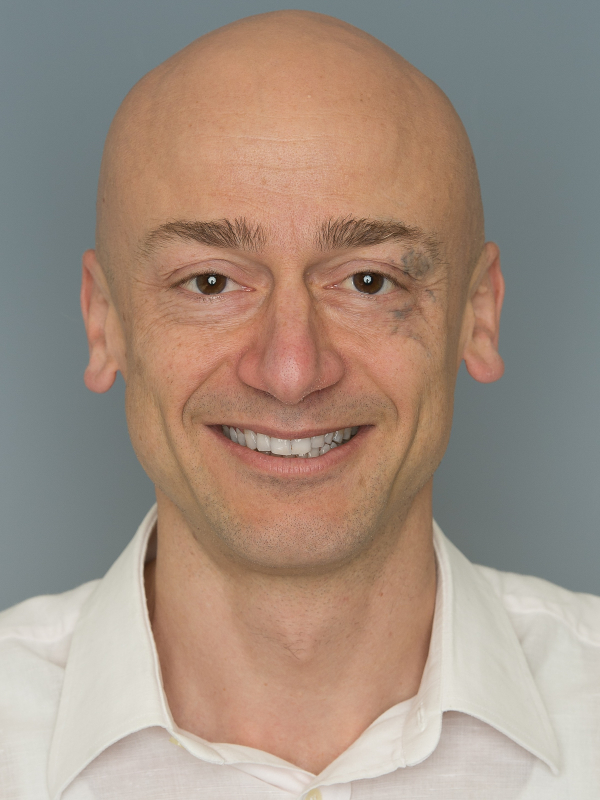 Ivan Martin合作论文数Department of Biomedicine, University of Basel;Gabriella - Gelato Artigianale13
 Benjamin Bellon合作论文数11
 Michel Dard合作论文数BioCellgraft10
 Rebecca Sandgren合作论文数6
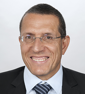 Anton Sculean合作论文数Department of Periodontology, School of Dental Medicine, Faculty of Medicine, University of Bern6
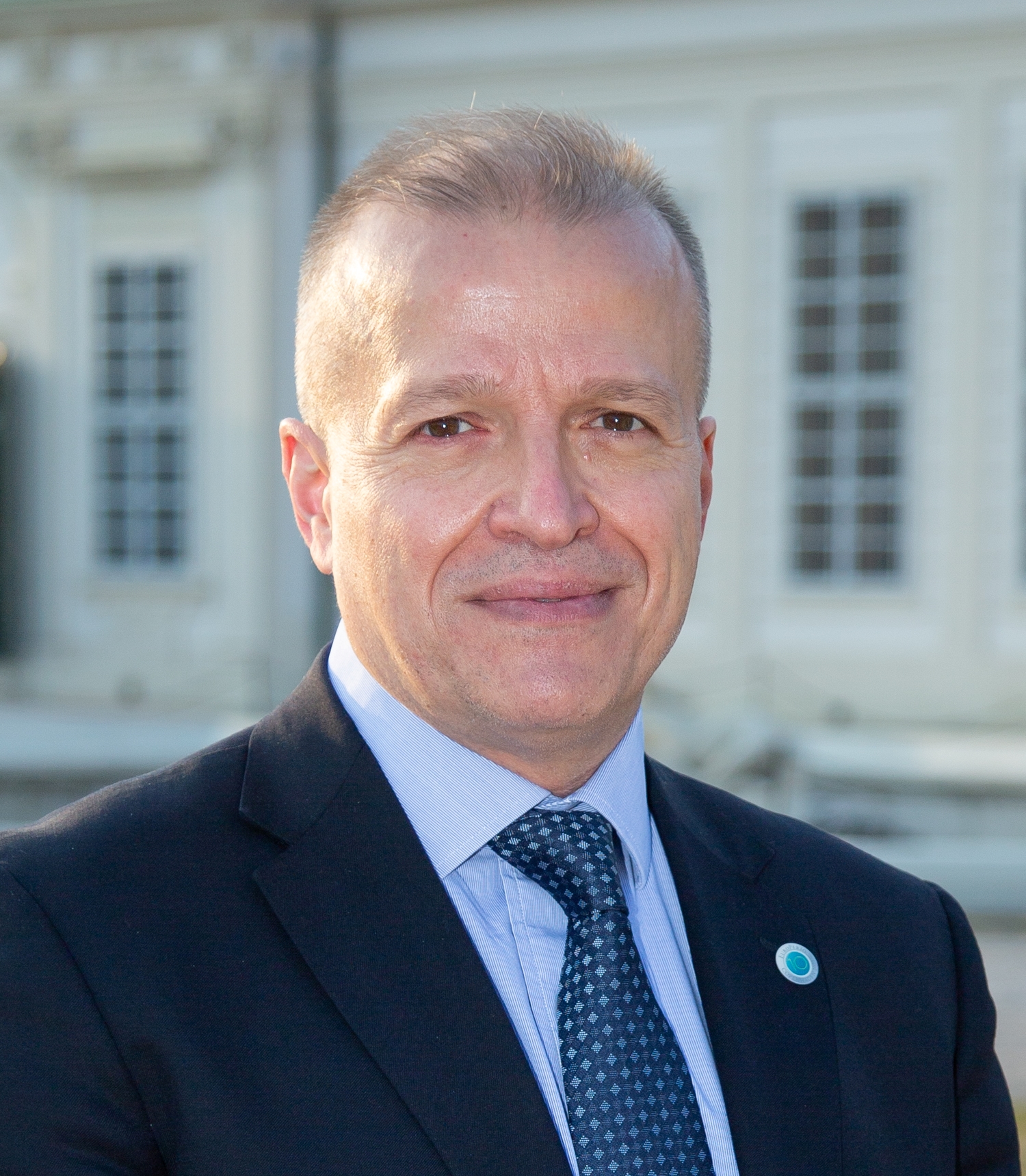 Andreas Stavropoulos合作论文数Department of Periodontology, Faculty of Odontology, University of Malmo6
 Richard J. Miron合作论文数Miron Lab;Department of Periodontology, Nova Southeastern University6
 Paul Bourgine合作论文数Complex Systems Digital Campus UniTwin, UNESCO5
 Arnaud Scherberich合作论文数5
 Sylvain Catros合作论文数Bordeaux University Hospital5
 Oleh Andrukhov合作论文数Division of Conservative Dentistry and Periodontology;Medical University of Vienna4
 Atanas Todorov合作论文数4Feliciano Sandra合作论文数4
 Marcel Obrecht合作论文数4
 Yufeng Zhang (张玉峰)合作论文数Hospital of Stomatology, Wuhan University4
 Jean-Christophe Fricain合作论文数Université de Bordeaux4
 Claude Jaquiery合作论文数4
 Dieter Bosshardt合作论文数Department of Oral Surgery and Stomatology, University of Bern;Robert K. Schenk Laboratory of Oral Histology, School of Dental Medicine, University of Bern4
 Jeroen Geurts合作论文数3
 Jean-Claude Imber合作论文数3
 Christian Behm合作论文数3Joëlle Amédée合作论文数3
 David L. Cochran合作论文数Department of Periodontics, School of Dentistry, UT Health San Antonio3
 Xiaohui Rausch-Fan合作论文数Medical University Vienna3Valderrabano Victor合作论文数3
 Karoliina Pelttari合作论文数3
 Fujioka-Kobayashi Masako合作论文数Graduate School of Medical and Dental Sciences, Tokyo Medical and Dental University3
 Fabien Guillemot合作论文数Universite Victor Segalen Bordeaux3
 Reine Bareille合作论文数3
 Andrea Barbero合作论文数Department of Biomedicine, University Hospital Basel3
 Y. Shirakata合作论文数3Emanuele Gaudiello合作论文数2Raphael S Wagner合作论文数2Maximilian Burger合作论文数2
 Fernando Munoz合作论文数2
 Alex Solderer合作论文数2Thomas Huegle合作论文数2
 Herber Valentin合作论文数2
 Alice Blufstein合作论文数2
 Andrea Roccuzzo合作论文数School of Dental Medicine, University of Bern2
 Andrea Banfi合作论文数USB2
 Lukasz Witek合作论文数Department of Biomaterials, College of Dentistry, New York University2
 Paulo G. Coelho合作论文数Biomaterials Division, New York University2
 Haberthür David合作论文数Institute for Anatomy, University of Bern2
 Buser Daniel合作论文数Department of Oral Surgery and Stomatology, School of Dental Medicine, University of Bern2
 Marianna Kruithof-de Julio合作论文数Departments of Medicine and Genetics and Development, Columbia University Medical Center;Herbert Irving Comprehensive Cancer Center, Columbia University Medical Center;Columbia University Medical Center, Columbia University Medical Center2
 Zoni Eugenio合作论文数2Schmidlin Patrick R合作论文数2Laurent A. Tchang合作论文数2




