Figure S5: Evaluation of SETDB1 knockdown in human melanoma cells, related to Figure 4:
Figure S3: Validation and characterization of Setdb1-/- cell lines, tumors, and microenvironments, related to Figure 2.
Ranked CRISPR screen dropout hits, related to Figure 1. Analysis was performed using MaGeCK.
Figure S4: Evaluation of differences in SKO tumor gene expression and microenvironment profiles, related to Figure 3:
Figure S1: Experimental validation of SETDB1 knockout and flow gating schemes, related to Figure 4
Despite recent advances in the treatment of melanoma, many patients with metastatic disease still succumb to their disease. To identify tumor-intrinsic modulators of immunity to melanoma, we performed a whole-genome CRISPR screen in melanoma and identified Setdb1 as well as all components of the human silencing hub complex. We found that loss of Setdb1 leads to increased immunogenicity and complete tumor clearance in a CD8(+) T cell-dependent manner. Mechanistically, loss of Setdb1 causes de-repression of endogenous retroviruses (ERV) in melanoma cells and triggers tumor cell-intrinsic type I IFN signaling, upregulation of MHC-I expression, and increased CD8(+) T-cell infiltration. Importantly, spontaneous immune clearance observed in Setdb1(-/-) tumors results in subsequent protection from other ERV-expressing tumor lines, supporting the functional antitumor role of ERV-specific CD8(+) T cells found in the Setdb1(-/-) microenvironment. Blocking the type I IFN receptor in mice grafted with Setdb1(-/-) tumors decreases immunogenicity by decreasing MHC-I expression, leading to decreased T-cell infiltration and increased melanoma growth, comparable with Setdb1(wt) tumors. Together, these results provide key in vivo evidence of a critical role for Setdb1 and type I IFNs in generating an inflamed tumor microenvironment and potentiating tumor cell-intrinsic immunogenicity in melanoma. This study further emphasizes regulators of ERV expression and type I IFN expression as potential therapeutic targets for augmenting anticancer immune responses.
The protein kinase ERK2 is recurrently mutated in human squamous cell carcinomas and other tumors. ERK2 mutations cluster in an essential docking recruitment site that interacts with short linear motifs found within intrinsically disordered regions of ERK substrates and regulators. Cancer-associated mutations do not disrupt ERK2 docking interactions altogether but selectively inhibit some interactions while sparing others. However, the full scope of disrupted or maintained interactions remains unknown, limiting our understanding of how these mutations contribute to cancer. We recently defined the docking interactome of wild-type ERK2 by screening a yeast two-hybrid library of proteomic short linear motifs. Here, we apply this approach to the two most recurrent cancer-associated mutants. We find that most sequences binding to WT ERK2 also interact with both mutant forms. Analysis of differentially interacting sequences revealed that ERK2 mutants selectively lose the ability to bind sequences conforming to a specific motif. We solved the co-crystal structure of ERK2 in complex with a peptide fragment of ISG20, a screening hit that binds exclusively to the WT kinase. This structure demonstrated the mechanism by which cancer hotspot mutations at Glu81, Arg135, Asp321, and Glu322 selectively impact peptide binding. Finally, we found that cancer-associated ERK2 mutations had decreased activity in phosphorylating GEF-H1/ARHGEF2, a known ERK substrate harboring a WT-selective docking motif. Collectively, our studies provide a structural rationale for how a broad set of interactions are disrupted by ERK2 hotspot mutations, suggesting mechanisms for pathway rewiring in cancers harboring these mutations.
Accurate chromosome segregation during mitosis requires that all chromosomes establish stable bi-oriented attachments with the spindle apparatus. Kinetochores form the interface between chromosomes and spindle microtubules and as such are under tight control by complex regulatory circuitry. As part of the chromosomal passenger complex (CPC), the Aurora B kinase plays a central role within this circuitry by destabilizing improper kinetochore-microtubule attachments and relaying the attachment status to the spindle assembly checkpoint, a feedback control system that delays the onset of anaphase by inhibiting the anaphase-promoting complex/cyclosome. Intriguingly, Aurora B is conserved even in kinetoplastids, an evolutionarily divergent group of eukaryotes, whose kinetochores are composed of a unique set of structural and regulatory proteins. Kinetoplastids do not have a canonical spindle checkpoint and it remains unclear how their kinetochores are regulated to ensure the fidelity and timing of chromosome segregation. Here, we show in Trypanosoma brucei, the kinetoplastid parasite that causes African sleeping sickness, that inhibition of Aurora B using an analogue-sensitive approach arrests cells in metaphase, with a reduction in properly bi-oriented kinetochores. Aurora B phosphorylates several kinetochore proteins in vitro, including the N-terminal region of the divergent Bub1-like protein KKT14. Depletion of KKT14 partially overrides the cell cycle arrest caused by Aurora B inhibition, while overexpression of a non-phosphorylatable KKT14 protein results in a prominent delay in the metaphase-to-anaphase transition. Finally, we demonstrate using a nanobody-based system that re-targeting the catalytic module of the CPC to the outer kinetochore is sufficient to promote mitotic exit but causes massive chromosome mis-segregation in anaphase. Our results indicate that the CPC and KKT14 are involved in an unconventional pathway controlling mitotic exit and error-free chromosome segregation in trypanosomes.
Phosphorylation of proteins on tyrosine (Tyr) residues evolved in metazoan organisms as a mechanism of coordinating tissue growth1. Multicellular eukaryotes typically have more than 50 distinct protein Tyr kinases that catalyse the phosphorylation of thousands of Tyr residues throughout the proteome1-3. How a given Tyr kinase can phosphorylate a specific subset of proteins at unique Tyr sites is only partially understood4-7. Here we used combinatorial peptide arrays to profile the substrate sequence specificity of all human Tyr kinases. Globally, the Tyr kinases demonstrate considerable diversity in optimal patterns of residues surrounding the site of phosphorylation, revealing the functional organization of the human Tyr kinome by substrate motif preference. Using this information, Tyr kinases that are most compatible with phosphorylating any Tyr site can be identified. Analysis of mass spectrometry phosphoproteomic datasets using this compendium of kinase specificities accurately identifies specific Tyr kinases that are dysregulated in cells after stimulation with growth factors, treatment with anti-cancer drugs or expression of oncogenic variants. Furthermore, the topology of known Tyr signalling networks naturally emerged from a comparison of the sequence specificities of the Tyr kinases and the SH2 phosphotyrosine (pTyr)-binding domains. Finally we show that the intrinsic substrate specificity of Tyr kinases has remained fundamentally unchanged from worms to humans, suggesting that the fidelity between Tyr kinases and their protein substrate sequences has been maintained across hundreds of millions of years of evolution.
The Ras–mitogen-activated protein kinase (MAPK) pathway is a major target for cancer treatment. To better understand the genetic pathways that modulate cancer cell sensitivity to MAPK pathway inhibitors, we performed a CRISPR knockout screen with MAPK pathway inhibitors on a colorectal cancer (CRC) cell line carrying mutant KRAS. Genetic deletion of the catalytic subunit of protein phosphatase 6 (PP6), encoded by PPP6C , rendered KRAS - and BRAF -mutant CRC and BRAF -mutant melanoma cells more resistant to these inhibitors. In the absence of MAPK pathway inhibition, PPP6C deletion in CRC cells decreased cell proliferation in two-dimensional (2D) adherent cultures but accelerated the growth of tumor spheroids in 3D culture and tumor xenografts in vivo. PPP6C deletion enhanced the activation of nuclear factor κB (NF-κB) signaling in CRC and melanoma cells and circumvented the cell cycle arrest and decreased cyclin D1 abundance induced by MAPK pathway blockade in CRC cells. Inhibiting NF-κB activity by genetic and pharmacological means restored the sensitivity of PPP6C -deficient cells to MAPK pathway inhibition in CRC and melanoma cells in vitro and in CRC cells in vivo. Furthermore, a R264 point mutation in PPP6C conferred loss of function in CRC cells, phenocopying the enhanced NF-κB activation and resistance to MAPK pathway inhibition observed for PPP6C deletion. These findings demonstrate that PP6 constrains the growth of KRAS - and BRAF -mutant cancer cells, implicates the PP6–NF-κB axis as a modulator of MAPK pathway output, and presents a rationale for cotargeting the NF-κB pathway in PPP6C -mutant cancer cells.
CD8 + T cells play an important role in anti-tumor immunity. Better understanding of their regulation could advance cancer immunotherapies. Here we identify, via stepwise CRISPR-based screening, that CUL5 is a negative regulator of the core signaling pathways of CD8 + T cells. Knocking out CUL5 in mouse CD8 + T cells significantly improves their tumor growth inhibiting ability, with significant proteomic alterations that broadly enhance TCR and cytokine signaling and their effector functions. Chemical inhibition of neddylation required by CUL5 activation, also enhances CD8 effector activities with CUL5 validated as a major target. Mechanistically, CUL5, which is upregulated by TCR stimulation, interacts with the SOCS-box-containing protein PCMTD2 and inhibits TCR and IL2 signaling. Additionally, CTLA4 is markedly upregulated by CUL5 knockout, and its inactivation further enhances the anti-tumor effect of CUL5 KO. These results together reveal a negative regulatory mechanism for CD8 + T cells and have strong translational implications in cancer immunotherapy.
 Lou Hua Jane合作论文数52
 Lewis Cantley合作论文数Department of Cell Biology, Harvard Medical School;Department of Cell Biology, Dana-Farber Cancer Institute22
 Miller Chad J合作论文数School of Medicine, Yale University16
 Stefan Knapp合作论文数Structural Genomics Consortium, Goethe-University Frankfurt16
 Gabriel J Escobar合作论文数Children’s Hospital and Harvard Medical School, Harvard School of Public Health;B.C. Research Institute for Children’s and Women’s Health, University of British Columbia and;Division of Research, Kaiser Permanente Medical Care Program;Department of Pediatrics, University of British Columbia;and Centre for Health Evaluation Research, B.C. Research Institute for Children’s and Women’s Health Vancouver15
 William Damsky合作论文数Department of Dermatology, Yale School of Medicine, Yale University13Huet-Calderwood Clotilde合作论文数13
 Micevic Goran合作论文数School of Medicine, Yale University13
 Akiko Iwasaki合作论文数Iwasaki Lab, School of Medicine, Yale University;Howard Hughes Medical Institute13
 Song Eric合作论文数Dept Immunobiol, Yale Sch Med13
 Qin Yan (严钦)合作论文数Department of Pathology, School of Medicine, Yale University12
 Titus Boggon合作论文数Biochemistry, Quantitative Biology, Biophysics, and Structural Biology, Biological & Biomedical Sciences, Yale University12David A. Calderwood合作论文数12
 Susan Kaech合作论文数Department of Immunobiology, Yale School of Medicine, Yale University;The Salk Institute for Biological Studies12
 Vincent X. Liu合作论文数Division of Research, Kaiser Permanente;Stanford Health Care12Parker Sirlester A合作论文数11Patricia Kipnis合作论文数11
 Grace R Jeschke合作论文数9
 Rune Linding合作论文数Molecular Logic Team;Section of Cell and Molecular Biology;Institute of Cancer Research;Molecular Logic Team, Institute of Cancer Research9
 David Calderwood合作论文数Biochemistry, Quantitative Biology, Biophysics, and Structural Biology, Biological & Biomedical Sciences, Yale University8
 Michael B. Yaffe合作论文数Koch Institute for Integrative Cancer Research, Massachusetts Institute of Technology;Beth Israel Deaconess Medical Center8
 Jun Liu合作论文数The Sidney Kimmel Comprehensive Cancer Center, Johns Hopkins University School of Medicine7Pau Creixell合作论文数The Institute of Cancer Research (ICR), London, UK.;Center for Biological Sequence Analysis (CBS),, Technical University of Denmark;The Institute of Cancer Research (ICR),, Technical University of Denmark6
 Alexey G. Ryazanov合作论文数Department of Pharmacology, Robert Wood Johnson Medical School, Rutgers, The State University of New Jersey;School of Arts and Sciences, Rutgers, The State University of New Jersey6Elvira An合作论文数5Kevin Dalby合作论文数5Arona Ragins合作论文数5Thorkell Andresson合作论文数Protein Characterization Laboratory, Cancer Research Technology Program, Frederick National Laboratory for Cancer Research, Leidos Biomedical Research, Inc., Frederick, MD 21702, USA5
 Philip M. Kim合作论文数Terrence Donnelly Center for Cellular and Biomolecular Research, University of Toronto;Department of Molecular Genetics, Temerty Faculty of Medicine, University of Toronto;Department of Computer Science, Temerty Faculty of Medicine, University of Toronto5Ha Byung Hak合作论文数5
 Pedro Torres-Ayuso合作论文数Department of Immunology and Oncology, Centro Nacional de Biotecnología (CSIC),5
 Mark B Gerstein合作论文数School of Engineering & Applied Science, Yale University5
 Sudipto Das合作论文数Dept Mol & Cellular Therapeut, Royal Coll Surgeons Ireland5
 Daniel Ritt合作论文数5
 Deborah K. Morrison合作论文数Laboratory of Cell and Developmental Signaling, Center for Cancer Research, National Cancer Institute, National Institutes of Health5John Brognard合作论文数Department of Pharmacology;University of California at San Diego;Department of Pharmacology, University of California at San Diego5Guangda Shi合作论文数4Eidarus Salah合作论文数Univ Oxford, Chem Res Lab, 12 Mansfield Rd, Oxford OX1 3TA, England4Chen Catherine合作论文数4Karim Baktiar O合作论文数4Robinson Christina M合作论文数4
 Katherine M Nyswaner合作论文数4
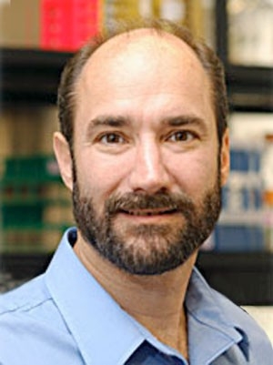 Michael Snyder合作论文数Department of Genetics, School of Medicine, Stanford University;Center for Genomics and Personalized Medicine;Karolinska Institute4
 Erwin M Schoof合作论文数Cellular Signal Integration Group (C-SIG),, Technical University of Denmark (DTU),4LaGuardia Juan Carlos合作论文数4
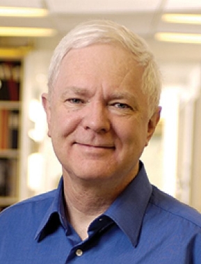 Tony Pawson (安东尼·鲍森)合作论文数Department of Molecular Genetics, Temerty Faculty of Medicine, University of Toronto4
 Panagis Filippakopoulos合作论文数Botnar Research Centre, University of Oxford4Simone Difilippantonio合作论文数4Jeffrey R. Peterson合作论文数3




