The quest for a non-dopaminergic approach to treating Parkinson's disease (PD) has been quietly progressing over the past several decades, and is now finding its momentum. Here, in what is more a memoir than a comprehensive review, we discuss work carried out over the past 50 years to show that adenosine acts as a critical signaling molecule via actions against a specific family of receptors. Importantly for PD, adenosine A2A receptors have a selective localization to the basal ganglia and specifically to the indirect output pathway, offering a targeted, non-dopaminergic opportunity to modulate basal ganglia output.
In this randomized blinded study, we investigated caffeine 5 mg/kg treatment given directly after neonatal brain hypoxia ischemia. Brain morphology, behavior and key brain infiltrating immune populations were examined. Caffeine treatment significantly improves outcome when compared to phosphate buffered saline. Flow cytometric analysis of immune responses revealed no persistent immunological alterations. Given its safety caffeine emerges as a candidate for neuroprotective intervention after neonatal brain injury.
The classic endogenous somnogen adenosine promotes sleep via A1 and A2A receptors. In this chapter, we present an overview of the current knowledge regarding the regulation of adenosine levels, adenosine receptors, and available pharmacologic and genetic tools to manipulate the adenosine system. This is followed by a summary of current knowledge of the role of adenosine and its receptors in the regulation of sleep and wakefulness. Despite strong data implicating numerous brain areas, including the basal forebrain, the tuberomammillary nucleus, the lateral hypothalamus, and the nucleus accumbens, in the adenosinergic control of sleep, the complete neural circuitry in the brain involved in the sleep-promoting effects of adenosine remains unclear. Moreover, the popular demand for natural sleep aids has led to a search for natural compounds that can promote sleep via adenosine receptor activation. Finally, we discuss the effects of caffeine in man and the possible use of more selective adenosine receptor drugs for the treatment of sleep disorders.
Adenosine receptor signaling plays important roles in normal physiology, but is also known to modulate the development or progression of several different diseases. The design of new, efficient, and safe pharmacological approaches to target the adenosine system may have considerable therapeutic potential, but is also associated with many challenges. This review summarizes the main challenges of adenosine receptor targeted treatment including tolerance, disease stage, cell type-specific effects, caffeine intake, adenosine level assessment and receptor distribution in vivo. Moreover, we discuss several potential ways to overcome these obstacles (i.e., the use of partial agonists, indirect receptor targeting, allosteric enhancers, prodrugs, non-receptor-mediated effects, neoreceptors, conditional knockouts). It is important to address these concerns during development of new and successful therapeutic approaches targeting the adenosine system.
BackgroundEarly‐life reduction in nephron number (uninephrectomy [UNX]) and chronic high salt (HS) intake increase the risk of hypertension and chronic kidney disease. Adenosine signaling via its different receptors has been implicated in modulating renal, cardiovascular, and metabolic functions as well as inflammatory processes; however, the specific role of the A3 receptor in cardiovascular diseases is not clear. In this study, gene‐modified mice were used to investigate the hypothesis that lack of A3 signaling prevents the development of hypertension and attenuates renal and cardiovascular injuries following UNX in combination with HS (UNX‐HS) in mice. Methods and ResultsWild‐type (A3+/+) mice subjected to UNX‐HS developed hypertension compared with controls (mean arterial pressure 106±3 versus 82±3 mm Hg; P<0.05) and displayed an impaired metabolic phenotype (eg, increased adiposity, reduced glucose tolerance, hyperinsulinemia). These changes were associated with both cardiac hypertrophy and fibrosis together with renal injuries and proteinuria. All of these pathological hallmarks were significantly attenuated in the A3−/− mice. Mechanistically, absence of A3 receptors protected from UNX‐HS–associated increase in renal NADPH oxidase activity and Nox2 expression. In addition, circulating cytokines including interleukins 1β, 6, 12, and 10 were increased in A3+/+ following UNX‐HS, but these cytokines were already elevated in naïve A3−/− mice and did not change following UNX‐HS. ConclusionsReduction in nephron number combined with chronic HS intake is associated with oxidative stress, chronic inflammation, and development of hypertension in mice. Absence of adenosine A3 receptor signaling was strongly protective in this novel mouse model of renal and cardiovascular disease.
Neonatal brain hypoxic ischemia (HI) often results in long-term motor and cognitive impairments. Post-ischemic inflammation greatly effects outcome and adenosine receptor signaling modulates both HI and immune cell function. Here, we investigated the influence of adenosine A(1) receptor deficiency (A(1)R(-/-)) on key immune cell populations in a neonatal brain HI model. Ten-day-old mice were subjected to HI. Functional outcome was assessed by open locomotion and beam walking test and infarction size evaluated. Flow cytometry was performed on brain-infiltrating cells, and semi-automated analysis of flow cytometric data was applied. A(1)R(-/-) mice displayed larger infarctions (+33 %, p < 0.05) and performed worse in beam walking tests (44 % more mistakes, p < 0.05) than wild-type (WT) mice. Myeloid cell activation after injury was enhanced in A(1)R(-/-) versus WT brains. Activated B lymphocytes expressing IL-10 infiltrated the brain after HI in WT, but were less activated and did not increase in relative frequency in A(1)R(-/-). Also, A(1)R(-/-) B lymphocytes expressed less IL-10 than their WT counterparts, the A(1)R antagonist DPCPX decreased IL-10 expression whereas the A(1)R agonist CPA increased it. CD4(+) T lymphocytes including FoxP3(+) T regulatory cells, were unaffected by genotype, whereas CD8(+) T lymphocyte responses were smaller in A(1)R(-/-) mice. Using PCA to characterize the immune profile, we could discriminate the A(1)R(-/-) and WT genotypes as well as sham operated from HI-subjected animals. We conclude that A(1)R signaling modulates IL-10 expression by immune cells, influences the activation of these cells in vivo, and affects outcome after HI.
Rationale: Accumulating studies suggest that nitric oxide (NO) deficiency and oxidative stress are central pathological mechanisms in type 2 diabetes (T2D). Recent findings demonstrate therapeutic effects by boosting the nitrate-nitrite-NO pathway, which is an alternative pathway for NO formation. This study aimed at investigating the acute effects of inorganic nitrate on glucose and insulin signaling in adenosine A2B receptor knockout mice (A(2B)(-/-), a genetic mouse model of impaired metabolic regulation.Methods: Acute effects of nitrate treatment were investigated in aged wild-type (WT) and A(2B)(-/-) mice. One hour after injection with nitrate (0.1 mmol/kg, i.p.) or placebo, metabolic regulation was evaluated by intraperitoneal glucose and insulin tolerance tests. NADPH oxidase-mediated superoxide production and AMPK phosphorylation were measured in livers obtained from non-treated or glucose-treated mice, with or without prior nitrate injection. Plasma was used to determine insulin resistance (HOMA-IR) and NO signaling.Results: A(2B)(-/-) displayed increased body weight, reduced glucose clearance, and attenuated overall insulin responses compared with age-matched WT mice. Nitrate treatment increased circulating levels of nitrate, nitrite and cGMP in the A(2B)(-/-), and improved glucose clearance. In WT mice, however, nitrate treatment did not influence glucose clearance. HOMA-IR increased following glucose injection in the A(2B)(-/-), but remained at basal levels in mice pretreated with nitrate. NADPH oxidase activity in livers from A(2B)(-/-), but not WT mice, was reduced by nitrate treatment. Livers from A(2B)(-/-) displayed reduced AMPK phosphorylation compared with WT mice, and this was increased by nitrate treatment. Finally, injection with the anti-diabetic agent metformin induced similar therapeutic effects in the A(2B)(-/-) as observed with nitrate.Conclusion: The A(2B)(-/-) mouse is a genetic mouse model of metabolic syndrome. Acute treatment with nitrate improved the metabolic profile in it, at least partly via reduction in oxidative stress and improved AMPK signaling in the liver.
A careful examination of the potency of caffeine (and also of its metabolite theophylline) showed that blockade of the actions of adenosine was achieved at considerably lower concentrations than other known actions, including blockade of phosphodiesterase, interaction with gamma amino butyric acid receptors, and increased calcium mobilization. Indeed, significant blockade of three of the four adenosine receptors occurs at caffeine concentrations achieved after ingestion of a single cup of coffee. The importance of adenosine receptors (especially A1 and A2A receptors) as targets for the actions of caffeine in normal doses has been demonstrated using genetic methods. They have also revealed that higher doses of caffeine have actions that are independent of adenosine receptor blockade. The individual sensitivity to the adenosine receptor blocking actions of caffeine that generally produce alerting effects, and the nonadenosine-dependent effects that are depressant and anxiogenic, determines individual caffeine tolerance.
Adenosine is an important regulator of metabolism; however, the role of the A1 receptor during ageing and obesity is unclear. The aim of this study was to investigate the effects of A1 signalling in modulating metabolic function during ageing.
BACKGROUND: Adenosine A1 and A2 receptors are known to regulate renal autoregulation and blood pressure, but the role of A3 signaling is unknown. We previously demonstrated early life reduction in nephron number (UNX) combined with high salt diet (HS) induced renal oxidative stress and hypertension in rats. This study aimed at investigating the role of A3 receptor in modulating renal and cardiovascular function in this disease model. METHODS: Wild-type (WT) and A3 knockout (A3KO) mice underwent UNX or sham-operation at 3-week-age followed by HS treatment. Blood pressure and renal function were measured in conscious aged mice and renal oxidative stress as well as inflammatory properties was characterized. RESULTS: WT but not A3KO with UNX+HS developed hypertension and cardiac hypertrophy (WT: 4.5±0.1 vs 4.0±0.1; A3KO: 4.7±0.2 vs 4.6±0.1 mg/gBW), characterized by impaired renal plasma flow (RPF), glomerular hyperfiltration and increased renal NADPH oxidase activity (Fig. 1A-C). UNX+HS increased plasma IL-6 (36.1±5.7 vs 16.3±2.4 pg/ml) and IL-10 (37.8±3.9 vs 25.4±2.4 pg/ml) levels in WT, but not in A3KO (39.7±3.9 vs 38.5±5.9 pg/ml and 46.5±3.7 vs 43.9±6.0 pg/ml respectively). However, A3KO displayed higher baseline cytokine levels. Furthermore, bone marrow-derived macrophages from A3KO mice expressed higher M1 (PDL-1 and CD86) and M2 (PDL2 and CD206) markers under LPS stimulation compared with WT. These suggested an enhanced innate immune response in A3KO. CONCLUSION: UNX followed by HS intake lead to renal and cardiovascular dysfunction, which importantly depends on A3 receptor-mediated regulation of oxidative stress and inflammation.
This minireview briefly summarizes the evidence that adenosine, acting on four G-protein coupled receptors, can play physiological roles, but is also critically involved in pathological processes. The factors that decide which of these is the more important in a specific cell or organ are briefly summarized. The fact that drugs that target adenosine receptors in disease will also hit the physiological processes will make drug development more tricky.
Adenosine signalling has long been a target for drug development, with adenosine itself or its derivatives being used clinically since the 1940s. In addition, methylxanthines such as caffeine have profound biological effects as antagonists at adenosine receptors. Moreover, drugs such as dipyridamole and methotrexate act by enhancing the activation of adenosine receptors. There is strong evidence that adenosine has a functional role in many diseases, and several pharmacological compounds specifically targeting individual adenosine receptors--either directly or indirectly--have now entered the clinic. However, only one adenosine receptor-specific agent--the adenosine A2A receptor agonist regadenoson (Lexiscan; Astellas Pharma)--has so far gained approval from the US Food and Drug Administration (FDA). Here, we focus on the biology of adenosine signalling to identify hurdles in the development of additional pharmacological compounds targeting adenosine receptors and discuss strategies to overcome these challenges.
There is excellent evidence that adenosine plays a role in physiology and pathophysiology to modulate neural activity in the brain. For example, adenosine is known to act on adenosine A1 receptors to decrease neuronal firing and neurotransmitter release (1). This mechanism is of importance in limiting excessive neuronal activity and thereby, epileptic seizures (2, 3). Although the role of adenosine is rather uncontroversial, the origin of the adenosine mediating this anticonvulsant effect has been somewhat contentious. The work by Lovatt et al. (4) in PNAS not only clarifies that adenosine released from neurons is the important source, but it also raises more fundamental issues regarding the interactions between signaling through adenine nucleotides and adenosine and the role of astrocytes.
Mind–Brain dualism is still a hotly debated issue.1,2 I am neither qualified nor inclined to outline the intricacies of these philosophical controversies. However, one aspect deserves attention by a pharmacologist: some forms of very strict dualism have problems in dealing with the fact that drugs can alter cognitive states. It also seems to me that the challenge would become even stronger if one could demonstrate not only that a drug can alter a mental state, but also provide a clear cellular mechanism of how this is brought about. This is what I will try to do in this brief essay. I will not, however, even try to consider how the effect on specific brain cells is translated into something that we perceive as mental.
OBJECTIVEAdenosine (Ado) mediates tubuloglomerular feedback, whereas AngII and NO are important modulators. A1‐deficiency abolish TGF and diminish effectiveness of L‐NAME and AngII to constrict renal resistance vessels. We used A1‐knockout (A1−/−) and wild‐type (A1+/+) mice to investigate the synergism between AngII and low‐dose Ado in regulation of arteriolar responses.METHODSContractions were measured in renal afferent arterioles from nontreated mice and those with 14‐days pretreatment with L‐NAME (10‐4M) or AngII (400 ng/kg/min). Concentration response curves were obtained by Ado (10‐8M; 15 min) alone, or in combination with cumulative application of AngII (10–12 to 10‐6M).RESULTSAdo contracted arterioles from A1+/+ (11%), but had no significant effect in A1−/−. Ado significantly enhanced AngII‐mediated contraction in both genotypes, however, the contractile response was stronger in A1+/+ (56%) than in A1−/− (40%). Prolonged treatment with L‐NAME or AngII did not change the responses to Ado alone. However, arteriolar response to combination with Ado+AngII was enhanced in A1+/+, but was attenuated in A1−/−.CONCLUSIONLow‐dose Ado augments AngII‐induced constriction in both A1+/+ and A1−/−, by non‐receptor‐mediated actions. Underlying mechanisms may involve modulation of NO bioavailability or intracellular effects of Ado, introduced by Ado‐transporters.
 Per Svenningsson合作论文数Department of Clinical Neuroscience, Karolinska Institutet;Department of Neurology, Karolinska University Hospital42
 Kjell Fuxe合作论文数Department of Neuroscience, Karolinska Institutet27
 Gunnar Schulte合作论文数Department of Physiology and Pharmacology, Karolinska Institutet25
 Christer Nordstedt合作论文数22
 Bjorn Kull合作论文数21Mikael Jondal合作论文数17I VANDERPLOEG合作论文数16Per Hedqvist合作论文数16Bror Jonzon合作论文数16
 Linda Halldner合作论文数15
 Bengt Persson合作论文数Department of Cell and Molecular Biology, Uppsala University;National Bioinformatics Infrastructure Sweden;ELIXIR14
 Boo Johansson合作论文数Department of Psychology, Faculty of Social Sciences, University of Gothenburg14Karin Lindström合作论文数14
 Ulrika Ådén合作论文数Karolinska Institutet;Linköping University13
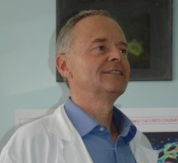 Luigi Agnati合作论文数Università Degli Studi di Modena e Reggio Emilia13Thomas V. Dunwiddie合作论文数Veterans Administration Medical Research Service, Denver, CO 80220 U.S.A.13AEG Persson合作论文数12
 Hugo Lagercrantz合作论文数Department of Women's and Children's Health, Karolinska Institutet;Karolinska University Hospital12
 F. Parkinson合作论文数12Pär Gerwins合作论文数12
 Olle Larsson合作论文数11
 Jiangfan Chen (陈江帆)合作论文数Eye Hospital, Wenzhou Medical University;Boston University School of Medicine11Anders Kvanta合作论文数11
 Mattias Carlström合作论文数Department of Physiology and Pharmacology, Karolinska Institutet;School of Medical Sciences, UNSW Sydney11Ps Hu合作论文数10NO LUNELL合作论文数10
 Johan Fastbom合作论文数10Elisabetta Daré合作论文数10G Arslan合作论文数10
 Maria Lindskog合作论文数Uppsala University9Sven Ove Ogren合作论文数9Paul Hjemdahl合作论文数9
 Alf Sollevi合作论文数9
 Kenneth A. Jacobson合作论文数Molecular Recognition Section, Laboratory of Bioorganic Chemistry, National Institute of Diabetes and Digestive and Kidney Diseases, National Institutes of Health8J. Wager合作论文数8M DUNERENGSTROM合作论文数8Fw Schildberg合作论文数7U Haglund合作论文数7
 Bruce N. Cronstein合作论文数Department of Medicine, Grossman School of Medicine, New York University;Department of Pathology, Grossman School of Medicine, New York University;Langone Medical Center, New York University7
 Paul Greengard合作论文数Laboratory of Molecular and Cellular Neuroscience, The Rockefeller University7Sune Rosell合作论文数7Ingeborg Van Der Ploeg合作论文数7Nedret Altiok合作论文数7
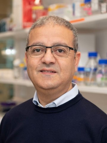 Abdel El Manira合作论文数Department of Neuroscience, Karolinska Institutet7R A Cunha合作论文数6C Lovdahl合作论文数6eva lindgren合作论文数6Jm Lundberg合作论文数6
 Joaquim Alexandre Ribeiro合作论文数Institute of Pharmacology and Neurosciences, University of Lisbon6




