Background:Screening efforts for Barrett's Esophagus (BE) predominantly focus on performing upper endoscopy (EGD) on patients with gastroesophageal reflux disease (GERD) symptoms who have additional risk factors for BE. However, cost and invasiveness preclude EGD in those who have no prior GERD symptoms, despite having other risk factors, representing missed opportunities for BE screening in individuals who account for approximately 40% of the patients who eventually develop esophageal adenocarcinoma (EAC). Aim:The aim of this study was to evaluate if non-endoscopic methods can enable BE detection in an at-risk population without GERD symptoms. Methods:Patients presenting for colonoscopy who had not undergone previous EGD plus had ≥3 BE risk factors (from among age ≥50 years, male sex, white race, smoking history, family history of BE/EAC, or central obesity) without chronic GERD symptoms were prospectively recruited for non-endoscopic screening. Trained nurses administered the EsoCheck (Lucid Diagnostics) encapsulated balloon. Samples were assayed with the EsoGuard BE detection methylated DNA marker panel (Lucid Diagnostics). Patients with a positive result were offered standard-of-care EGD, while patients with a negative EG result were offered free of cost research EGD. Positive predictive value (PPV), negative predictive value (NPV), and BE prevalence were calculated. Results:The mean age of the 132 study subjects was 60.7 years, 129 (98%) were white, 124 (94%) were male, 71 (54%) had a prior smoking history, 46 (35%) were centrally obese, and 5 (4%) reported a family history. EsoCheck was successfully administered in 124 (94%) and the EsoGuard methylated DNA marker panel could be assayed in 120 (97%) of the samples. Thirty-four assays were positive of which 27 underwent a follow-up EGD and BE was identified in 9, PPV = 33% [17%, 54%] subjects. EGD was also performed in 22 of the 86 subjects whose assays were negative and none of them had BE, NPV = 100% [85%, 100%]. A logistic regression model fitted to impute the presence of BE estimated the PPV as 27% [13%, 44%], NPV as 98% [92%, 100%], and BE prevalence as 8.4% [4.5%, 14.3%]. Conclusion:Patients without chronic GERD who have ≥3 BE risk factors have a moderately high prevalence of BE. Non-endoscopic detection can effectively identify BE, enabling expansion of screening to this larger at-risk population. Those with a negative EG assay have a low likelihood of BE.
INTRODUCTION:Upper endoscopy (EGD) is generally recommended in those with chronic gastroesophageal reflux disease (GERD). To evaluate nonendoscopic screening in those without GERD symptoms. METHODS:EsoCheck/Esoguard (LucidDx) was performed in recruited patients without chronic GERD who had ≥3 other BE risk factors. RESULTS:The EsoGuard assay was positive in 34 of 120 patients. BE was identified in 9 of 27 who had follow-up EGD, positive predictive value = 33% (17%-54%). EGD performed in 22 of 86 subjects with negative assays found no BE, negative predictive value = 100% (85%-100%). DISCUSSION:Nonendoscopic BE detection is effective in patients without chronic GERD.
Background Despite wide use of adjuvanted influenza vaccine in nursing home residents (NHR), little immunogenicity data exist for this population. Methods We collected blood from NHR (n = 85) living in nursing homes participating in a cluster randomized clinical trial comparing MF59-adjuvanted trivalent inactivated influenza vaccine (aTIV) with nonadjuvanted vaccine (TIV) (parent trial, NCT02882100). NHR received either vaccine during the 2016-2017 influenza season. We assessed cellular and humoral immunity using flow cytometry and hemagglutinin inhibition, antineuraminidase (enzyme-linked lectin assay), and microneutralization assays. Results Both vaccines were similarly immunogenic and induced antigen-specific antibodies and T cells, but aTIV specifically induced significantly larger 28 days after vaccination (D28) titers against A/H3N2 neuraminidase than TIV. Conclusions NHRs respond immunologically to TIV and aTIV. From these data, the larger aTIV-induced antineuraminidase response at D28 may help explain the increased clinical protection observed in the parent clinical trial for aTIV over TIV in NHR during the A/H3N2-dominant 2016-2017 influenza season. Additionally, a decline back to prevaccination titers at 6 months after vaccination emphasizes the importance of annual vaccination against influenza. We find that standard and adjuvanted influenza vaccines both induce increases in humoral and cellular immunity that contract to baseline by D180. In the 2016-2017 influenza season, the primary difference between vaccines was humoral antineuraminidase immunity to A/H3N2.
BACKGROUND:Vitamin D deficiency may increase esophageal cancer risk. Vitamin D affects genes regulating proliferation, apoptosis, and differentiation and induces the tumor suppressor 15-hydroxyprostaglandin dehydrogenase (PGDH) in other cancers. This nonrandomized interventional study assessed effects of vitamin D supplementation in Barrett's esophagus (BE). We hypothesized that vitamin D supplementation may have beneficial effects on gene expression including 15-PGDH in BE.METHODS:BE subjects with low grade or no dysplasia received vitamin D3 (cholecalciferol) 50,000 international units weekly plus a proton pump inhibitor for 12 weeks. Esophageal biopsies from normal plus metaplastic BE epithelium and blood samples were obtained before and after vitamin D supplementation. Serum 25-hydroxyvitamin D was measured to characterize vitamin D status. Esophageal gene expression was assessed using microarrays.RESULTS:18 study subjects were evaluated. The baseline mean serum 25-hydroxyvitamin D level was 27 ng/mL (normal ≥30 ng/mL). After vitamin D supplementation, 25-hydroxyvitamin D levels rose significantly (median increase of 31.6 ng/mL, p<0.001). There were no significant changes in gene expression from esophageal squamous or Barrett's epithelium including 15-PGDH after supplementation.CONCLUSION:BE subjects were vitamin D insufficient. Despite improved vitamin D status with supplementation, no significant alterations in gene expression profiles were noted. If vitamin D supplementation benefits BE, a longer duration or higher dose of supplementation may be needed.
BACKGROUND & AIMS:Central adiposity is a risk factor for Barrett's esophagus (BE). Serum levels of adiponectin and leptin are deregulated in obese states and are implicated as putative mediators in the pathophysiology of esophageal columnar metaplasia. We describe associations between serum adiponectin and leptin levels with BE.METHODS:Patients were recruited prospectively for a case-control study. Fasting serum levels of adiponectin and leptin were measured in 135 patients with BE and compared with 2 separate control groups: 133 subjects with gastroesophageal reflux disease (GERD) and 1157 colon screening controls.RESULTS:Multivariate analyses adjusted for age, race, and waist-to-hip ratio showed that patients within the highest tertile of serum adiponectin level had decreased odds of BE compared with screening colonoscopy controls (odds ratio [OR], 0.42; 95% confidence interval [CI], 0.22-0.80). This effect was more pronounced in men (OR, 0.35; 95% CI, 0.17-0.74) compared with women (OR, 0.71; 95% CI, 0.17-3.03). In comparisons of BE cases with GERD controls, subjects within the highest tertile of serum adiponectin level showed decreased odds of BE (OR, 0.65; 95% CI, 0.31-1.36), however, this was not statistically significant. Patients in the highest tertile of serum leptin level did not have a significantly increased risk of BE in comparison with GERD (OR, 1.32; 95% CI, 0.61-2.88) or screening colonoscopy controls (OR, 1.57; 95% CI, 0.82-3.04) in analyses including both sexes. Based on sex-specific analyses, sex did not significantly alter the association of leptin with odds of BE.CONCLUSIONS:Serum adiponectin was associated inversely with BE and this effect was more pronounced in men, whereas serum leptin showed no evidence of association with BE in comparisons with multiple control groups. The exact mechanism, if any, by which these adipokines promote metaplasia in the esophagus needs to be explored further.
Introduction: Barrett's esophagus (BE) is a precancerous condition caused by gastroesophageal reflux disease.The principle of molecular endoscopy is to visualize disease specific fluorescence labeled probes using confocal laser endomicroscopy (CLE).Aims: Main study objective was to assess the potential of molecular imaging for diagnosis of BE using a novel fluorescence labeled probe highly specific for BE.Material & Methods: Patients with long segment BE were included.Random biopsies were taken for histologic and subsequent molecular imaging.The goblet cell specific Muc2 antibody was labeled with Fluorescein isothiocyanate (FITC) and then topically applied to the esophageal biopsies.Subsequently, CLE was performed and confocal images were digitally stored.Confocal images were than scored by two investigators according to BE specific fluorescence signals.Histopathology served as the reference standard.Results: Overall, 300 confocal images were recorded.Molecular imaging achieved an overall sensitivity, specificity and accuracy of 98.9%, 88.2%, and 97.5%, for the detection of BEspecific signals.Positive and negative predictive values were 98.3% and 91.8%.Interobserver and intraobserver agreement for prediction of BE were excellent with kappa values of 0.8597 and 0.9521, respectively.Conclusion: Molecular imaging with the novel highly-specific fluorescent Muc2 antibody enabled excellent results for prediction of BE.The technique should now be transferred to an in vivo setting in order to improve diagnosis and surveillance of BE-patients.
FIGURE 1: (A) Cross-sectional view using VLE of a segment of BE previously treated with RFA.(B) Neosquamous epithelium is characterized by a layered structure that is absent in (C) specialized intestinal metaplasia.Measurements of NSE thickness (red double arrow) were taken at four quadrants (Q1-4) along each centimeter of the pre-RFA BE segment.Quadrants that contained both NSE and SIM were excluded from measurement (gray rectangle involving Q2 and Q3).
There is a critical need to identify molecular markers that can reliably aid in stratifying esophageal adenocarcinoma (EAC) risk in patients with Barrett's esophagus. MicroRNAs (miRNA/miR) are one such class of biomolecules. In the present cross‐sectional study, we characterized miRNA alterations in progressive stages of neoplastic development, i.e., metaplasia–dysplasia–adenocarcinoma, with an aim to identify candidate miRNAs potentially associated with progression. Using next generation sequencing (NGS) as an agnostic discovery platform, followed by quantitative real‐time PCR (qPCR) validation in a total of 20 EACs, we identified 26 miRNAs that are highly and frequently deregulated in EACs (≥4‐fold in >50% of cases) when compared to paired normal esophageal squamous (nSQ) tissue. We then assessed the 26 EAC‐derived miRNAs in laser microdissected biopsy pairs of Barrett's metaplasia (BM)/nSQ ( n = 15), and high‐grade dysplasia (HGD)/nSQ ( n = 14) by qPCR, to map the timing of deregulation during progression from BM to HGD and to EAC. We found that 23 of the 26 candidate miRNAs were deregulated at the earliest step, BM, and therefore noninformative as molecular markers of progression. Two miRNAs, miR‐31 and −31*, however, showed frequent downregulation only in HGD and EAC cases suggesting association with transition from BM to HGD. A third miRNA, miR‐375, showed marked downregulation exclusively in EACs and in none of the BM or HGD lesions, suggesting its association with progression to invasive carcinoma. Taken together, we propose miR‐31 and −375 as novel candidate microRNAs specifically associated with early‐ and late‐stage malignant progression, respectively, in Barrett's esophagus. © 2012 Wiley Periodicals, Inc.
BACKGROUND:It is postulated that high serum levels of insulin and insulin growth factor 1 (IGF-1) mediate obesity-associated carcinogenesis. The relationship of insulin, IGF-1 and IGF binding proteins (IGFBP) with Barrett's oesophagus (BO) has not been well examined.METHODS:Serum levels of insulin and IGFBPs in patients with BO were compared with two separate control groups: subjects with gastro-oesophageal reflux disease (GORD) and screening colonoscopy controls. Fasting insulin, IGF-1 and IGFBPs were assayed in the serum of BO cases (n = 135), GORD (n = 135) and screening colonoscopy (n = 932) controls recruited prospectively at two academic hospitals. Logistic regression was used to estimate the risk of BO.RESULTS:Patients in the highest tertile of serum insulin levels had an increased risk of BO compared with colonoscopy controls (adjusted OR 2.02, 95% CI 1.15 to 3.54) but not compared with GORD controls (adjusted OR 1.55, 95% CI 0.76 to 3.15). Serum IGF-1 levels in the highest tertile were associated with an increased risk of BO (adjusted OR 4.05, 95% CI 2.01 to 8.17) compared with the screening colonoscopy control group but were not significantly different from the GORD control group (adjusted OR 0.57, 95% CI 0.27 to 1.17). IGFBP-1 levels in the highest tertile were inversely associated with a risk of BO in comparison with the screening colonoscopy controls (adjusted OR 0.11, 95% CI 0.05 to 0.24) but were not significantly different from the GORD control group (adjusted OR 1.04, 95% CI 0.49 to 2.16). IGFBP-3 levels in the highest tertile were inversely associated with the risk of BO compared with the GORD controls (OR 0.36, 95% CI 0.16 to 0.81) and also when compared with the colonoscopy controls (OR 0.40, 95% CI 0.20 to 0.79).CONCLUSIONS:These results provide support for the hypothesis that the insulin/IGF signalling pathways have a role in the development of BO.
Purpose: The use of capsule endoscopy (CE) for the evaluation of Crohn's disease (CD) has become increasingly common. However, how well CE findings predict CD clinical activity remains uncertain. This pilot study aims to correlate CE findings, using the Lewis score, with the Crohn's disease activity index (CDAI) in patients starting therapy with an anti-tumor necrosis factor (anti-TNF) antibody. Methods: Patients with active CD starting anti-TNF therapy underwent CE at initiation of treatment, at 10 wks, and at 26 wks. The Lewis score, based on CE evaluation of small bowel villous edema, ulceration, and stenosis, was calculated by an expert capsule endoscopist blinded to the patient's clinical status. At each study visit, a CDAI was calculated, H&P performed, and lab studies obtained. Results were analyzed with students paired t-test. Results: Five patients have completed the study to date. Average CDAI before treatment was 308, at 10 wks 203, and at 26 wks 287. The decrease in CDAI score after 10 wks and 26 wks were not statistically significant (p=0.10 and 0.12, respectively). Average Lewis scores before treatment was 1,157, at 10 wks 1,854, and at 26 wks 1490. Changes in the Lewis score were also not statistically significant (p=0.11 and 0.49, respectively). No correlation between Lewis score and CDAI was found. (Figures 1 and 2) Conclusion: In our study, Lewis scores did not correlate with the CDAI in subjects managed with anti-TNF therapy. In fact, Lewis scores were worse at 10 wks even though CDAI scores improved. These results suggest that the Lewis score may not correlate with clinical disease activity in CD patients receiving anti-TNF therapy. More patients need to be studied to confirm these preliminary results. Acknowledgements: This study is supported by the ASGE Given Capsule Endoscopy Research Award.FigureFigure
a delay in subspecialist referral once cholestasis is identified.This study suggests that a more uniform approach to the management of prolonged neonatal jaundice is needed to expedite the diagnosis of BA.
Aim: Many studies reported that gastric acid and bile acid could affect the development of Barrett's esophagus (BE), but the pathogenesis of BE has not been clarified.It has been demonstrated that cytotoxic concentration of nitric oxide (NO) is generated luminally at gastroesophageal junction through the entero-salivary re-circulation of dietary nitrate in human.Furthermore, the site of luminal NO generation shifts to the lower esophagus when gastric acid refluxes into the esophagus.In the present study, using a rat model, we investigated whether NO generated luminally could affect the development of BE.Method: We made a rat model of BE by performing an esophagojejunostomy and gastrojejunostomy as previously described (Tao Zhang, et.al.DDS 2007).In this model, it was proved that both gastric and duodenal contents refluxed into the esophagus with the esophagus maintained at acidic pH.After the surgery, rats were divided into two groups.One group was administrated 0.05% of sodium-nitrite (NaNO2) in tap water and 1.0% of ascorbic acid in powdered diet.(Ascorbic acid converts NaNO2 to NO in the acidic condition.)The other group was administrated a tap water and conventional diet as controls.Four and eight weeks later, rats were sacrificed and the esophagus was taken.Severity of inflammation was assessed macroscopically and incidence of BE was assessed microscopically in each group, as well.Furthermore, CDX2, MUC2, MUC5AC and MUC6 expression were investigated by immunohistochemistry in order to elucidate histogenity of Barrett's esophagus.Results: The severity of inflammation tended to be increased in NaNO2 group compared with controls.At the 4 weeks, BE was more frequently emerged significantly in NaNO2-administered group compared with controls with the incidence of 40% and 5%, respectively (P<0.05).Subsequently, at 8 weeks, BE was observed in substantial parts of both groups (77.8% in NaNO2 group v.s.62.5% in controls).These findings suggest that administration of NaNO2 could accelerate the development of BE in the rat model.CDX2, MUC2, MUC6 immunostaining were positive and MUC5AC immunostaining was negative in the emerged columnar epithelium in the rat model, being consistent with immunohistochemical property of BE in human.Conclusion: In this study, we found that administration of nitrite accelerates the development of BE in the rat model, suggesting that NO generated luminally in the esophagus may be involved in columnar transformation of squamous epithelium of the esophagus.
Background: Little is known regarding the role of mucosal and submucosal glands in the development of Barrett's esophagus (BE). Some studies have suggested that mucosal and/ or submucosal glands, and their ducts, may contain precursor stem cells for BE. Unfortunately, previous studies that have evaluated glands in BE did not always specify the type or location of these structures. The aim of this study was to evaluate the pathologic features of mucosal and submucosal glands in BE in order to gain insight into their role in the pathogenesis of this condition. Design: Routinely processed tissue sections from 112 patients with BE (64 patients with biopsies only, 48 with resection specimens) and 32 non-BE controls (9 esophageal resections, 23 autopsies) were evaluated for the number and location of glands, and for their association with the type of overlying epithelium. BE patients with biopsies were separated into no (i.e. esophageal columnar metaplasia without goblet cells), low, high, and very high density goblet cell groups based on predetermined criteria. Immunostaining for p63, a peptide known to be present in basal squamous cells, including stem cells, was performed on a subset of BE biopsies. Results: Submucosal glands were nearly significantly increased in prevalence in non-BE controls (resections and autopsies) versus BE resection specimens (mean # glands per tissue section: 2.5 vs. 1.4, p=0.08). Mucosal glands were present in 86% of BE resections, but in none of the control esophageal resections or autopsies without BE, except at the distal GEJ. In BE biopsies, mucosal glands were present in 66% of cases, but showed a highly significant decrease in number from no (89%), to low (44%), to high (21%), and very high (7%) goblet cell density BE subgroups (p<0.001). P63 was positive in 43 % of glands overall, and it was significantly more common in glands of “BE” cases without goblet cells (p<0.01). Finally, a strong significant association was also noted between the presence of non-goblet columnar epithelium including multilayered epithelium overlying mucosal glands in all patients, regardless of the patients' goblet cell density (p<0.001). Conclusions: Increased submucosal glands may play a protective role in the prevention of BE. Our data also suggests that mucosal glands are metaplastic in origin, may be derived from the adjacent basal cell layer of squamous epithelium, and probably give rise to early non-goblet columnar metaplasia in patients developing BE.
Enteric nematode infection features increased mucosal permeability and decreased glucose transport.Multiple transporters are involved in epithelial cell glucose transport: SGLT1, the main transporter; GLUT2 transports glucose in and out of cells; and GLUT5 transports fructose.Infection also induces infiltration of AAMΦ that express unique markers.Macrophages express GLUT1 and GLUT5 and the enhanced association of macrophages and adipocytes in obesity is linked to insulin resistance.Aim: To determine the role of AAMΦ in nematode infection-induced changes in epithelial cell glucose transport.Methods: BALB/ c mice were infected with N. brasiliensis (Nb) and concurrently given daily IV injections of clodronate (Cl 2 MDP) to deplete macrophages or PBS containing liposomes as control or S-(2-boronoethyl)-1-cysteine (BEC) in water to inhibit arginase activity or Vehicle (VEH).Nine days later, muscle-free sections of jejunum were mounted in micro-snapwells to determine transepithelial electrical resistance (TEER), an index of mucosal permeability, or Ussing chambers to assess responses to glucose.Real-time PCR was used to measure mRNA expression of cytokines, AAMΦ markers and glucose transporters.Results: Nb-infection significantly up regulated Th2 cytokines (IL-13, 10.3±3.8 fold) and markers of AAMΦ (arginase-1, 8.5±3.1 fold; CD206, 3.2±0.5 fold) and decreased TEER (32±3 vs 4±1 Ω•cm 2 ) and glucose absorption (236±38 vs 21±10 µA/cm 2 ).Infection did not alter SGLT1 expression, but the decrease in glucose absorption reflects reduced activity.Infection increased GLUT1 expression, but reduced GLUT2 and GLUT5.Cl 2 MDP or BEC alone had no effect on glucose transport or transporter expression and did not prevent Nb-induced drop in resistance.In contrast, the Nb-induced change in glucose transport was attenuated by Cl 2 MDP (Table ) or BEC and was associated with normalization of GLUT1 and GLUT5 expression.Cl 2 MDP or BEC did not affect Nb-induced upregulation of IL-4 or IL-13.Conclusion: Nb-induced accumulation of AAMΦ creates a "lean" epithelial cell with reduced SGLT1 activity and decreased GLUT2 expression.Nb induced changes in GLUT1 and GLUT5 exhibit macrophage dependence.These changes in glucose transport may explain the ability of nematode infection to prevent diabetes in NOD mice.
 Amitabh Chak合作论文数Department of Medicine, School of Medicine, Case Western Reserve University;Digestive Health Research Institute, School of Medicine, Case Western Reserve University22
 Bettegowda Chetan合作论文数The Johns Hopkins University10
 Katarina Brenkusova Greer合作论文数9
 Gary W. Falk合作论文数Department of Medicine, Perelman School of Medicine, University of Pennsylvania8
 Lacie Brenner合作论文数8
 Gregory Cooper合作论文数Department of Biomedical Informatics, University of Pittsburgh7
 Sanford Markowitz合作论文数Department of Medicine, School of Medicine, Case Western Reserve University;Department of Genetics and Genome Sciences, School of Medicine, Case Western Reserve University;Department of Molecular Biology and Microbiology, School of Medicine, Case Western Reserve University;Case Comprehensive Cancer Center, Case Western Reserve University6Dawn Dawson合作论文数5
 William M. Grady合作论文数Translational Science and Therapeutics Division, Fred Hutchinson Cancer Center;Public Health Sciences Division, Fred Hutchinson Cancer Center;Division of Gastroenterology, University of Washington School of Medicine5Kayode Olowe合作论文数5Helen R Moinova合作论文数4
 John Dumot合作论文数Digestive Health Institute, University Hospitals4Linda C. Cummings合作论文数4
 Cheryl Thompson合作论文数School of Performance, Creative School, Toronto Metropolitan University;Laboratory for Black Creativity4Suman Verma合作论文数3Curtis Tatsuoka合作论文数3Gupta, R.合作论文数3Joseph Willis合作论文数3lawrence a miller合作论文数2Jennifer S. Kimble合作论文数2
 Ashley L. Faulx合作论文数2Gi-Ming Wang合作论文数2Patrick Leahy合作论文数2Komal S Keerthy合作论文数2Jason Taylor合作论文数2
 Mimi Canto合作论文数Department of Pathology, School of Medicine, The Johns Hopkins University;Johns Hopkins Hospital2
 Jean S Wang合作论文数2
 Jill S. Barnholtz-Sloan合作论文数Winship Cancer Institute, Emory University;Department of Biostatistics and Bioinformatics, Rollins School of Public Health, Emory University2Yanwen Chen合作论文数Case Western Reserve University2
 Sanjay R. Patel合作论文数Department of Medicine, University of Pittsburgh School of Medicine;Center for Sleep and Cardiovascular Outcomes Research, University of Pittsburgh2
 Susan Redline合作论文数Division of Sleep Medicine, Harvard Medical School;Division of Sleep and Circadian Disorders, Departments of Medicine and Neurology, Brigham and Women's Hospital;Division of Pulmonary Medicine, Department of Medicine, Beth Israel Deaconess Medical Center2
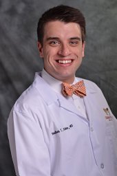 Nathan T. Cohen合作论文数Case Western Reserve University, Case Western Reserve University2Maimone Santo合作论文数2Prashanthi N. Thota合作论文数2Vijay S. Khiani合作论文数2Anokh Kondru合作论文数2
 Sapna Thomas合作论文数School of Medicine, Case Western Reserve University;Gastroenterology, St John Medical Center, University Hospitals;Gastroenterology, Cleveland Medical Center, University Hospitals2Jennifer Cutter合作论文数2Wendy Brock合作论文数2
 Kishore Guda合作论文数2Cynthia Calley合作论文数1Emily Ching合作论文数1Nadim S. Jafri合作论文数1Kevin M Cronley合作论文数1Carson L. Smith合作论文数1Laurie Logan合作论文数1Abey Thomas合作论文数1Dennis Wilk合作论文数1




