Sensitivity analysis for handling residual CAR19 qPCR data below limit of quantification (LOQ) in leukapheresis material
The impact of chimeric antigen receptor (CAR)-T cell expansion and persistence on clinical outcomes and treatment-related morbidity in patients with relapsed/refractory multiple myeloma (RRMM) remains incompletely defined, in part due to limited availability of standardized CAR-T cell quantification assays. We evaluated CAR-T cell kinetics and their association with efficacy and toxicity in RRMM patients treated with idecabtagene vicleucel (ide-cel) or ciltacabtagene autoleucel (cilta-cel). Using a uniform flow cytometry-based platform (N=90; cilta-cel, n=54; ide-cel, n=36), we observed significantly greater CAR-T cell expansion with cilta-cel than with ide-cel (median 106 vs 49 cells/uL). Peak CAR-T cell expansion was associated with clinical response in the ide-cel cohort but not with cilta-cel, where rapid and excessive expansion was instead associated with an increased risk of delayed neurotoxicities (DNTs), a complication with potential long-term functional consequences (median peak 1,009 vs 96 cells/uL). To identify clinically accessible biomarkers of CAR-T cell expansion, we analyzed absolute lymphocyte count (ALC) as a surrogate biomarker in a larger multicenter cohort (N=532; cilta-cel, n=256; ide-cel, n=276). Higher peak ALC was significantly associated with the development of DNTs, particularly Parkinsonism after cilta-cel. A peak ALC ³3000/uL - or ³2500/uL following a daily twofold increase - predicted elevated DNT risk (sensitivity 81%, specificity 59%). Together, these findings delineate distinct expansion-toxicity relationships in cilta-cel and ide-cel therapy, establish ALC as a practical, uniformly available surrogate for CAR-T cell expansion, and define quantitative thresholds that may enable early recognition of patients at risk for DNT, informing preemptive strategies to mitigate morbidity following cilta-cel.
Supplemental Figure 2 shows lab values over time (A) and at peak/nadir (B) in grade 2+ vs no IECHS
CD39 expression on CD4+ and CD8+ CAR22, stratified by median expression levels (High/Low) or using a 20% cutoff, and its correlation with CAR22 expansion as measured by flow cytometry and qPCR, treatment response, and survival outcomes. (A) The CD39 expression level on CD4+ or CD8+ CAR22 in the product, stratified by median (> median or < median) or using a 20% cutoff, shows no significant difference in CAR22 maximal expansion (CMax) measured by flow cytometry in peripheral blood (PB) after infusion. (B) CD39 expression on CD4+ or CD8+ CAR22, stratified by median, shows no significant difference in CAR22 area under the curve from day 0 to 28 (AUC0-28) post-infusion as measured by flow cytometry in PB. (C) CD39 expression on CD4+ or CD8+ CAR22, stratified by median, shows no significant difference in CAR22 CMax and AUC0-28 post-infusion as measured by qPCR in PB. (D) CD39 expression on CD4+ or CD8+ CAR22, stratified by median, shows no significant difference in complete response rate, progression-free survival, or overall survival. Dotted lines represent the 95% confidence interval.
Introduction BCMA targeted CAR-T therapy in multiple myeloma (MM) results in deeper, more durable responses and a higher frequency of minimal residual disease (MRD) negativity when compared to standard of care. Many patients, however, will relapse after initially achieving a response. We evaluated outcomes of MRD positive patients following ciltacabtagene autoleucel (cilta-cel) to determine the expected outcomes in this population. Objectives To compare outcomes based on MRD status, and dynamics of CAR-T expansion in these patients. Methods In this multicenter study, we evaluated 317 patients receiving cilta-cel for MM who achieved a VGPR or better response to cilta-cel and had MRD results available at 3 months post cilta-cel infusion using next generation sequencing or next generation flow cytometry with sensitivity of 1 × 10⁻⁵ or better. Efficacy outcomes based on MRD status were examined. Median follow-up from the time of CAR-T infusion was 11.93 months. We also evaluated a cohort of cilta-cel patients (n=38) from one center where data on CAR-T expansion by flow cytometry was available. CAR expansion was determined via peripheral blood cilta-cel concentration at day 7, 14, 21, and 28 post-infusion. Results Amongst the 317 patients with VGPR or better response, only 32 (10%) were MRD positive at 3 months post cilta-cel. Median age of the population was 65 years. 52% were male and 14% were black. 33% had high-risk cytogenetics and 34% had EMD. Median prior lines of therapy was 5. Patients who were MRD positive were more likely to have high risk disease (41% vs 32%) and higher baseline ferritin, but no other differences in baseline characteristics were noted. PFS was significantly different based on MRD status (p=0.002), with median PFS of the MRD+ patients at 12.6 months compared to 26.4 months for MRD- patients. 1-year PFS estimates were 51% (95% CI: 31%, 68%) and 81% (95% CI: 75%, 86%), respectively.In the single center cohort with CAR expansion data, 32% (10/31) of patients were MRD+. MRD data was not available for 7 patients. We observed similar trends in PFS with the larger, multi-site cohort. When evaluating the interaction of CAR-T expansion and persistence with MRD negativity, we observed no significant differences in CAR-T expansion at peak (around day 14) or at other timepoints based on MRD status. CAR-T persistence at day 90 was known in 21 patients and was not significantly different based on MRD status. Conclusion Overall, our data confirms that patients who remain MRD positive at 3 months post cilta-cel are at significantly higher risk of early relapse with median PFS of 12.6 months. This is likely independent of CAR-T expansion or persistence, but should be confirmed in a larger dataset. Importantly, there is an urgent need for clinical trials of bridging and consolidation strategies to prevent early relapse in patients who remain MRD positive following cilta-cel.
Comparison of CAR19 transgene level, CAR22 product characteristic and cell kinetics across different CAR19 co-stimulatory domain
There is limited systemic data on the dynamics of BCMA-target antigen expression with BCMA CAR-T at relapse. We analyzed 76 patients receiving standard-of-care BCMA-directed CAR-T who underwent real-time BCMA expression evaluation at baseline (n = 50), relapse (6), or both (20) using flow cytometry (FC) and/or immunohistochemistry (IHC). BCMA was universally expressed at baseline with significant heterogeneity in expression level. No concordance was seen between FC and IHC in categorizing high vs. low expression (Spearman: 0.07, Cohen kappa: 0). Plasma cell BCMA expression by FC correlated with clinical outcomes, whereas IHC did not. High BCMA expression by FC was associated with increased likelihood for VGPR/CR (p = 0.007) and longer time to progression (p = 0.005), including the ciltacabtagene autoleucel cohort (median: 23.0 vs. 7.7 months, p = 0.02). Relapsed patients retained BCMA expression by FC, though 29% (5/16) had BCMA loss by IHC, with 4/5 showing concurrent positive BCMA expression by FC. BCMA expression at relapse by FC was significantly lower than baseline (p = 0.04); downregulation (≥25% decrease) occurred in 50% (8/16) with paired samples. Higher BCMA expression by FC correlated with higher likelihood of deep, durable responses following BCMA-directed CAR-T. While BCMA loss is rare, decreased expression is common at relapse, with implications for sequencing BCMA-directed therapies.
Immune effector cell-associated hemophagocytic lymphohistiocytosis-like syndrome (IEC-HS) is a poorly characterized inflammatory toxicity of chimeric antigen receptor (CAR) T cells with high risk of mortality. In this study, we describe IEC-HS manifestations in patients with large B-cell lymphoma and B-cell acute lymphoblastic leukemia after CD22-directed CAR T cells. IEC-HS occurred in 19 of 54 patients (35%), including 11 grade 1 and 8 grade 2 or higher. IEC-HS was associated with higher nonrelapse mortality (NRM) yet lower relapse rates. CAR T-cell expansion in peripheral blood was significantly associated with IEC-HS severity. Cytokine profiling identified 41 cytokines primarily related to the IFNγ, TNFα, and IL1 families that significantly correlated with IEC-HS severity. We developed a parsimonious model composed of IFNγ, IL10, and IL1RA that correlated with grade 2+ IEC-HS on day 14, outperforming the full signature (AUC 0.93 vs. 0.75, P = 0.038). Thus, a cytokine signature with potential prognostic utility helps distinguish IEC-HS from inflammatory toxicities with overlapping symptoms. SIGNIFICANCE:IEC-HS is a serious inflammatory toxicity of CAR T cells. We demonstrate that IEC-HS after CD22-directed CAR T-cell therapy is associated with lower rates of relapse yet higher NRM. CAR T-cell expansion and a 41-cytokine signature are associated with IEC-HS, and a simplified signature of IFNγ, IL10, and IL1RA precedes severe disease. See related commentary by Rocco and Shah, p. 163.
The CAR T-cell maximal expansion (CMax) and area under the curve from day 0 to 28 (AUC0-28) for CAR19 and CAR22 following CAR19 or CAR22 infusion in the same patient. (A) Analysis by flow cytometry. (B) Analysis by qPCR assay. No statistical comparison was performed due to the limited number of patients.
Abstract Introduction: Brexucabtagene autoleucel (BA) is an effective chimeric antigen receptor T cell (CAR-T) therapy for B cell acute lymphoblastic leukemia (ALL); however, it causes high-grade immune toxicities. Preclinical models show that short dasatinib (D) pulses given after CAR-T transiently disrupt CAR signaling, promoting a reversible “rest” state that improves T cell function. We hypothesized that D given soon after BA in vivo could induce transient rest during rapid expansion, thereby mitigating toxicities while preserving efficacy. We therefore designed a trial to evaluate safety and feasibility of D pulses after BA in relapsed/refractory (r/r) ALL patients (pts). Methods: Adult r/r ALL pts meeting treatment criteria for BA were eligible for this open-label Phase 1b trial at Stanford University (BA+D group). After BA infusion, D 100mg daily was started between Day +4 to +10 and continued on a 3 days on/4 days off pulse schedule during month 1. Primary endpoints were feasibility (≥ 2 D pulses in month 1) and safety. Clinical outcomes were compared to a cohort of 11 Stanford pts who received BA for r/r ALL from 2022-2024 (BA-only group). Results: Eleven pts were enrolled; 4 withdrew prior to receiving BA; 7 were evaluable and received BA+D. At time of BA infusion, pt median age was 44 (range 35-56), median prior lines of therapy was 2 (1-3), 4 pts (57%) had morphologic disease, and 3 (43%) had measurable residual disease (MRD). D pulses began at median Day +5 after BA (range 4-13). Feasibility was met as 6/7 pts (86%) completed ≥ 2 D pulses following BA. D was well-tolerated without excess toxicity.For BA+D vs BA-only, respectively, rates of cytokine release syndrome [CRS, all grade (AG): 100% vs 73%; G3+: 0% vs 0%], were similar; rates of immune effector cell-associated (IEC) neurotoxicity syndrome (ICANS, AG: 29% vs 55%; G3+: 29% vs 36%), and IEC hemophagocytic syndrome (IECHS, AG: 29% vs 36%; G3+: 0% vs 18%) were numerically lower. Rates of complete remission (CR, 85% vs 73%), MRD-negative CR (57% vs 45%), 1-year relapse-free survival (57% vs 44%) and 1-year overall survival (85% vs 73%) were similar. Pts in BA+D who had immune toxicity trended towards lower total steroid exposure relative to similar pts in BA-only (median 2 vs 6 days). For BA+D, CAR-T expansion was assessed by flow assisted cell sorting. 5/7 pts (71%) had CAR-T cells that expanded despite D pulses, but with lower median peak (34 CAR-Ts/uL) than reported with BA-only. All 5 pts with CAR expansion had detectable circulating CAR-T at D+28. Further comparative correlative data will be presented at AACR. Conclusion: This direct translation of preclinical work shows that 3-day pulses of D after BA are feasible and safe. While we could not clearly determine if D pulses reduced CAR-T toxicity in this small cohort, BA does expand in the setting of D. Citation Format: Nikeshan Jeyakumar, Parveen Shiraz, Evan Weber, Alyssa Kanegai, Caroline Wagner, Arvind Ramakrishnan, Bita Sahaf, Matthew Frank, Saurabh Dahiya, Melody Smith, Surbhi Sidana, Crystal Mackall, David Miklos, Lori Muffly. Brexu-cel + dasatinib: Safety and feasibility of dasatinib pulses after brexucabtagene autoleucel to modulate CAR T cell activity in relapsed/refractory B cell acute lymphoblastic leukemia [abstract]. In: Proceedings of the American Association for Cancer Research Annual Meeting 2026; Part 2 (Late-Breaking, Clinical Trial, and Invited Abstracts); 2026 Apr 17-22; San Diego, CA. Philadelphia (PA): AACR; Cancer Res 2026;86(8_Suppl):Abstract nr CT190.
CAR T-cell therapy has emerged as a transformative treatment for hematological malignancies, yet its potential to drive lymphomagenesis poses significant clinical concerns. In this study, we investigated the mechanisms underlying CAR T-cell-associated lymphomagenesis in the gastrointestinal (GI) tract on a single case, focusing specifically on the role of integrin a4b7 expression and a predisposing somatic SH2B3 mutation. We observed oligoclonal CAR T-cells homing to and clonally expanding in the GI tract, with the dominant expanded clone harboring both a pathogenic SH2B3 mutation and a CAR transgene integration within a TFCP2 locus. The clonal CAR T-cells subsequently transitioned beyond the GI tract into the peripheral blood, suggesting a potential pathway for systemic dissemination. We found clinical, histological, and molecular evidence demonstrating the efficacy of cyclosporine in reducing the expanded malignant clone and achieving durable clinical remission for more than a year. Our findings highlight the complex interplay between CAR T-cell therapy, pre-existing genetic vulnerabilities, and the GI microenvironment, emphasizing the need for vigilant monitoring and tailored therapeutic strategies to address the risks associated with CAR-T lymphomagenesis.
CD19-targeted CAR T-cell therapy (CAR-T) has transformed treatment for elderly patients with relapsed or refractory large B-cell lymphoma (rrLBCL), yet data on its safety and efficacy in octogenarians (aged ≥80 years) often with more comorbidities and frailty remain limited. In this single-institution retrospective study, 24 octogenarians were identified and matched with 48 patients aged 65-79 years receiving standard-of-care CAR-T using 2:1 propensity score matching. Baseline characteristics were similar, except for more use of lisocabtagene maraleucel and tisagenlecleucel (P<0.001) and bridging therapy (P=0.024) in the octogenarian cohort. Median follow-up was 15.8 and 36.7 months for the octogenarian and comparator cohorts, respectively. The octogenarian cohort had a higher rate of grade ≥3 immune effector cell-associated neurotoxicity syndrome (ICANS) (25% vs 21%; age treatment effect (ATE) P=0.027). Among those experiencing grade ≥3 cytokine release syndrome (CRS) or ICANS, hospital stays were significantly longer in the octogenarian cohort (30 days vs. 16 days, ATE P<0.001). CAR T-cell expansion was reduced in octogenarians, with lower peak counts (ATE P=0.038) and expansion over 28 days post-infusion (ATE P<0.01). Overall and complete response rates were comparable between cohorts. Six- and 12-month progression-free survival rates in octogenarians were 57% and 45%, and overall survival rates were 88% and 81%, respectively, with no significant differences from those of the comparator cohort. These findings suggest that selected octogenarians tolerate and benefit from CAR T-cell therapy and underscore the need for strategies to mitigate severe toxicities through close monitoring and supportive care.
Introduction Obecabtagene autoleucel (obe-cel) and brexucabtagene autoleucel (brexu-cel) are CD19 targeted chimeric antigen receptor T cell (CAR-T) therapies, approved for adults with relapsed/refractory (r/r) B-cell acute lymphoblastic leukemia (ALL). Mechanistic differences between obe-cel and brexu-cel including differing costimulatory domains (4-1BB vs. CD28), CD19 binding domains (intermediate vs. high affinity) and split dose (Days 1 and 10) vs. single infusion may impact in-vivo cellular kinetics that translate into variant clinical outcomes. Real-world utilization and outcomes with obe-cel are unknown given the relatively recent approval. Methods The ROCCA database, comprising real world data from patients (pts) with r/r ALL treated at 40 North American institutions was used in this analysis. Pts with r/r ALL were eligible if they were apheresed for obe-cel since its approval (11/8/2024) or brexu-cel over a comparable period (since 8/1/24) and had at least 30 days of follow up. Data cut off was 7/15/2025. CRS/ICANS were graded per ASTCT criteria. Measurable residual disease (MRD) was assessed by flow cytometry and/or next generation sequencing per institutional standards. Results 38 pts have undergone apheresis for obe-cel (36 infused, all received both infusions) and 54 (53 infused) for brexu-cel over the study period. Baseline characteristics are shown in Table 1.CAR-mediated toxicity differed significantly between the cohorts (Table 2). CRS occurred in 56% of obe-cel pts compared to 94% of brexu-cel pts (p < 0.0001). There were no Gr3+ CRS events among the obe-cel pts; 3 (6%) brexu-cel pts had Gr3+ CRS (p = 0.27). ICANS occurred in 17% of obe-cel pts vs. 51% of brexu-cel pts (p = 0.001). Gr3+ ICANS occurred in 6% of obe-cel vs. 32% of brexu-cel pts (p = 0.0027). Among the obe-cel pts, CRS occurred in 31% after the first infusion and 46% after the second; ICANS occurred in 3% after the first infusion and 15% after the second. Prolonged Gr4 neutropenia (ANC < 500 cells/uL beyond day 30 from infusion) occurred in 24% of obe-cel vs. 28% of brexu-cel pts (p = 0.73). Deaths within the first 28 days of infusion occurred in 0 obe-cel pts and 4 brexu-cel pts (2 of infection, 1 of infection/brain bleed, and 1 of liver failure in the setting of Gr4 CRS and HLH).Response rates were high and did not significantly differ between cohorts (p = 0.85) (Table 3). 81% of obe-cel pts vs. 80% of brexu-cel pts achieved an MRD- CR/CRi. Conclusion Pts selected for obe-cel apheresis were similar to those for brexu-cel over the study period (noting that not all centers had access to obe-cel during this time). Similar to clinical trial results, obe-cel was associated with lower rates of CRS/ICANS. Rates of MRD-negative CR were high and did not differ between cohorts. A larger sample and longer follow up are required for further analyses; we anticipate a cohort of ∼75 obe-cel treated pts by the annual meeting and will provide updated data.
More than half of patients with relapsed/refractory large B-cell lymphoma experience disease progression after CD19-directed chimeric antigen receptor (CAR19) T-cell therapy. We investigated CAR22 therapy in 38 patients with CAR19-refractory disease, achieving 68% overall response rate (ORR) and 53% complete response rate (CR)(NCT04088890). Long-term follow-up (median 43.5 months), revealed sustained clinical benefit among complete responders, who achieved a median progression-free survival of 52 months and a 75% estimated 3-year overall survival rate. Using multi-omics analyses, we identified that intrinsic T-cell stemness characteristics present at apheresis associated with therapeutic success. Responding patients exhibited elevated TCF7 and LEF1 transcription factor activity and enhanced chromatin accessibility at TCF/LEF binding motifs. CAR T-cell products from CR patients demonstrated higher T-cell receptor diversity. Immune effector cell-associated hemophagocytic lymphohistiocytosis-like syndrome (IEC-HS) was associated with interferon-stimulated gene expression and STAT2 activity. These findings challenge the paradigm that T cells are irreversibly compromised after CAR therapy failure and provide mechanistic insights for optimizing sequential CAR therapies.
Linear regression analysis of CAR22 transduction efficiency, vector copy number (VCN) and T cell subsets in CD4/CD8 enrichment material and CAR22 product (A) Linear regression of CAR22 transduction efficiency versus T cell subsets in CD4/CD8 enrichment material. (B) Linear regression of CAR22 transduction efficiency versus T cell subsets in the products. (C) Linear regression of CAR22 VCN versus T cell subsets in CD4/CD8 enrichment material. (D) Linear regression of CAR22 VCN versus T cell subsets in the products.
Quantifiable CAR19 transgene levels in leukapheresis material and CAR22 products, and their correlation with CAR22 complete response (CR) rate and survival outcomes. (A) The CR rate is similar between patients with and without quantifiable CAR19 transgene levels in leukapheresis material and CAR22 products. (B) Progression-free survival (PFS) and overall survival (OS) are similar between patients with and without quantifiable CAR19 transgene levels in leukapheresis material. (C) PFS and OS are similar between patients with and without quantifiable CAR19 transgene levels in CAR22 products.
Comparison of CRS, ICANS, and IEC-HS between different leukapheresis interval groups, CAR19 transgene level, and CAR22 product transduction rate cut off by median
GD2-CAR T cell therapy has demonstrated clinical benefit in patients with H3K27M + diffuse midline glioma (DMG), but the durability of response has been limited in many patients 1,2 . To identify mechanisms of therapeutic resistance, we conducted longitudinal single-cell RNA and TCR sequencing of cerebrospinal fluid (CSF) lymphocytes from DMG patients receiving intravenous followed by sequential intracerebral GD2-CAR therapy, with lymphodepleting chemotherapy administered once prior to the start of CAR T cell therapy ( NCT04196413 ). CSF GD2-CAR T cells manifested limited persistence and clonal expansion, while non-engineered CSF lymphocytes underwent significant clonal expansion and repertoire stabilization, ultimately dominating the CSF immune compartment. Concurrently, peripheral blood CD4 + and CD8 + T cells manifested anti-CAR immune reactivity targeting epitopes enriched within murine-derived and engineered junctional regions of the CAR construct. This was associated with appearance of circulating Human Anti-CAR Antibodies (HACAs) that bound cells expressing the GD2-CAR, as well as clonal expansion of CSF B cells which produced HACA which impeded the cytotoxic activity of GD2-CAR T cells. In several cases, appearance of circulating HACA temporally correlated with disease progression and across the patient population, and levels of circulating HACA inversely correlated with circulating CAR T cell persistence. These findings reveal robust induction of systemic and CNS adaptive T cell and B cell responses to GD2-CAR T cells following intravenous then sequential intracerebroventricular GD2-CAR therapy and provide strong evidence that anti-CAR immunity is a significant contributor to therapeutic resistance in this setting.
 David Miklos合作论文数Blood and Marrow Transplantation & Cellular Therapy Division, School of Medicine, Stanford University;Stanford Cancer Institute, School of Medicine, Stanford University109
 Crystal Mackall合作论文数School of Medicine, Stanford University100
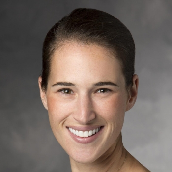 Lori Muffly合作论文数Blood & Marrow Transplantation & Cellular Therapy Division, Department of Medicine, School of Medicine, Stanford University;Stanford Cancer Institute, School of Medicine, Stanford University67
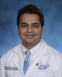 Dahiya Saurabh合作论文数Baystate Medical Center, Tufts University School of Medicine62
 Matthew Frank合作论文数Medicine Department, Stanford University61Sushma Bharadwaj合作论文数55
 Good Zinaida合作论文数51
 Sneha Ramakrishna合作论文数Pediatrics Department, Stanford University48Melody Smith合作论文数47
 Sally Arai合作论文数Blood and Marrow Transplantation and Cellular Therapy Division, School of Medicine, Stanford University45
 Kara Davis合作论文数Pediatrics Department, Stanford University43Steven A Feldman合作论文数40
 Surbhi Sidana合作论文数Cleveland Clinic Foundation, Medicine Institute40
 Patel Shabnum合作论文数39
 Wen-Kai Weng合作论文数Blood and Marrow Transplantation & Cellular Therapy Division, School of Medicine, Stanford University;Department of Dermatology, School of Medicine, Stanford University38
 Anne Marijn Kramer合作论文数36
 Robert Negrin合作论文数Blood & Marrow Transplantation & Cellular Therapy Division, School of Medicine, Stanford University35
 Robert Lowsky合作论文数Blood and Marrow Transplantation & Cellular Therapy Division, Stanford School of Medicine, Stanford University33Liora Schultz合作论文数Pediatrics Department, Stanford University31
 Laura Johnston合作论文数Department of Medicine, Stanford University29
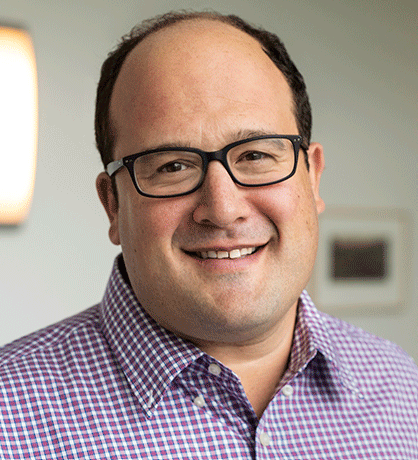 Robbie Majzner合作论文数Dana-Farber Cancer Institute29
 Everett Meyer合作论文数Department of Medicine, Stanford University27
 Michelle Monje合作论文数Department of Psychiatry and Behavioral Sciences, School of Medicine, Stanford University26
 Jasia Mahdi合作论文数25Sharon A. Mavroukakis合作论文数25Rezvani Andrew R合作论文数24
 Hamilton Mark P合作论文数Department of Molecular and Cellular Biology, Baylor College of Medicine24Emily Egeler合作论文数23Warren Reynolds合作论文数23Courtney Erickson合作论文数22
 Elena Sotillo合作论文数Stanford Canc Inst, Stanford Univ22Jay Y. Spiegel合作论文数Department of Medicine, University of Miami Health System22
 Christina R Baggott合作论文数22
 Snehit Prabhu合作论文数Columbia University22Jennifer Moon合作论文数21
 Judith Shizuru合作论文数Pediatrics Department, Stanford University21Zachary Ehlinger合作论文数20
 Sonia Partap合作论文数20Hitomi Hosoya合作论文数19Agnes Reschke合作论文数19Harshini Chinnasamy合作论文数18
 Yiyun Chen (陈以昀)合作论文数State Key Laboratory of Bioorganic and Natural Products Chemistry, Shanghai Institute of Organic Chemistry, Chinese Academy of Sciences/University of Chinese Academy of Sciences18
 John Harvey Baird合作论文数17
 Jean S. Oak合作论文数Department of Pathology, Stanford University17
 Sean Bendall合作论文数School of Medicine, Stanford University17Valentin Barsan合作论文数16John S Tamaresis合作论文数15Michael Kunicki合作论文数15Ashley Jacobs合作论文数14




