Neurogenic bladder is a debilitating consequence of spinal cord injury (SCI), with limited treatments that restore voluntary voiding. Although in vivo glial reprogramming has been achieved in the injured spinal cord, its effect on bladder function remains unclear. Here, we show that SOX2-mediated reprogramming of NG2 glia enhances bladder function in a clinically relevant mouse model of contusive SCI. The reprogramming process induces new neurons, attenuates scarring, and significantly improves urinary performance, as assessed by voiding assays and conscious cystometry. Functional recovery correlates positively with neurogenesis and inversely with scarring. These findings reveal that in vivo glial reprogramming promotes autonomic circuit repair and provides a regenerative strategy for treating neurogenic bladder after SCI.
ABSTRACT Neurogenic bladder is one of the most disabling consequences of spinal cord injury (SCI), yet it remains unclear whether residual brain–bladder communication persists after injury and can be therapeutically strengthened. Here, we integrated analysis of a clinical SCI cohort with studies in a mouse model that recapitulates key features of human neurogenic bladder. SCI markedly impaired both descending and ascending limbs of the spinobulbospinal micturition reflex, reducing bladder responses evoked by stimulation of the pontine micturition center (PMC) and bladder filling-induced activation of the periaqueductal gray. Despite this marked functional impairment, pseudorabies virus tracing, optogenetics, and machine learning-assisted three-dimensional imaging revealed persistent bladder-connected neurons within the spared T8–T9 interlesion region. Neurons within this region remained responsive to descending PMC input, identifying the interlesion network as a candidate substrate for residual brain–bladder communication. Early continuous intrathecal bumetanide improved urinary storage and emptying, enhanced descending PMC-to-bladder and ascending bladder-to-brain signaling, and increased the functional engagement of bladder-connected T8–T9 neurons without increasing urine production. Bumetanide also increased trans-synaptic labeling between the bladder and PMC, consistent with enhanced polysynaptic brain–bladder connectivity. Together, these findings indicate that brain–bladder communication is not completely lost after SCI and may be supported by a functionally compromised residual spinal network that remains amenable to therapeutic reinforcement. Targeting spared autonomic circuitry may therefore provide a strategy for improving bladder function after SCI. Highlights SCI disrupts bidirectional communication within the brain–bladder circuit. Spared T8–T9 interlesion neurons remain engaged in residual brain–bladder signaling. Intrathecal bumetanide strengthens brain–bladder signaling and improves urinary function. Graphic abstract
Chronic low back pain (LBP), predominantly driven by intervertebral disc degeneration (IVDD) is a major public health burden, yet the molecular mechanisms linking disc pathology to inflammation and pain remain poorly understood. Here, we identify a fibrotic, late-stage population of nucleus pulposus (NP) cells in degenerating intervertebral discs that aberrantly upregulate Colony Stimulating Factor 1 (CSF1). NP-derived CSF1 activates the CSF1 Receptor (CSF1R) on infiltrating macrophages, triggering a sustained pro-inflammatory cascade within the microenvironment. Using a murine model of disc injury, we demonstrate that either genetic ablation of Csf1r or pharmacologic inhibition with the selective CSF1R antagonist GW2580 markedly attenuates disc degeneration, confirmed by longitudinal MRI and histopathology, and significantly reduces associated neuropathic pain behaviors. These findings establish the CSF1–CSF1R axis as a central mechanistic link between fibrotic NP cell remodeling and macrophage-driven inflammation, highlighting it as a promising therapeutic target for mitigating disc degeneration and alleviating chronic LBP. One Sentence Summary: Blocking CSF1 signaling alleviates intervertebral disc degeneration and neuropathic pain
Background Recent evidence suggests extra-cortical adaptations within the cerebellum may contribute to motor recovery in patients with cortical ischemic strokes. The molecular/cellular adaptations enabling this effect to have not been identified. Chloride transport proteins (NKCC1 and KCC2) are important regulators of neuronal transmission and may underlie adaptive changes following ischemic stroke. Objective Examine changes in cerebellar NKCC1 and KCC2 protein expression following cortical ischemic stroke. Methods Adult C57BL/6J male mice underwent sham or the left middle cerebral artery occlusion (tMCAo)-induced ischemic stroke. Changes of NKCC1 and KCC2 proteins within the deep cerebellar nuclei (DCN) were assessed by immunofluorescence staining. Results tMCAo induced selective infarct lesion in the left striatum and cortex of the stroke mice but not in other brain regions including cerebellum. The inwardly directed chloride transporter NKCC1 was equivocally expressed within bi-hemispheric DCN of both sham control and stroke mice. In contrast, the outwardly directed chloride transporter KCC2 protein expression was significantly higher in the bi-hemispheric DCN of stroke brains, compared to sham controls. Double immunostaining analysis revealed a statistically significant increase in KCC2 intensity within VGLUT-1+ neurons of the ipsilateral DCN of the stroke mice, but not in the VGAT+ neurons. Conclusions Ischemic cortical stroke stimulates KCC2 protein expression in the DCN VGLUT-1+ neurons, without a change in NKCC1 protein expression.
Spinal cord injury (SCI) often results in permanent sensory deficits, significantly impairing the quality of life. These deficits are poorly addressed due to a lack of valid animal models with translational relevance. Here, we utilized a thoracic Level 8 lateral hemisection SCI mouse model (including both male and female mice) and applied a battery of behavioral assays requiring supraspinal transmission of sensory information. We also assessed ascending spinal circuits from the lumbar spinal cord to the brain. By 28 d post-SCI, sensory assessments revealed distinct deficits: reduced innocuous sensation in the ipsilateral hindpaw and enhanced sensation in the contralateral hindpaw. Both hindlimbs exhibited disrupted nocifensive behaviors, with chronic neuropathic dysesthesia observed only in the contralateral hindlimb. We provided anatomical evidence to elucidate the neural substrates responsible for these sensory discrepancies. This SCI mouse model mimics key features of human lateral hemisection conditions (Brown-Séquard syndrome) and offers a robust platform to explore underlying mechanisms and develop new therapeutic strategies.
Spinal cord injury (SCI) results in acute damage and triggers secondary injury responses with sustained neuronal loss and dysfunction. However, the underlying mechanisms for these delayed neuronal pathologies are not entirely understood. SCI results in the swelling of spinal neurons, but the contribution of cell swelling to neuronal loss and functional deficits after SCI has not been systematically characterized. In this study, we devised a three-dimensional image analysis pipeline to evaluate spinal neurons, examining their types, quantities, volumes, and spatial distribution in a double-lateral hemisection SCI mouse model. We found that both excitatory and inhibitory neurons swell and are lost, albeit with distinct temporal patterns. Inhibitory neurons demonstrated marked swelling and decline in number on day 2 after SCI, which resolved by day 14. In contrast, excitatory neurons maintained persistent swelling and continued cell loss for at least 35 days after SCI in mice. Excitatory neurons exhibited sustained expression of the Na+-K+-Cl- cotransporter 1 (NKCC1), whereas inhibitory neurons down-regulated the protein by day 14 after SCI. Treatment with a Food and Drug Administration-approved NKCC1 inhibitor, bumetanide, mitigated swelling of excitatory neurons and reduced their loss in the secondary injury phase after SCI. The administration of bumetanide after SCI in mouse improved locomotor recovery, with functional benefits persisting for at least 4 weeks after treatment cessation. This study advances our understanding of SCI-related pathology and introduces bumetanide as a potential treatment to mitigate sustained neuronal swelling and enhance recovery after SCI.
Intervertebral disc (IVD) degeneration is one of the major causes of low back pain. Inflammation has been implicated in discogenic back pain and disc degeneration, however, the detailed molecular mechanisms remain unclear. Herein we demonstrate that Colony Stimulating Factor 1 Receptor (CSF1R) signaling plays an essential role in the development of IVD degeneration and discogenic back pain. Genetic deletion of CSF1R from microglia/macrophages or oral administration of a CSF1R competitive inhibitor, GW2580, decreased IVD degeneration as evidenced by serial magnetic resonance imaging (MRI) and histopathological analyses in adult mice following disc injury. CSF1R deletion or GW2580 administration inhibited pro-inflammatory cytokine release from injured discs and blocked dorsal root ganglion (DRG) macrophage and spinal cord dorsal horn microglia activation and in so doing, eliminated neuropathic pain secondary to disc injury. These results suggest a novel therapeutic strategy for the treatment of chronic low back pain secondary to IVD degeneration.
Spinal interneurons (SpINs) facilitate local, intersegmental, and long-distance neuronal connections and are crucial for coordinated motor and sensory activity within the spinal cord. Spinal cord injuries (SCIs) damage direct descending pathways but spare many SpINs. However, traumatic injury and inflammation can cause maladaptive plasticity and excitability in SpINs, which may be detrimental to spinal relay circuits. Recent studies in human patients and SCI animal models have revealed several strategies for modulating SpIN function to promote functional recovery. Here we summarize recent progress in our understanding of SpIN function in intact and spinal cord lesioned mammals and describe the cellular mechanisms of SpIN adaptations and their contribution to adaptive and maladaptive plasticity in post-SCI relay circuits.
The Na-K-2Cl cotransporter (NKCC1) is considered an attractive drug target in the Central nervous system (CNS) for treating various CNS disorders. However, the specific role of NKCC1 in different types following injury within the CNS is not well understood due to its expression in multiple cell types. Additionally, there is a lack of a robust method for knocking down NKCC1 transcripts. In this study, we utilized Cas13 nucleases, a type of programmable RNA-targeting CRISPR enzyme, to effectively degrade NKCC1 mRNA in cultured cells. We developed a versatile pipeline for crRNA screening and validation in vitro and demonstrated the successful knockdown of NKCC1 using RfxCas13d. Our findings establish RfxCas13d as a powerful tool for targeting specific transcripts in vitro. By demonstrating the successful in vitro application of RfxCas13d-mediated NKCC1 RNA knockdown, we have laid the groundwork for future investigations into the therapeutic potential of NKCC1 modulation in CNS disorders.
>From the days of Ramon y Cajal's first sketches, neuroscientists have recognized the importance of visualizing the complex architecture of the central nervous system. In the past century, we have come to appreciate how the rich structural and functional complementarity of axons and cell types in the spinal cord make it uniquely suited for information transfer between the periphery and the brain.
Precisely measuring the number and somatic volume of neurons in the central nervous system at single-cell resolution is technically challenging. Here, we combine multiple techniques to address this challenge in optically cleared mouse spinal cords. We describe in vivo neuron labeling approaches, tissue-clearing technology, light sheet fluorescence microscopy, and machine learning-guided imaging analysis. This combination provides a precise determination of the cell number and somatic volume of any neuron population in the spinal cords.
After complete spinal cord injuries (SCI), spinal segments below the lesion maintain inter-segmental communication via the intraspinal propriospinal network. However, it is unknown whether selective manipulation of these circuits can restore locomotor function in the absence of brain-derived inputs. By taking advantage of the compromised blood-spinal cord barrier following SCI, we optimized a set of procedures in which AAV9 vectors administered via the tail vein efficiently transduce neurons in lesion-adjacent spinal segments after a thoracic crush injury in adult mice. With this method, we used chemogenetic actuators to alter the excitability of propriospinal neurons in the thoracic cord of the adult mice with a complete thoracic crush injury. We showed that activating these thoracic neurons enables consistent and significant hindlimb stepping improvement, whereas direct manipulations of the neurons in the lumbar spinal cord led to muscle spasms without meaningful locomotion. Strikingly, manipulating either excitatory or inhibitory propriospinal neurons in the thoracic levels leads to distinct behavioural outcomes, with preferential effects on standing or stepping, two key elements of the locomotor function. These results demonstrate a strategy of engaging thoracic propriospinal neurons to improve hindlimb function and provide insights into optimizing neuromodulation-based strategies for treating SCI.
Spinal cord injury in mammals is thought to trigger scar formation with little regeneration of axons1–4. Here we show that a crush injury to the spinal cord in neonatal mice leads to scar-free healing that permits the growth of long projecting axons through the lesion. Depletion of microglia in neonatal mice disrupts this healing process and stalls the regrowth of axons, suggesting that microglia are critical for orchestrating the injury response. Using single-cell RNA sequencing and functional analyses, we find that neonatal microglia are transiently activated and have at least two key roles in scar-free healing. First, they transiently secrete fibronectin and its binding proteins to form bridges of extracellular matrix that ligate the severed ends of the spinal cord. Second, neonatal—but not adult—microglia express several extracellular and intracellular peptidase inhibitors, as well as other molecules that are involved in resolving inflammation. We transplanted either neonatal microglia or adult microglia treated with peptidase inhibitors into spinal cord lesions of adult mice, and found that both types of microglia significantly improved healing and axon regrowth. Together, our results reveal the cellular and molecular basis of the nearly complete recovery of neonatal mice after spinal cord injury, and suggest strategies that could be used to facilitate scar-free healing in the adult mammalian nervous system. In neonatal mice, scar-free healing after spinal cord injury is organized by microglia, and transplantation of neonatal microglia or peptidase-inhibitor-treated adult microglia into adult mice after injury improves healing and axon regrowth.
Many human spinal cord injuries are anatomically incomplete but exhibit complete paralysis. It is unknown why spared axons fail to mediate functional recovery in these cases. To investigate this, we undertook a small-molecule screen in mice with staggered bilateral hemisections in which the lumbar spinal cord is deprived of all direct brain-derived innervation, but dormant relay circuits remain. We discovered that a KCC2 agonist restored stepping ability, which could be mimicked by selective expression of KCC2, or hyperpolarizing DREADDs, in the inhibitory interneurons between and around the staggered spinal lesions. Mechanistically, these treatments transformed this injury-induced dysfunctional spinal circuit to a functional state, facilitating the relay of brain-derived commands toward the lumbar spinal cord. Thus, our results identify spinal inhibitory interneurons as a roadblock limiting the integration of descending inputs into relay circuits after injury and suggest KCC2 agonists as promising treatments for promoting functional recovery after spinal cord injury.
Current models of somatosensory perception emphasize transmission from primary sensory neurons to the spinal cord and on to the brain1-4. Mental influence on perception is largely assumed to occur locally within the brain. Here we investigate whether sensory inflow through the spinal cord undergoes direct top-down control by the cortex. Although the corticospinal tract (CST) is traditionally viewed as a primary motor pathway5, a subset of corticospinal neurons (CSNs) originating in the primary and secondary somatosensory cortex directly innervate the spinal dorsal horn via CST axons. Either reduction in somatosensory CSN activity or transection of the CST in mice selectively impairs behavioural responses to light touch without altering responses to noxious stimuli. Moreover, such CSN manipulation greatly attenuates tactile allodynia in a model of peripheral neuropathic pain. Tactile stimulation activates somatosensory CSNs, and their corticospinal projections facilitate light-touch-evoked activity of cholecystokinin interneurons in the deep dorsal horn. This touch-driven feed-forward spinal-cortical-spinal sensitization loop is important for the recruitment of spinal nociceptive neurons under tactile allodynia. These results reveal direct cortical modulation of normal and pathological tactile sensory processing in the spinal cord and open up opportunities for new treatments for neuropathic pain.
A major hurdle for functional recovery after both spinal cord injury and cortical stroke is the limited regrowth of the axons in the corticospinal tract (CST) that originate in the motor cortex and innervate the spinal cord. Despite recent advances in engaging the intrinsic mechanisms that control CST regrowth, it remains to be tested whether such methods can promote functional recovery in translatable settings. Here we show that post-lesional AAV-assisted co-expression of two soluble proteins, namely insulin-like growth factor 1 (IGF1) and osteopontin (OPN), in cortical neurons leads to robust CST regrowth and the recovery of CST-dependent behavioral performance after both T10 lateral spinal hemisection and a unilateral cortical stroke. In these mice, a compound able to increase axon conduction, 4-aminopyridine-3-methanol, promotes further improvement in CST-dependent behavioral tasks. Thus, our results demonstrate a potentially translatable strategy for restoring cortical dependent function after injury in the adult.
Efficient transport of stem/progenitor cells without affecting their survival and function is a key factor in any practical cell-based therapy. However, the current approach using liquid nitrogen for the transfer of stem cells requires a short delivery time window is technically challenging and financially expensive. The present study aims to use semipermeable alginate hydrogels (crosslinked by strontium) to encapsulate, store, and release stem cells, to replace the conventional cryopreservation method for the transport of therapeutic cells within worldwide distribution time frame. Human mesenchymal stem cell (hMSC) and mouse embryonic stem cells (mESCs) were successfully stored inside alginate hydrogels for 5 days under ambient conditions in an air-tight environment (sealed cryovial). Cell viability, of the cells extracted from alginate gel, gave 74% (mESC) and 80% (hMSC) survival rates, which compared favorably to cryopreservation. More importantly, the subsequent proliferation rate and detection of common stem cell markers (both in mRNA and protein level) from hMSCs and mESCs retrieved from alginate hydrogels were also comparable to (if not better than) results gained following cryopreservation. In conclusion, this new and simple application of alginate hydrogel encapsulation may offer a cheap and robust alternative to cryopreservation for the transport and storage of stem cells for both clinical and research purposes.
 Che John Connon合作论文数Department of Ophthalmology, Kyoto Prefectural University of Medicine13
 Shengli Mi (弥胜利)合作论文数清华大学深圳研究生院9
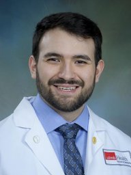 Alfredo Sandoval合作论文数8
 Zhigang He合作论文数Boston Children's Hospital;Department of Ophthalmology, Harvard Medical School, Harvard University6
 Junjie Zhu合作论文数5
 Bernice Wright合作论文数5
 Zicong Zhang (张子聪)合作论文数Department of Rehabilitation Sciences, The Hong Kong Polytechnic University4Yiming Zhang合作论文数3
 Yuanyuan Liu合作论文数3Zhiyun Yang合作论文数3John Moth合作论文数Department of Neurobiology, University of Texas Medical Branch3
 Songlin Zhou (周松林)合作论文数Jiangxi Chest Hospital3Wright B合作论文数3Dunn Tiffany J合作论文数3Yi Li合作论文数3Yu Zhang合作论文数2Melissa Henwood合作论文数2Junfeng Su合作论文数2
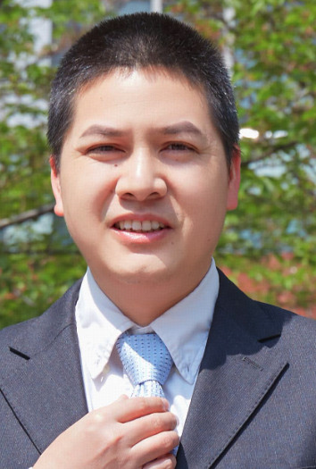 Xuhua Wang (王绪化)合作论文数School of Medicine, Zhejiang University;School of Brain Science and Brain Medicine, MOE Frontier Science Center for Brain Science and Brain-Machine Integration, Zhejiang University2
 Jones Roanne R合作论文数School of Chemistry, Pharmacy and Food Biosciences, University of Reading2
 Philip Michael Williams合作论文数School of Pharmacy, University of Nottingham2
 James Foster合作论文数School of Biological Sciences, University of Bristol2
 Yuan Subo合作论文数2Bin Yu合作论文数2
 Xiaosong Gu (顾晓松)合作论文数Nantong University;School of Medicine, Tianjin University;School of Medicine, Nanjing University of Chinese Medicine2
 Ian Hamley合作论文数Department of Chemistry, School of Chemistry, Food & Pharmacy, University of Reading2
 Qiang Li合作论文数2
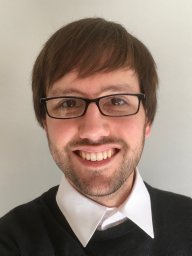 Benedikt Brommer合作论文数Department of Ophthalmology, Harvard Medical School, Boston, MA, USA2
 Philip R. Williams合作论文数1
 Shamseldin Metwally合作论文数1Okan Capuk合作论文数1Richard A Cave合作论文数1Wei Li合作论文数1Daryl Fields合作论文数1Jin-Yong Zhou合作论文数1Wenlei Li合作论文数1Junkui Shang合作论文数1Yi Lu合作论文数1
 Anne Jacobi合作论文数Institute of Clinical Neuroimmunology, University Hospital LMU1
 Kathiravan Kaliyappan合作论文数Dept Rehabil Sci, Univ Buffalo State Univ New York1Zhongju Shi合作论文数1
 Zhongyang Gao (高中洋)合作论文数The Second Affiliated Hospital, Xi’an Jiaotong University1Chungui Xu合作论文数1
 Holly Catherine Gibbs合作论文数Microscopy and Imaging Center, Texas A&M University1
 Mengying Chen合作论文数1
 Mohammad Iqbal H Bhuiyan合作论文数Univ Pittsburgh, Dept Neurol, Pittsburgh, PA 15213 USA1T. Nakamura合作论文数1
 Hajira Elahi合作论文数Center for Advanced Pain Studies and Department of Neuroscience, The University of Texas at Dallas1Jing Tang合作论文数1




