Abstract Pancreatic ductal adenocarcinoma (PDAC) has a prominent fibrotic stroma, which is a result of interactions between tumor, immune and pancreatic stellate cells (PSC), or cancer-associated fibroblasts (CAF). Targeting inflammatory pathways present within the stroma may improve access of effector immune cells to PDAC and response to immunotherapy. Heat shock protein-90 (Hsp90) is a chaperone protein and a versatile target in pancreatic cancer. Hsp90 regulates a diverse array of cellular processes of relevance to both the tumor and the immune system. However, to date the role of Hsp90 in PSC/CAF has not been explored in detail. We hypothesized that Hsp90 inhibition would limit inflammatory signals, thereby reprogramming the PDAC tumor microenvironment to enhance sensitivity to PD-1 blockade. Treatment of immortalized and primary patient PSC/CAF with the Hsp90 inhibitor XL888 decreased IL6, a key cytokine that orchestrates immune changes in PDAC at the transcript and protein level in vitro. XL888 directly limited PSC/CAF growth and reduced Jak/STAT and MAPK signaling intermediates and alpha-SMA expression as determined via immunoblot. Combined therapy with XL888 and anti–PD-1 was efficacious in C57BL/6 mice bearing syngeneic subcutaneous (Panc02) or orthotopic (KPC-Luc) tumors. Tumors from mice treated with both XL888 and anti–PD-1 had a significantly increased CD8+ and CD4+ T-cell infiltrate and a unique transcriptional profile characterized by upregulation of genes associated with immune response and chemotaxis. These data demonstrate that Hsp90 inhibition directly affects PSC/CAF in vitro and enhances the efficacy of anti–PD-1 blockade in vivo.
HLA-A2/DR1 sarcoma (A2/Sarcoma) cell line transfected to express either GFP or SSX2. T2 and LNCaP cells do not upregulate levels of PD-L1 expression when cultured with recombinant IFNγ. Tumor infiltrating CD8+ T cells express detectable levels of PD-1 by flow cytometry.
T cell and macrophage depletion studies in mice bearing Myc-CaPWT or Myc-CaPΔRb tumors.
Androgen deprivation increases the frequency of infiltrating T cells and M1 tumor-associated macrophages in the tumor microenvironment.
Loss of Rb results in altered gene expression that promotes immune suppression as well as increased Tmem173/STING expression.
JQ-1 does not induce apoptosis at 1 µM concentration that activates non-canonical STING/NF-κB signaling.
Generation and characterization of growth kinetics of Rb-deficient Myc-CaP cell line.
Enrichment of Rb1 or CDKN2A loss in non-T cell-inflamed primary prostate cancer and across multiple primary tumor histologies
JQ-1 does not induce p-IRF3 translocation to the nucleus in wild-type or Rb-deficient Myc-CaP tumor cells.
Decreased Rb1 expression correlates with predicted decreased immune infiltration across multiple disease histologies.
BACKGROUND: Pancreatic ductal adenocarcinoma (PDAC) is the most malignant neoplasm of the pancreas, and is refractory to most therapeutic strategies, including immunotherapy. We have previously demonstrated that loss of CDKN2A in PDAC tumor cells promotes an immunologically-cold tumor microenvironment, which can be counteracted through the use of bromodomain and extraterminal domain inhibitors (BETi). The immunosuppressive PDAC tumor microenvironment is also characterized by a dense desmoplastic stroma, rich in cancer associated fibroblasts (CAFs). These CAFs are a highly plastic population of cells, comprised of myofibroblastic, inflammatory, and antigen-presenting CAFs (myCAFs, iCAFs, and apCAFs, respectively). Previously research has suggested BETi can alter the function of CAFs, suggesting this therapeutic strategy may be effective at targeting both the tumor and stromal compartments of PDAC. Here we hypothesize that BETi enhances anti-tumor immunity in CDKN2A-deficient PDAC by modulating the phenotype and function of PDAC CAFs. METHODS: Isogenic wild-type and CDKN2A-deficient KPCluc pancreatic cancer cell lines were used in conjunction with KPCluc-derived murine CAF cells. CAF spheroids were used to evaluate the effects of BETi on CAF phenotype (as measured by flow cytometry) and function (as measured by NanoString analysis, checkpoint ligand expression by flow cytometry, and T cell migration assays). Additionally, the impact of BETi-induced modulation of CAF phenotype and function on wild-type and CDKN2A-deficient tumor cell spheroids was measured by flow cytometry. RESULTS: Treatment of pancreatic CAFs with BETi induced expression of genes associated with DNA damage responses and type I IFN expression. Additionally, while CAF supernatants suppressed T cell migration in vitro, treatment with BETi restored T cell migration across a transwell. This was due in part to DNA damage/STING/NF-κB signaling induced in PDAC CAF following BETi. Supernatants from BETi-treated PDAC CAFs were found to modulate PD-L1 expression on PDAC tumor cells. Direct treatment of CAFs with BETi induced a dramatic dose- and time-dependent polarization of CAF spheroids towards a myCAF phenotype. However, while BETi induced DNA damage/STING/NF-κB signaling in CAF cells, this signaling did not contribute towards this myCAF polarization, as treatment of CAF spheroids with BETi and STING or NF-κB inhibitors did not impact myCAF polarization. Additional mechanisms of polarization are currently being evaluated and will be updated at presentation. CONCLUSIONS: BET inhibition modulates CAF cell phenotype and function to promote anti-tumor immune responses. Taken together with the impact of BET inhibition on CDKN2A-deficient PDAC tumor cells, these data suggest BETi is a rational treatment approach to promote anti-tumor immunity in this genetically defined immunotherapy resistant patient population. Citation Format: Trisha Minocha, Alison Thomas, Brian M. Olson. BET inhibition reprograms pancreatic cancer associated fibroblasts to promote anti-tumor immunity. [abstract]. In: Proceedings of the American Association for Cancer Research Annual Meeting 2023; Part 1 (Regular and Invited Abstracts); 2023 Apr 14-19; Orlando, FL. Philadelphia (PA): AACR; Cancer Res 2023;83(7_Suppl):Abstract nr 5173.
Background Semaphorin4D (SEMA4D) regulates immune cell infiltration and suppressive features of myeloid cells in the tumor via engaging with receptors, PLXNB1/2 and CD72.1 In preclinical and clinical studies, SEMA4D blockade enhances efficacy of immune checkpoint blockade (ICB) by invigorating lymphocyte infiltration and limiting suppressive myeloid cell properties in tumors.2 3 We hypothesized pepinemab (a SEM4D-blocking Ab), when combined with ICB, enhances crosstalk between B and T cells present as part of tertiary lymphoid structures (TLS) within the tumor microenvironment (TME) of melanoma patients. Methods Consenting patients with resectable stage III melanoma received standard-of-care, neoadjuvant nivolumab alone, pepinemab/nivolumab, pepinemab/ipilimumab, or pepinemab/nivolumab/ipilimumab (NCT03769155). Following six weeks of therapy, patients underwent surgical resection. Patients also provided peripheral blood draws pre-treatment, two weeks into treatment, and at time of surgery. Surgical tissue and blood underwent high dimensional immune analysis using 32-color flow cytometry. Pretreatment archival tissue was also used in conjunction with on study surgical resection tissue to evaluate spatial distribution of immune populations using multiplex immunohistochemistry and Nanostring CosMx single cell imaging. Surgical resection tissue and matched blood was used for TCR and BCR sequencing. Results Our work reveals that neoadjuvant combination treatments were well tolerated and resulted in pathologic and durable clinical responses. Moreover, patients given pepinemab/nivolumab/ipilimumab have not experienced tumor recurrence after treatment more than 2 years later (ongoing response in 8/8 patients). Pepinemab in combination with nivolumab (either alone or with ipilimumab) significantly increased tumor-infiltrating B cells (CD19+) and CD4+ T cells within surgical tissue, compared to surgery or nivolumab alone. An increase in the frequency of CD4+T cells that expressed the novel costimulatory molecule CD26 was also detected in responsive patients. Multiplex IHC revealed that pepinemab with nivolumab (alone or with ipilimumab) led to generation of tertiary lymphoid structures (TLS) comprised of B cells and T cells within the tumor bed. More tumor-infiltrating B cells and CD4+CD26hi T cells were associated with clinical response in patients receiving pepinemab and nivolumab (with or without ipilimumab). An increase in TCR and BCR clonal diversity was observed in tumors from patients receiving pepinemab-containing regimens, as compared to those receiving nivolumab alone. Nanostring CosMX transcriptomic analysis will be updated at presentation. Conclusions Pepinemab in combination with nivolumab and ipilimumab robustly modulates immune responses in tumors from patients with resectable metastatic melanoma. Given the encouraging clinical activity and tolerability of these regimens, future studies should evaluate the use of pepinemab combined with ICB in other tumor indications. Trial Registration NCT03769155 References 1. Nkyimbeng-Takwi, E. & Chapoval, S. P. Biology and function of neuroimmune semaphorins 4A and 4D. Immunol. Res. 50, 10–21 (2011). 2. Evans, E. E. et al. Antibody blockade of semaphorin 4D promotes immune infiltration into tumor and enhances response to other immunomodulatory therapies. Cancer Immunol. Res. 3, 689–701 (2015). 3. Clavijo, P. E. et al. Semaphorin4D inhibition improves response to immune-checkpoint blockade via attenuation of MDSC recruitment and function. Cancer Immunol. Res. 7, 282–291 (2019).
Increased CXCL12 within tumor cells by RNA in situ hybridization following cabozantinib treatment.
S1. Phenotypic validation of HLA-A2-transfected 22Rv1/FCS and 22Rv1/CSS cells. S2. AR-specific T cells obtained following peptide-immunization have increased recognition and lysis of androgen-deprived prostate tumor cells. S3. Immunization with pTVG-AR delays tumor growth, in a CD8-dependent fashion, in the presence or absence of ADT. S4. Immunization with pTVG-AR delays tumor growth in the presence or absence of ADT, and results in increased tumor-infiltrating T cells.
Supplementary Figure 3: IgG immunological response: IgG responses to AR LBD protein (panel A), PSA protein (panel B), and tetanus toxoid (panel C) were evaluated by Luminex. Sera samples were assessed at a 1:100 dilution from the time points indicated. Data were plotted over time, subtracting the pre-treatment value to evaluate change in post-treatment specimens.
Background Preclinical studies showed metformin reduces exhaustion of tumor-infiltrating lymphocytes and potentiates programmed cell death protein-1 (PD-1) blockade. We hypothesized that metformin with nivolumab would elicit potent antitumor and immune modulatory activity in metastatic microsatellite stable (MSS) colorectal cancer (CRC). We evaluated this hypothesis in a phase II study. Methods Nivolumab (480 mg) was administered intravenously every 4 weeks while metformin (1000 mg) was given orally, two times per day following a 14-day metformin only lead-in phase. Patients ≥18 years of age, with previously treated, stage IV MSS CRC, and Eastern Cooperative Oncology Group 0–1, having received no prior anti-PD-1 agent were eligible. The primary endpoint was overall response rate with secondary endpoints of overall survival (OS) and progression-free survival (PFS). Correlative studies using paired pretreatment/on-treatment biopsies and peripheral blood evaluated a series of immune biomarkers in the tumor microenvironment and systemic circulation using ChipCytometry and flow cytometry. Results A total of 24 patients were enrolled, 6 patients were replaced per protocol, 18 patients had evaluable disease. Of the 18 evaluable patients, 11/18 (61%) were women and the median age was 58 (IQR 50–67). Two patients had stable disease, but no patients had objective response, hence the study was stopped for futility. Median OS and PFS was 5.2 months (95% CI (3.2 to 11.7)) and 2.3 months (95% CI (1.7 to 2.3)). Most common grade 3/4 toxicities: Anemia (n=2), diarrhea (n=2), and fever (n=2). Metformin alone failed to increase the infiltration of T-cell subsets in the tumor, but combined metformin and nivolumab increased percentages of tumor-infiltrating leukocytes (p=0.031). Dual treatment also increased Tim3+ levels in patient tissues and decreased naïve CD8+T cells (p=0.0475). Conclusions Nivolumab and metformin were well tolerated in patients with MSS CRC but had no evidence of efficacy. Correlative studies did not reveal an appreciable degree of immune modulation from metformin alone, but showed trends in tumorous T-cell infiltration as a result of dual metformin and PD-1 blockade despite progression in a majority of patients.
 Douglas McNeel合作论文数Department of Medicine Directory, School of Medicine and Public Health, University of Wisconsin-Madison34
 Akash Patnaik合作论文数Committee on Cancer Biology, The University of Chicago19
 Gregory B Lesinski合作论文数Arthur G. James Cancer Hospital and Richard J. Solove Research Institute, Ohio State University19Alison Thomas合作论文数16Marguerite Li合作论文数14Christina Hong合作论文数14Srikrishnan Rameshbabu合作论文数14
 Chaudagar Kiranj K合作论文数L.M. College of Pharmacy, Opp. Gujarat University14Laura E. Johnson合作论文数13Raymond Chen合作论文数13
 Sweta Sharma Saha合作论文数<label>*</label><p>Equal Contribution of Authors</p>12E. Evans合作论文数9
 Crystal Mallow合作论文数9
 Christina Wu合作论文数8Terrence L. Fisher合作论文数8
 Leigh Ellis合作论文数Genitourinary Program, Roswell Park Cancer Institute8Maurice Zauderer合作论文数7
 Bassel El-Rayes合作论文数Department of Hematology and Medical Oncology, Emory University School of Medicine;O'Neal Comprehensive Cancer Center, School of Medicine, The University of Alabama at Birmingham7
 Michael Lowe合作论文数7
 Jens Eickhoff合作论文数Data Coordinating Center, Department of Biostatistics and Medical Informatics, School of Medicine and Public Health, University of Wisconsin–Madison7
 Huihui Ye合作论文数Cedars-Sinai6
 Becker Jordan T合作论文数Department of Medicine, University of Wisconsin Carbone Comprehensive Cancer Center6Reilly Christine合作论文数6Melinda L Yushak合作论文数5Jacklyn Hammons合作论文数5Chrystal M. Paulos合作论文数5
 Keith Delman合作论文数Division of Surgical Oncology, Department of Surgery, School of Medicine, Emory University;Winship Cancer Institute, Emory University5Thomas Sawicki合作论文数4Brian T Rekoske合作论文数4Michael B. Ware合作论文数4Brian Burns合作论文数4Ali Mokhtari合作论文数4Sukanya Panja合作论文数4
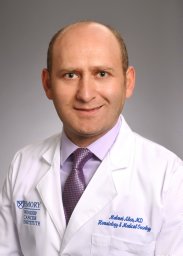 Mehmet Akce合作论文数Winship Canc Inst, Emory Univ4Deborah L Burkhart合作论文数4Carla Calagua合作论文数4Amanda N Ruggieri合作论文数4
 David P. Labbé合作论文数Division of Urology, Department of Surgery, Faculty of Medicine and Health Sciences, McGill University4
 Adam Stuart Kibel合作论文数Department of Urology, Brigham and Women’s Hospital;Dana-Farber Brigham Cancer Center4Kudchadkar Ragini R合作论文数4
 Antonina Mitrofanova合作论文数4
 Joseph Seliski合作论文数4
 Sowalsky Adam G合作论文数Department of Medicine, Beth Israel Deaconess Medical Center and Harvard Medical School4
 Olatunji B. Alese合作论文数Winship Cancer Institute, Emory University;Department of Hematology and Medical Oncology, Emory University School of Medicine4
 Morel Katherine L合作论文数Flinders Centre for Innovation in Cancer, Flinders University and Medical Centre4
 Walid L. Shaib合作论文数Cartersville Cancer Center, Northwest Georgia Oncology Centers;Marietta Cancer Center, Northwest Georgia Oncology Centers4
 Anjali Sheahan合作论文数Dana-Farber Cancer Institute4
 Massimo Loda合作论文数Department of Pathology and Laboratory Medicine, Weill Cornell Medicine, Cornell University;New York-Presbyterian Hospital;Harvard Medical School4
 Stephanie K. Dougan合作论文数Department of Cancer Immunology and Virology, Dana-Farber Cancer Institute;Harvard Medical School4




