Chirality conferral is fundamental for understanding the origin of life, and it is of direct importance for synthesizing new pharmaceuticals in the face of growing antibiotic resistance. Human-made, self-assembling nanostructures replicate the biological chirality conferral processes utilizing covalent and non-covalent bonds. However, chirality conferral from one form of matter to another via electromagnetic fields is more subtle and less explored. Here we report chirality conferral between gold nanohelices and achiral molecules (crystal violet). This conferral enables the experimental observation of a physical effect predicted in 1979-hyper-Raman optical activity. To benefit from Fermi's golden rule, the chirality conferral system was designed as doubly resonant, with the nanohelices and molecules resonating at the fundamental frequency and at the second-harmonic, respectively. We provide a theoretical framework for our results that expands the original mathematical formalism to include surface-enhanced hyper-Raman scattering and the chirality conferral process. Our results demonstrate that field-driven chirality conferral mechanisms are opening up entire fields of research, as exemplified by the discovery of a physical phenomenon. Theoretically predicted in 1979, hyper-Raman optical activity is now experimentally observed through chirality conferral from the electromagnetic field of chiral plasmonic gold nanohelices to crystal violet molecules that are achiral, sparking new science at the organic-inorganic interface.
Against the background of the current healthcare and climate emergencies, surface enhanced Raman scattering (SERS) is becoming a highly topical technique for identifying and fingerprinting molecules, e.g., within viruses, bacteria, drugs, and atmospheric aerosols. Crucial for SERS is the need for substrates with strong and reproducible enhancements of the Raman signal over large areas and with a low fabrication cost. Here, dense arrays of plasmonic nanohelices (≈100 nm in length), which are of interest for many advanced nanophotonics applications, are investigated, and they are shown to present excellent SERS properties. As an illustration, two new ways to probe near‐field enhancement generated with circular polarization at chiral metasurfaces are presented, first using the Raman spectra of achiral molecules (crystal violet) and second using a single, element‐specific, achiral molecular vibrational mode (i.e., a single Raman peak). The nanohelices can be fabricated over large areas at a low cost and they provide strong, robust and uniform Raman enhancement. It is anticipated that these advanced materials will find broad applications in surface enhanced Raman spectroscopies and material science.
Optical biopsy describes a range of medical procedures in which light is used to investigate disease in the body, often in hard-to-reach regions via optical fibres. Optical biopsies can reveal a multitude of diagnostic information to aid therapeutic diagnosis and treatment with higher specificity and shorter delay than traditional surgical techniques. One specific type of optical biopsy relies on Raman spectroscopy to differentiate tissue types at the molecular level and has been used successfully to stage cancer. However, complex micro-optical systems are usually needed at the distal end to optimise the signal-to-noise properties of the Raman signal collected. Manufacturing these devices, particularly in a way suitable for large scale adoption, remains a critical challenge. In this paper, we describe a novel fibre-fed micro-optic system designed for efficient signal delivery and collection during a Raman spectroscopy-based optical biopsy. Crucially, we fabricate the device using a direct-laser-writing technique known as ultrafast laser-assisted etching which is scalable and allows components to be aligned passively. The Raman probe has a sub-millimetre diameter and offers confocal signal collection with 71.3% ± 1.5% collection efficiency over a 0.8 numerical aperture. Proof of concept spectral measurements were performed on mouse intestinal tissue and compared with results obtained using a commercial Raman microscope.
Early detection of (pre-)cancerous changes improves prognosis, therefore in the UK patients at high risk of developing gastrointestinal cancers are enrolled on endoscopic surveillance programmes or the Bowel Cancer Screening Programme. The current gold standard technique for the detection of pre-cancerous changes in the gastrointestinal tract is histopathological analysis of biopsy tissue collected at endoscopy. This relies upon subjective assessment of morphological changes within the excised tissue samples and poor targeting of pre-malignant lesions. Raman spectroscopy offers a number of potential advantages for in vivo assessment of tissue at endoscopy. The performance of a custom built Raman probe as a biopsy targeting tool has been evaluated using excised biopsy material. Multivariate classification models have been used to demonstrate the likely ability of a miniature, confocal, fibre optic Raman probe to be used as an optical biopsy tool at endoscopy to provide spectral information in clinically practicable timescales. This technique could facilitate improved targeting of excisional biopsy with associated clinical benefits.
Raman spectroscopy has proved to be a highly sensitive tool for differentiating between normal, cancerous or pre-cancerous tissues. To date, histological application of Raman mapping has been limited due to lengthy mapping times. StreamlineTM Raman imaging is a novel mapping technique that has reduced total mapping times to a level that is becoming clinically practicable. Raman Streamline mapping was carried out on a 20μm frozen section of an oesophageal biopsy. A contiguous 7μm section was stained with haematoxylin and eosin (H&E) with histpathology analysed by a pathologist. The step size and acquisition times were varied and the resulting spectra, principal component score maps and loads were compared. The signal to noise for the raw spectra and a relative 'signal to noise' of the principal component loads were determined. The Streamline mapping technique was also compared to traditional point Raman mapping. The principal component loads were similar despite varying the acquisition time and number of spectra, with the fifth load used for comparison of the noise levels. Gross biochemical information was extracted showing good correlation with the H&E section even for short overall mapping times as low as 30-90 minutes for a biopsy ~2mm in diameter (0.5s acquisition time per 25.3μm Raman pixel). Streamline mapping was of the order of 3-7 times faster than traditional point mapping with the greatest improvement made for high resolution maps. Further optimization of the system is still possible which will reduce this mapping time further making implementation in a clinical environment a future possibility.
Routine clinical microbiological identification of pathogenic microorganisms is largely based on nutritional and biochemical tests. In the case of severely ill patients, the unavoidable time delay associated with such identification procedures can be fatal. We present a novel identification method based on confocal Raman microspectroscopy. With this approach it is possible to obtain Raman spectra directly from microbial microcolonies on the solid culture medium, which have developed after only 6 h of culturing for the most commonly encountered organisms. Due to the limited thickness of microcolonies, some of the underlying culture medium is sampled together with the bacteria. Spectra measured at different depths in a microcolony contain different amounts of the medium signal. A mathematical routine, involving vector algebra, is described for the nonsubjective correction of spectra for variable signal contributions of the medium. To illustrate the possibilities of our approach for the identification of microorganisms, Raman spectra were collected from 6-h microcolonies of five bacterial strains on solid culture medium. The classification results show that confocal Raman microspectroscopy has great potential as a powerful new tool in clinical diagnostic microbiology.
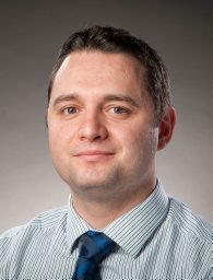 Robin Jones合作论文数Ctr Therapeut Innovat, Univ Bath2
 Hyunah Kwon合作论文数Max Planck Inst Intelligent Syst2
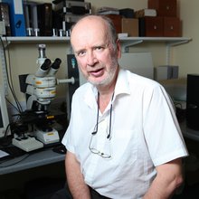 Hugh Barr合作论文数Royal College of Surgeons of Ireland/Gloucestershire Royal Hospital2
 Nick Stone合作论文数Department of Biomedical Imaging and Biosensing, School of Physics and Astronomy, University of Exeter2Neil A Shepherd合作论文数Cheltenham General Hospital2
 Liwu Zhang (张立武)合作论文数Department of Environmental Science and Engineering, Fudan University2
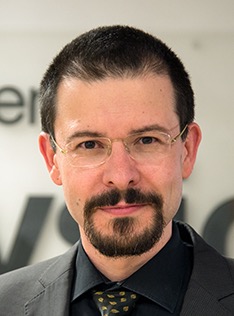 Ventsislav K. Valev合作论文数Department of Physics, University of Bath2
 Daniel Wolverson合作论文数School of Physics, University of East Anglia2
 Tim Batten合作论文数2
 Catherine A Kendall合作论文数2
 Peer Fischer合作论文数Institute for Molecular Systems Engineering and Advanced Materials, Heidelberg University;Max Planck Institute for Medical Research2Joanne Hutchings合作论文数2K Maquelin合作论文数1Samuel R. Clowes合作论文数1Cornelia Miksch合作论文数1
 Emilija Petronijevic合作论文数1Ha Bruining合作论文数1Coosje Pothoven合作论文数1
 Gong Kedong合作论文数Shanghai Key Laboratory of Atmospheric Particle Pollution and Prevention, Fudan University1david g maclachlan合作论文数1
 Kristina Rusimova合作论文数Univ Bath, Dept Phys, Bath BA2 7AY, Avon, England1
 Calum Ross合作论文数Computer Science and Engineering Department, Edge Hill University1Choo-Smith L-P合作论文数1john c c day合作论文数1
 David L. Andrews合作论文数Centre for Photonics and Quantum Science, School of Chemistry, Faculty of Science, University of East Anglia1
 Jonathan D. Shephard合作论文数School of Engineering and Physical Sciences, Heriot-Watt University1
 Gerwin J Puppels合作论文数1
 Robert Thomson合作论文数Institute of Photonics and Quantum Sciences, School of Engineering & Physical Sciences, Heriot-Watt University;Royal Academy of Engineering1
 Hubert P. Endtz合作论文数Department of Medical Microbiology and Infectious Diseases, Erasmus Medical Centre1
 Alejandro V. Silhanek合作论文数Department of Physics, Université de Liège1
 N J Weston合作论文数School of Engineering & Physical Sciences, Heriot-Watt University1Rainer J Beck合作论文数1T. Van Vreeswijk合作论文数1
 G. Dan Pantos合作论文数Department of Chemistry and Biochemistry, University of Texas at Austin, 1 University Station A5300, Austin, TX 78712, USA1




