Genetics has become a critical component of medicine over the past five to six decades. Alongside genetics, a relatively new discipline, dysmorphology, has also begun to play an important role in providing critically important diagnoses to individuals and families. Both have become indispensable to unraveling rare diseases. Almost every medical specialty relies on individuals experienced in these specialties to provide diagnoses for patients who present themselves to other doctors. Additionally, both specialties have become reliant on molecular geneticists to identify genes associated with human disorders. Many of the medical geneticists, dysmorphologists, and molecular geneticists traveled a circuitous route before arriving at the position they occupied. The purpose of collecting the memoirs contained in this article was to convey to the reader that many of the individuals who contributed to the advancement of genetics and dysmorphology since the late 1960s/early 1970s traveled along a journey based on many chances taken, replying to the necessities they faced along the way before finding full enjoyment in the practice of medical and human genetics or dysmorphology. Additionally, and of equal importance, all exhibited an ability to evolve with their field of expertise as human genetics became human genomics with the development of novel technologies.
The delineation of disease entities is complex, yet recent advances in the molecular characterization of diseases provide opportunities to designate diseases in a biologically valid manner. Here, we have formalized an approach to the delineation of Mendelian genetic disorders that encompasses two distinct but inter-related concepts: (1) the gene that is mutated and (2) the phenotypic descriptor, preferably a recognizably distinct phenotype. We assert that only by a combinatorial or dyadic approach taking both of these attributes into account can a unitary, distinct genetic disorder be designated. We propose that all Mendelian disorders should be designated as “GENE-related phenotype descriptor” (e.g., “CFTR-related cystic fibrosis”). This approach to delineating and naming disorders reconciles the complexity of gene-to-phenotype relationships in a simple and clear manner yet communicates the complexity and nuance of these relationships.
We report on a 26-year-old male with extreme short stature, microcephaly, macroglossia, other dysmorphic features, severe intellectual disability, and a bone dysplasia. The patient had an extensive genetic and biochemical evaluation that was all normal or noninformative. Recently, the proband died following a period of not eating. He likely had a previously undescribed syndrome of unknown etiology.
American Journal of Medical Genetics Part AVolume 182, Issue 7 p. 1541-1546 ISSUE INFORMATIONFree Access Table of Contents, Volume 182A, Number 7, July 2020 First published: 20 June 2020 https://doi.org/10.1002/ajmg.a.61236AboutPDF ToolsRequest permissionExport citationAdd to favoritesTrack citation ShareShare Give accessShare full text accessShare full-text accessPlease review our Terms and Conditions of Use and check box below to share full-text version of article.I have read and accept the Wiley Online Library Terms and Conditions of UseShareable LinkUse the link below to share a full-text version of this article with your friends and colleagues. Learn more.Copy URL Share a linkShare onEmailFacebookTwitterLinked InRedditWechat Volume182, Issue7July 2020Pages 1541-1546 RelatedInformation
A diagnostic journey began in 1966 when a male was born with a lethal hyperkeratosis of undetermined etiology, only to be followed by three additional siblings with the same unknown disorder. All four siblings had unique circumferential skin constrictions on all of their digits. They died within 5 days after birth with no diagnosis or etiology established. The first author (BDH) maintained notes, partial medical records, photographs, and comments about one autopsy report. This information was regularly revisited in the hope of finding a literature match, but no etiological diagnosis was forthcoming. However, in 2017, Rush et al. reported two siblings with similar phenotype in whom they found dolichol kinase deficiency (DOLK). Ultimately, our family was relocated and DNA isolated from the pathology slides of the third affected infant showed compound heterozygous pathogenic variants in the DOLK gene. The variants were in trans, with different missense variants from the mother and father. This 52-year diagnostic pursuit, culminated in an answer that gave the family an explanation for their losses.
In addition to the major generalized overgrowth syndromes described in this text, there is a rich case report literature on less common and less well-defined disorders in which overgrowth has been noted. The latter disorders are described concisely in this chapter. In some cases there have been multiple cases reported and the causative gene or genomic alterations have been identified. Although others are suspected to be genetic or genomic disorders, no specific cause has been implicated. These disorders are sufficiently uncommon that a body of literature has not accumulated and the manifestations are sufficiently variable that most are not clinically recognized. In addition, the chapter takes note of those environmental influences that can produce overgrowth. Maternal diabetes is the most common of the environmental influences that cause overgrowth during fetal life and carries a several-fold increased risk of malformations.
Cornelia de Lange syndrome (CdLS) is a multisystem genetic disorder with distinct facies, growth failure, intellectual disability, distal limb anomalies, gastrointestinal and neurological disease. Mutations in NIPBL, encoding a cohesin regulatory protein, account for >80% of cases with typical facies. Mutations in the core cohesin complex proteins, encoded by the SMC1A, SMC3 and RAD21 genes, together account for ∼5% of subjects, often with atypical CdLS features. Recently, we identified mutations in the X-linked gene HDAC8 as the cause of a small number of CdLS cases. Here, we report a cohort of 38 individuals with an emerging spectrum of features caused by HDAC8 mutations. For several individuals, the diagnosis of CdLS was not considered prior to genomic testing. Most mutations identified are missense and de novo. Many cases are heterozygous females, each with marked skewing of X-inactivation in peripheral blood DNA. We also identified eight hemizygous males who are more severely affected. The craniofacial appearance caused by HDAC8 mutations overlaps that of typical CdLS but often displays delayed anterior fontanelle closure, ocular hypertelorism, hooding of the eyelids, a broader nose and dental anomalies, which may be useful discriminating features. HDAC8 encodes the lysine deacetylase for the cohesin subunit SMC3 and analysis of the functional consequences of the missense mutations indicates that all cause a loss of enzymatic function. These data demonstrate that loss-of-function mutations in HDAC8 cause a range of overlapping human developmental phenotypes, including a phenotypically distinct subgroup of CdLS.
Townes–Brocks syndrome is a recognizable variable pattern of malformation caused by mutations to the SALL1 gene located on chromosome 16q12.1. Only three known cases of Townes–Brocks syndrome with proven SALL1 gene mutation and concurrent endocrine abnormalities have been previously documented to our knowledge [Kohlhase et al., 1999; Botzenhart et al., 2005; Choi et al., 2010]. We report on two unrelated patients with Townes–Brocks syndrome who share an identical SALL1 mutation (c.3414_3415delAT), who also have endocrine abnormalities. Patient 1 appears to be the first known case of growth hormone deficiency, and Patient 2 extends the number of documented mutation cases with hypothyroidism to four. We suspect endocrine abnormalities, particularly treatable deficiencies, may be an underappreciated component to Townes–Brocks syndrome. © 2013 Wiley Periodicals, Inc.
LUMBAR syndrome (lower body congenital infantile hemangiomas and other skin defects; urogenital anomalies and ulceration; myelopathy; bony deformities; anorectal malformations and arterial anomalies; and rectal anomalies) is a rare association between infantile hemangiomas of the lower half of the body and regional congenital anomalies. Since 1986, 53 cases have been reported and no etiology has been identified. We report on the 54th case in a male infant and review the literature concerning the manifestations of the LUMBAR syndrome. © 2013 Wiley Periodicals, Inc.
Nicolaides-Baraitser syndrome (NBS) is characterized by sparse hair, distinctive facial morphology, distal-limb anomalies and intellectual disability. We sequenced the exomes of ten individuals with NBS and identified heterozygous variants in SMARCA2 in eight of them. Extended molecular screening identified nonsynonymous SMARCA2 mutations in 36 of 44 individuals with NBS; these mutations were confirmed to be de novo when parental samples were available. SMARCA2 encodes the core catalytic unit of the SWI/SNF ATP-dependent chromatin remodeling complex that is involved in the regulation of gene transcription. The mutations cluster within sequences that encode ultra-conserved motifs in the catalytic ATPase region of the protein. These alterations likely do not impair SWI/SNF complex assembly but may be associated with disrupted ATPase activity. The identification of SMARCA2 mutations in humans provides insight into the function of the Snf2 helicase family.
Basal cell nevus syndrome (BCNS), also known as Gorlin syndrome (OMIM #109400) is a well-described rare autosomal dominant condition due to haploinsufficiency of PTCH1. With the availability of comparative genomic hybridization arrays, increasing numbers of individuals with microdeletions involving this locus are being identified. We present 10 previously unreported individuals with 9q22.3 deletions that include PTCH1. While 7 of the 10 patients (7 females, 3 males) did not meet strict clinical criteria for BCNS at the time of molecular diagnosis, almost all of the patients were too young to exhibit many of the diagnostic features. A number of the patients exhibited metopic craniosynostosis, severe obstructive hydrocephalus, and macrosomia, which are not typically observed in BCNS. All individuals older than a few months of age also had developmental delays and/or intellectual disability. Only facial features typical of BCNS, except in those with prominent midforeheads secondary to metopic craniosynostosis, were shared among the 10 patients. The deletions in these individuals ranged from 352 kb to 20.5 Mb in size, the largest spanning 9q21.33 through 9q31.2. There was significant overlap of the deleted segments among most of the patients. The smallest common regions shared among the deletions were identified in order to localize putative candidate genes that are potentially responsible for each of the non-BCNS features. These were a 929 kb region for metopic craniosynostosis, a 1.08 Mb region for obstructive hydrocephalus, and a 1.84 Mb region for macrosomia. Additional studies are needed to further characterize the candidate genes within these regions.
Carbimazole (CMZ) and its active metabolite methimazole (MMI) are antithyroid medications, which can result in MMI/CMZ embryopathy in susceptible individuals. The incidence of birth defects related to MMI/CMZ embryopathy remains unclear as several epidemiologic studies failed to prove a correlation, despite positive case–control studies and numerous case reports. Malformations reported in exposed individuals and commonly recognized as MMI/CMZ embryopathy include cutis aplasia of the scalp, choanal atresia, esophageal atresia (EA), tracheo‐esophageal fistula (TEF), persistent vitelline duct, athelia/hypothelia, and subtle facial dysmorphisms including sparse or arched eyebrows. Here, we report on individuals with early pregnancy exposure to MMI, with microtia and various other anomalies associated with MMI embryopathy, suggesting that microtia is also seen with increased frequency after prenatal MMI exposure. Additional unusual malformations among our patients include a previously unreported type of TEF with three separate esophageal pouches and a fistula connecting the middle pouch to the trachea in one child, and absence of the gall bladder in another. An enlarged anterior fontanel was seen in three patients, and clinodactyly of the fifth finger was noted in three. The similarities between our three patients with microtia after MMI exposure and the two previously reported with microtia after CMZ exposure support the concept of microtia being related to the MMI/CMZ exposure. Recognition of microtia as a manifestation of MMI/CMZ embryopathy will likely increase the number of diagnosed cases and thus affect ascertainment. We propose diagnostic criteria for MMI/CMZ embryopathy, including the presence of at least one major characteristic finding. © 2011 Wiley‐Liss, Inc.
American Journal of Medical Genetics Part AVolume 155, Issue 3 p. fm i-fm iv Table of ContentsFree Access Table of Contents, Volume 155, Number 3, March 2011 First published: 24 February 2011 https://doi.org/10.1002/ajmg.a.33993AboutPDF ToolsRequest permissionExport citationAdd to favoritesTrack citation ShareShare Give accessShare full text accessShare full-text accessPlease review our Terms and Conditions of Use and check box below to share full-text version of article.I have read and accept the Wiley Online Library Terms and Conditions of UseShareable LinkUse the link below to share a full-text version of this article with your friends and colleagues. Learn more.Copy URL Share a linkShare onFacebookTwitterLinked InRedditWechat Volume155, Issue3March 2011Pages fm i-fm iv RelatedInformation
Noonan syndrome (NS) is a common, clinically and genetically heterogeneous condition characterized by distinctive facial features, short stature, chest deformity, congenital heart disease, and other comorbidities. Gene mutations identified in individuals with the NS phenotype are involved in the Ras/MAPK (mitogen-activated protein kinase) signal transduction pathway and currently explain ∼61% of NS cases. Thus, NS frequently remains a clinical diagnosis. Because of the variability in presentation and the need for multidisciplinary care, it is essential that the condition be identified and managed comprehensively. The Noonan Syndrome Support Group (NSSG) is a nonprofit organization committed to providing support, current information, and understanding to those affected by NS. The NSSG convened a conference of health care providers, all involved in various aspects of NS, to develop these guidelines for use by pediatricians in the diagnosis and management of individuals with NS and to provide updated genetic findings.
American Journal of Medical Genetics Part AVolume 152A, Issue 10 p. fm i-fm v Table of ContentsFree Access Table of Contents, Volume 152A, Number 10, October 2010 First published: 21 September 2010 https://doi.org/10.1002/ajmg.a.33732AboutPDF ToolsRequest permissionExport citationAdd to favoritesTrack citation ShareShare Give accessShare full text accessShare full-text accessPlease review our Terms and Conditions of Use and check box below to share full-text version of article.I have read and accept the Wiley Online Library Terms and Conditions of UseShareable LinkUse the link below to share a full-text version of this article with your friends and colleagues. Learn more.Copy URL Share a linkShare onFacebookTwitterLinked InRedditWechat Volume152A, Issue10October 2010Pages fm i-fm v RelatedInformation
In 1990, Petty et al. described two patients representing a novel syndrome with “congenital progeriod” features and neither had classical progeria nor Wiedemann–Rautenstrauch syndrome, though many findings were overlapping. One of the cases had previously been described by Dr. Wiedemann in 1948. The key features of Petty syndrome include pre and postnatal growth restriction, decreased subcutaneous fat with loose skin, enlarged fontanelle with underdeveloped calvarium, coronal synostosis, unruly hair pattern with non‐uniform distribution, prominent eyebrows, umbilical hernia, distal digital hypoplasia, and normal or near normal development. Significant overlap to other syndromes, particularly the Fontaine–Farriaux syndrome, is apparent. In 2004, Ardinger postulated that Petty syndrome, like classical progeria, might be secondary to a defect in the lamin A/C ( LMNA ) gene. The purpose of this paper is to describe two new unrelated cases of this unique syndrome that further delineate the phenotype, compare to phenotypically similar syndromes, and postulate that Petty syndrome could represent a new laminopathy. In addition, evidence suggesting that the Petty syndrome and Fontaine–Farriaux syndromes are variable expressions of the same condition is discussed. © 2010 Wiley‐Liss, Inc.
American Journal of Medical Genetics Part AVolume 152A, Issue 6 p. 1372-1373 Invited Comment Invited Comment regarding “Pilot Assessment of a Radiological Classification for Segmentation Defects of the Vertebrae” by Offiah et al.† Bryan D. Hall, Corresponding Author Bryan D. Hall [email protected] Department of Pediatrics, University of Kentucky, Lexington, KentuckyDepartment of Pediatrics, University of Kentucky, Lexington, KY.Search for more papers by this author Bryan D. Hall, Corresponding Author Bryan D. Hall [email protected] Department of Pediatrics, University of Kentucky, Lexington, KentuckyDepartment of Pediatrics, University of Kentucky, Lexington, KY.Search for more papers by this author First published: 14 May 2010 https://doi.org/10.1002/ajmg.a.33423 † How to Cite this Article: Hall BD. 2010. Invited Comment regarding “Pilot Assessment of a Radiological Classification for Segmentation Defects of the Vertebrae” by Offiah et al. Am J Med Genet Part A 152A:1372–1373. Read the full textAboutPDF ToolsRequest permissionExport citationAdd to favoritesTrack citation ShareShare Give accessShare full text accessShare full-text accessPlease review our Terms and Conditions of Use and check box below to share full-text version of article.I have read and accept the Wiley Online Library Terms and Conditions of UseShareable LinkUse the link below to share a full-text version of this article with your friends and colleagues. Learn more.Copy URL Share a linkShare onEmailFacebookTwitterLinkedInRedditWechat No abstract is available for this article. Volume152A, Issue6June 2010Pages 1372-1373 RelatedInformation
 David W Smith合作论文数Department of Discovery Chemistry, Bristol-Myers Squibb Pharmaceutical Research Institute8
 Mahin Golabi合作论文数7
 John C Carey合作论文数Division of Medical Ethics, University of Utah;Department of Pediatrics, School of Medicine, University of Utah7
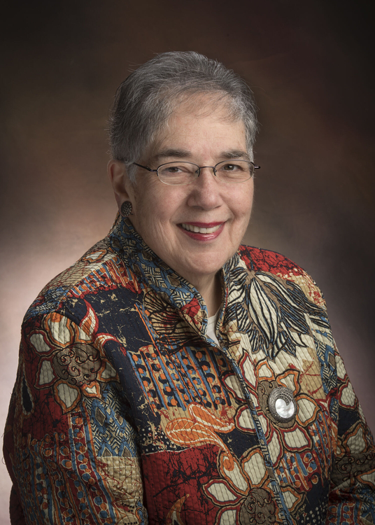 Elaine H. Zackai合作论文数Children's Hospital of Philadelphia;Perelman School of Medicine, University of Pennsylvania6
 Karen W. Gripp合作论文数Nemours Children's Health;FDNA5David D. Weaver合作论文数5
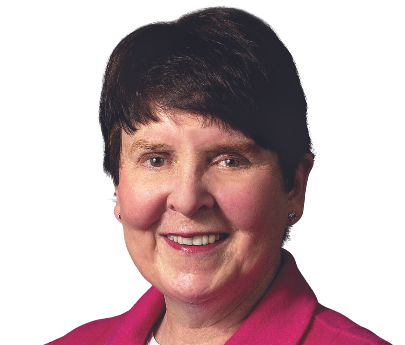 Cynthia Curry合作论文数Department of Pediatrics, School of Medicine, Stanford University;Department of Pediatrics, School of Medicine, University of California, San Francisco5Adam Margaret P合作论文数4
 John Graham合作论文数Cedars-Sinai Medical Center;Guerin Children's;School of Medicine, UCLA4
 Roger E. Stevenson (Roger Stevenson)合作论文数Greenwood Genetic Center;School of Health Studies, Clemson University4
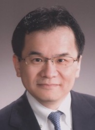 Kenjiro Kosaki (小崎健次郎)合作论文数Center for Medical Genetics, Keio University School of Medicine4Louise C. Wilson合作论文数3
 Robin Clark合作论文数3
 Emma L Wakeling合作论文数3
 Yuri Zarate合作论文数Division of Human Genetics, Cincinnati Children's Hospital Medical Center3
 Micheil Innes合作论文数Department of Medical Genetics, University of Calgary3Giampietro Philip F合作论文数3
 Raoul Hennekam合作论文数Amsterdam UMC;Department of Paediatrics, University of Amsterdam;Institute of Neurology, University College London3
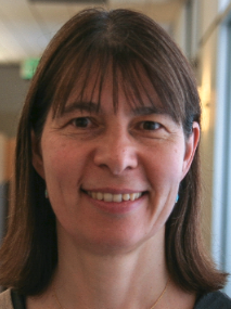 Anne Slavotinek合作论文数Sherr Lab, University of California, San Francisco3
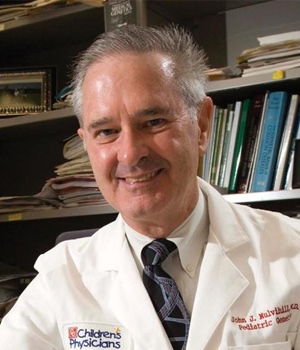 John J Mulvihill合作论文数Department of Pediatrics, College of Medicine, University of Oklahoma3
 Kenneth Jones合作论文数Center for Better Beginnings, University of California, San Diego3
 William Dobyns合作论文数Department of Pediatrics, Medical School, University of Minnesota;Division of Genetics and Metabolism, University of Minnesota3
 Michael J Bamshad合作论文数Division of Genetic Medicine, Department of Pediatrics, School of Medicine, University of Washington;Center for Clinical and Translational Research, Seattle Children’s Hospital3
 Anita E. Beck合作论文数3
 Louanne Hudgins合作论文数Pediatrics Department, Stanford University3Jacqueline A. Noonan合作论文数3
 David Stevenson合作论文数Department of Pediatrics, Stanford University3Joseph T. C. Shieh合作论文数3
 Kim M. Keppler‐Noreuil合作论文数3Wen-Hann Tan合作论文数2Vesna Martich Kriss合作论文数2Sally Ann Lynch合作论文数2Kerstin Reicherter合作论文数2Jennifer A Brault合作论文数2Kathy Culver合作论文数2
 Pedro A Sanchez-Lara合作论文数2
 Marc S Williams合作论文数2Mary Beth Dinulos合作论文数2Rhonda E Schnur合作论文数2Andrew K Sobering合作论文数2Małgorzata J M Nowaczyk合作论文数2Giovanni Neri合作论文数2
 Katta Girisha合作论文数2
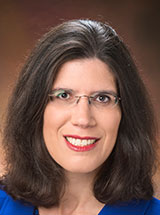 Jennifer M. Kalish合作论文数Department of Genetics, Perelman School of Medicine, University of Pennsylvania2
 Encarnacion Guillen Navarro合作论文数Departamento of Surgery, Pediatrics, Obstetrics and Gynecology, Faculty of Medicine, Universidad de Murcia;Hospital Clínico Universitario Virgen de la Arrixaca;IMIB-Arrixaca;CIBERER-ISCIII2
 James T Bennett合作论文数2Haldeman-Englert Chad合作论文数2
 Ghayda M Mirzaa合作论文数2
 Andrea Superti Furga合作论文数Centre Hospitalier Universitaire Vaudois2




