[This corrects the article DOI: 10.3389/fonc.2025.1695468.].
BACKGROUND:Intraoperative cone-beam computed tomography (CBCT) provides a valuable option for accurate three-dimensional applicator positioning in gynecologic brachytherapy, but is associated with radiation exposure and increased intervention time especially in case of repeated CBCT imaging being required for creating a sufficient implant arrangement. PURPOSE:To reduce the need for multiple CBCT scans for corresponding applicator verification, this work proposes two methods for needle path navigation, including corrections of potential bending in situ, by combining infrared tracking with planar x-ray imaging for enabling accurate intraoperative needle guidance. METHODS:An examined 200 mm brachytherapy needle was rigidly mounted on an infrared-reflective tracking tool to enable real time tracking. Two planar x-ray images, acquired from varying distinct angles, were used to determine the exact 3D position of the needle tip region via backprojection. A spline was fitted through the obtained coordinates to reconstruct the full needle path. Based on this, only a single initial CBCT scan was required to visualize the predicted needle path within this scan. Additionally, a second approach for needle prediction was presented focusing on only one planar x-ray image by incorporating prior needle bending information from the initial CBCT scan. Both methods were evaluated in preclinical studies and validated against a corresponding ground-truth obtained from CBCT. RESULTS:The proposed method considering two planar x-ray images successfully reconstructed the needle path with deviations of less than 1 mm from the CBCT reference scan, when using at least 20° offset between the x-ray image acquisitions. The single-scan approach, using prior bending information, yielded promising results with deviations at the tip of below 1.3 mm. CONCLUSIONS:Both described methods demonstrated their feasibility in preclinical studies, showing potential to improve and accelerate clinical implantation workflows by means of needle navigation in the future.
Background and purpose:Patient or treatment plan mix-ups are among the most serious patient-specific human errors in brachytherapy. However, many brachytherapy departments rely only on review by a second, independent person, which does not eliminate the risk of human failure. In this work, we developed and retrospectively evaluated an automated patient identification method based solely on the geometry of the interstitial implant in breast cancer patients. Materials and methods:The implant geometry is assessed using an electromagnetic tracking (EMT) system that provides real-time positional data of each catheter with sub-millimetre accuracy. The measured implant geometry is rigidly registered to the CT-based implant geometry associated with the clinical treatment plan. To quantitatively compare them, a similarity metric based on a distance-to-agreement (DTA) criterion (3-10 mm) and a pass rate threshold (50-95%) was used. The implants of 80 patients were included in the evaluation, resulting in 6400 patient-treatment plan combinations. Results:The classifier reliably identified patients with an area under the receiver operating characteristic (ROC) curve close to 1, highlighting an overall excellent discriminative performance. At the optimal decision threshold under the requirement of a false positive rate of 0%, it achieved a sensitivity between 94.8% and 97.5% depending on the DTA and pass rate thresholds, and an overall accuracy of 99.9%. Conclusion:Interstitial implants in breast brachytherapy are virtually unique, so determining their geometry prior to each fraction is a viable option for patient identification. The EMT-based automated technique has proven to be effective in detecting patient or treatment plan mix-ups with near-perfect accuracy.
BACKGROUND AND PURPOSE:Synthetic computed tomography (sCT) from magnetic resonance imaging (MRI) enables computed tomography (CT)-free cranial radiotherapy planning, yet no consensus exists on which head phantoms are usable for sCT quality assurance (QA). This study benchmarked head phantoms to identify the most promising candidates for cranial sCT QA and the design characteristics associated with favorable sCT-to-conventional CT agreement. MATERIALS AND METHODS:Ten head phantoms underwent clinical MRI (1.5 T) and CT imaging. Two vendor-provided sCT algorithms (2D slice-based, 3D volume-based) were applied to identical input. sCT and CT were compared using Dice similarity coefficient (DSC), 95th-percentile Hausdorff distance (HD95), CT number mean error (ME) within both a self-mask (sCT-derived) and a common-mask (CT-derived), and percentage dose deviations (%ΔD2%, %ΔD98%, %ΔDmean) across six beam configurations. Metrics were interpreted against a priori three-tier acceptance thresholds. RESULTS:Phantoms without internal skull-equivalent structures showed bone DSC ≤ 0.001 and common-mask bone ME≤ -1100 HU. Phantoms with skull-equivalent anatomy achieved bone DSC of 0.24-0.71, bone HD95 of 4.2-24.4 mm, and self-mask bone ME between -95 and +470 HU across both algorithms. Common-mask bone ME in skull-containing phantoms ranged from -198 to -1115 HU across both algorithms. Soft-tissue ME stayed within ±120 HU across skull-containing phantoms for both masks. Average absolute |%ΔDmean| was 3.2% (2D) and 3.7% (3D) without significant inter-algorithm difference (p = 0.49). CONCLUSIONS:Internal skull-equivalent anatomy is a necessary but not sufficient design characteristic; low-susceptibility and MR-compatible materials proved essential. Joint evaluation of geometric, CT number, and dose agreement was required to rank phantom performance. No phantom met all criteria simultaneously.
 Rainer Fietkau合作论文数Department of Radiation Oncology, Universitätsklinikums Erlangen143
 Florian Putz合作论文数88
 Marco Durante合作论文数Biophysics Division, GSI Helmholtzzentrum fur Schwerionenforschung GmbH;University of Darmstadt82
 Eike Rietzel合作论文数79
 Vratislav Strnad合作论文数Department of Radiation Oncology, Universitätsklinikum Erlangen67Andre Karius合作论文数Friedrich Alexander Univ Erlangen Nurnberg, Univ Klinikum Erlangen50
 Benjamin Frey合作论文数Department of Radiation Oncology, University of Erlangen-Nuremberg49
 Naved Chaudhri合作论文数GSI Helmholtz Centre for Heavy Ion Research GmbH40Thomas Weissmann合作论文数36
 Christian Graeff合作论文数36
 Yixing Huang (黄益星)合作论文数Institute of Medical Technology, Peking University Health Science Center36
 Ahmed Gomaa合作论文数34Juliane Szkitsak合作论文数34
 Naohito Saito合作论文数34
 Sabine Semrau合作论文数Department of Radiotherapy, University of Rostock33
 Udo Gaipl合作论文数Department of Radiation Oncology, Universitätsklinikum Erlangen32
 Oliver Ott合作论文数31F. Putz合作论文数30
 Andreas Maier合作论文数Pattern Recognition Lab, Department of Computer Science, University of Erlangen-Nuremberg27Michael Lotter合作论文数26
 Philipp Schubert合作论文数25Alexander Gemmel合作论文数25Nami Saito合作论文数25Sebastian Lettmaier合作论文数23Stephan Kreppner合作论文数23
 Siti Masitho合作论文数Universitätsklinikum Erlangen, Friedrich-Alexander-Universität Erlangen-Nürnberg (FAU),;CCC ER-EMN, Comprehensive Cancer Center Erlangen-EMN;Universitätsklinikum Erlangen, Friedrich-Alexander-Universität Erlangen-Nürnberg (FAU),22D. Schardt合作论文数21T. Weissmann合作论文数20Johanna Grigo合作论文数20Robert Kaderka合作论文数18
 Katia Parodi合作论文数Department of Medical Physics, Faculty of Physics, Ludwig-Maximilians-Universität München18
 Karoline Kallis合作论文数Department of Radiation Medicine and Applied Sciences, University of California17
 Arnd Dörfler合作论文数Department of Neuroradiology, University Hospital Erlangen, Friedrich-Alexander-Universität Erlangen-Nürnberg16S. Mansoorian合作论文数15Daniel Hoefler合作论文数15D. Richter合作论文数15
 Manuel Schmidt合作论文数15
 Thomas Haberer合作论文数Heidelberger Ionenstrahl-Therapiezentrum, Universitätsklinikum Heidelberg;ohann Wolfgang Goethe-Universität Frankfurt am Main;GSI15R. Lüchtenborg合作论文数14Wolfgang Enghardt合作论文数14
 Stefanie Corradini合作论文数13
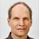 Luitpold Distel合作论文数13
 Anna Constantinescu合作论文数13
 Jürgen Debus合作论文数Universitätsklinikum Heidelberg;Nationales Centrum für Tumorerkrankungen;Heidelberger- Ionenstrahl- Therapiezentrum;Deutschen Krebsforschungszentrum;Radiologischen Klinik und Poliklinik, Universität Heidelberg13
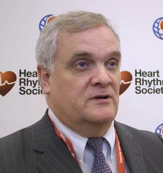 Douglas L Packer合作论文数Division of Cardiac Electrophysiology, Department of Cardiovascular Diseases, Mayo Clinic;Translational Electrophysiology Research Laboratory, Mayo Clinic12M. Schmidt合作论文数12S. Lettmaier合作论文数11Claudia Schweizer合作论文数11
 Markus Kellermeier合作论文数Department of Radiation Oncology, University Clinic Erlangen;Friedrich-Alexander-University Erlangen-Nürnberg;University Clinic Erlangen, Friedrich-Alexander-University Erlangen-Nürnberg11




