Sarcin is a member of a fungal toxin family that enters cells and specifically cleaves one of the thousands of RNA phosphodiester bonds in the ribosome. As a result, elongation factor binding is disrupted, translation is inhibited and apoptosis is triggered. The toxin targets a universal RNA structure in the ribosome called the sarcin/ricin loop (SRL). A 1.11 Angstrom resolution structure of a minimal SRL RNA substrate (similar to30-mer) shows that the loop portion of the substrate folds into two common building blocks of RNA structure: a bulged-G motif (recognition site) and a GAGA tetraloop (cleavage site). To elucidate the structural basis of toxin action, we determined two cocrystal structures of the sarcin homologue restrictocin bound to different analogs of a minimal SRL RNA substrate. Our studies argue that site selection by the toxin depends on direct base and shape recognition of the SRL RNA, and that cleavage by the toxin depends on a base flipping mechanism that positions the nucleophile for in-line attack on the scissile bond.
In eukaryotes, formation of short duplexes between the U3 small nucleolar RNA (snoRNA) and the precursor rRNA (pre-rRNA) at multiple sites is a prerequisite for three endonucleolytic cleavages that initiate small subunit biogenesis by releasing the 18S rRNA precursor from the pre-rRNA. The most likely role of these RNA duplexes is to guide the U3 snoRNA and its associated proteins, designated the small subunit processome, to the target cleavage sites on the pre-rRNA. Studies by others in Saccharomyces cerevisiae have identified the proteins Mpp10p, Imp3p, and Imp4p as candidates to mediate U3-pre-rRNA interactions. We report here that Imp3p and Imp4p appear to stabilize an otherwise unstable duplex between the U3 snoRNA hinge region and complementary bases in the external transcribed spacer of the pre-rRNA. In addition, Imp4p, but not Imp3p, seems to rearrange the U3 box A stem structure to expose the site that base-pairs with the 5' end of the 18S rRNA, thereby mediating duplex formation at a second site. By mediating formation of both essential U3-pre-rRNA duplexes, Imp3p and Imp4p may help the small subunit processome to dock onto the pre-rRNA, an event indispensable for ribosome biogenesis and hence for cell growth.
During translocation peptidyl-tRNA moves from the A-site to the P-site and mRNA is displaced by three nucleotides in the 3′ direction. This reaction is catalyzed by elongation factor-G (EF-G) and is associated with ribosome-dependent hydrolysis of GTP. The molecular basis of translocation is the most important unsolved problem with respect to ribosome function. A critical question, one that might provide a clue to the mechanism of translocation, is the precise identity of the contacts between EF-G and ribosome components. To make the identification, a covalent bond was formed, by ultraviolet irradiation, between EF-G and a sarcin/ricin domain (SRD) oligoribonucleotide containing 5-iodouridine. The cross-link was established, by mass spectroscopy and by Edman degradation, to be between a tryptophan at position 127 in the G domain in EF-G and either one of two 5-iodouridine nucleotides in the sequence UAG2655U in the SRD. G2655 is a critical identity element for the recognition of the factor's ribosomal binding site. The site of the cross-link provides the first direct evidence that the SRD is in close proximity to the EF-G catalytic center. The proximity suggests that the SRD RNA has a role in the activation of GTP hydrolysis that leads to a transition in the conformation of the factor and to its release from the ribosome.
Bulged-G motifs are ubiquitous internal RNA loops that provide specific recognition sites for proteins and RNAs. To establish the common and distinctive features of the motif we determined the structures of three variants and compared them with related structures. The variants are 27-nt mimics of the sarcin/ricin loop (SRL) from Escherichia coli 23S ribosomal RNA that is an essential part of the binding site for elongation factors (EFs). The wild-type SRL has now been determined at 1.04 A resolution, supplementing data obtained before at 1.11 A and allowing the first calculation of coordinate error for an RNA motif. The other two structures, having a viable (C2658U*G2663A) or a lethal mutation (C2658G*G2663C), were determined at 1.75 and 2.25 A resolution, respectively. Comparisons reveal that bulged-G motifs have a common hydration and geometry, with flexible junctions at flanking structural elements. Six conserved nucleotides preserve the fold of the motif; the remaining seven to nine vary in sequence and alter contacts in both grooves. Differences between accessible functional groups of the lethal mutation and those of the viable mutation and wild-type SRL may account for the impaired elongation factor binding to ribosomes with the C2658G*G2663C mutation and may underlie the lethal phenotype.
GNRA tetraloops (N is A, C, G, or U; R is A or G) are basic building blocks of RNA structure that often interact with proteins or other RNA structural elements. Understanding sequence-dependent structural variation among different GNRA tetraloops is an important step toward elucidating the molecular basis of specific GNRA tetraloop recognition by proteins and RNAs. Details of the geometry and hydration of this motif have been based on high-resolution crystallographic structures of the GRRA subset of tetraloops; less is known about the GYRA subset (Y is C or U). We report here the structure of a GUAA tetraloop determined to 1.4 A resolution to better define these details and any distinctive features of GYRA tetraloops. The tetraloop is part of a 27-nt structure that mimics the universal sarcin/ricin loop from Escherichia coli 23S ribosomal RNA in which a GUAA tetraloop replaces the conserved GAGA tetraloop. The adenosines of the GUAA tetraloop form an intermolecular contact that is a commonplace RNA tertiary interaction called an A-minor motif. This is the first structure to reveal in great detail the geometry and hydration of a GUAA tetraloop and an A-minor motif. Comparison of tetraloop structures shows a common backbone geometry for each of the eight possible tetraloop sequences and suggests a common hydration. After backbone atom superposition, equivalent bases from different tetraloops unexpectedly depart from coplanarity by as much as 48 degrees. This variation displaces the functional groups of tetraloops implicated in protein and RNA binding, providing a recognition feature.
The sarcin-ricin domain (SRD) of Escherichia coli 23S rRNA, which includes a near-universal, purine rich sequence of 12 nucleotides, is the site of action of the eponymous ribotoxins. The chapter focuses on the structure of SRD. There is an evidence that the SRD constitutes an important part of the elongation factor G (EF-G) binding site. The three SRD purines that are protected from chemical modification by EF-G bound to E. coli ribosomes are presumptive EF-G identity elements. A number of mutations have been made in the SRD in an E. coli 23S rRNA gene. The mutations have been constructed in a plasmid carrying an E. coli rrnB operon. This operon has a transcription unit with the genes for 16S, 23S, and 5S rRNAs. There is biochemical and genetic evidence that the flexible region in the SRD affects the binding of the elongation factors. There are differences in the structures of the flexible regions of the rat and E. coli SRDs. There are three nucleotides in the SRD that are protected from chemical modification by both EF-G and the EF-Tu ternary complex. The EF-Tu ternary complex may bind to the ribosome only when peptidyl-tRNA is in the P site (posttranslocation), and EF-G may bind only when it is in the A site.
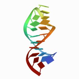
The sarcin/ricin domain of 23 S - 28 S ribosomal RNA is essential for protein synthesis because it forms a critical part of the binding site for elongation factors. A crystal structure of an RNA of 27 nucleotides that mimics the domain in Escherichia coli 23 S rRNA was determined at 1.11 A resolution. The domain folds into a hairpin distorted by four non-canonical base-pairs and one base triple. The fold is stabilized by cross-strand and intra-stand stacking; no intramolecular stabilizing metal ions are observed. This is the first structure to reveal in great detail the geometry and the hydration of two common motifs that are conserved in this rRNA domain, a GAGA tetraloop and a G-bulged cross-strand A stack. Differences in the region connecting these motifs to the stem in the E. coli and in the rat sarcin/ricin domains may contribute to the species-specific binding of elongation factors. Correlation of nucleotide protection data with the structure indicates that the domain has two surfaces. One surface is accessible, lies primarily in the major groove, and is likely to bind the elongation factors. The second lies primarily in the minor groove, and is likely to be buried in the ribosome. This minor groove surface includes the Watson-Crick faces of the cytosine bases in the unusual A2654.C2666 and U2653.C2667 water-mediated base-pairs.
The structure of a 29-nucleotide RNA containing the sarcin/ricin loop (SRL) of rat 28 S rRNA has been determined at 2.1 A resolution. Recognition of the SRL by elongation factors and by the ribotoxins, sarcin and ricin, requires a nearly universal dodecamer sequence that folds into a G-bulged cross-strand A stack and a GAGA tetraloop. The juxtaposition of these two motifs forms a distorted hairpin structure that allows direct recognition of bases in both grooves as well as recognition of nonhelical backbone geometry and two 5'-unstacked purines. Comparisons with other RNA crystal structures establish the cross-strand A stack and the GNRA tetraloop as defined and modular RNA structural elements. The conserved region at the top is connected to the base of the domain by a region presumed to be flexible because of the sparsity of stabilizing contacts. Although the conformation of the SRL RNA previously determined by NMR spectroscopy is similar to the structure determined by x-ray crystallography, significant differences are observed in the "flexible" region and to a lesser extent in the G-bulged cross-strand A stack.
Two important challenges confronting RNA crystallographers are producing crystals and finding isomorphous heavy-atom derivatives. Non-isomorphism can be addressed by determining the phases using the multiwavelength anomalous dispersion (MAD) method. These phases can be greatly improved by combining phases from MAD experiments done on different heavy-atom derivatives. Heavy-atom derivatives can be created by chemically modifying the RNA through covalent attachment of bromine or mercury to C5 of pyrimidines or [Pt(NH3)3](2+) to N7 of guanine. While phosphorothioates can provide mercury binding sites, disorder can reduce their value for phase determination. The location of these chemical modifications is critical since crystallization of these derivatized RNAs is sensitive to heavy atom induced conformational alterations and crystal packing.
 Thomas Steitz合作论文数Department of Molecular Biophysics and Biochemistry, Yale University6
 T Gérczei合作论文数Department of Theoretical Chemistry, Eötvös University6
 Ira G. Wool合作论文数Departments of Physiology and Molecular Biology, University of Chicago6
 Peter B. Moore合作论文数Department of Chemistry, Yale Faculty of Arts and Sciences, Yale University5M.J. Plantinga合作论文数4
 Yuen-Ling Chan合作论文数Department of Biochemistry and Molecular Biology, The University of Chicago4Betty Freeborn合作论文数4Alexander Munishkin合作论文数3K.K. Swinger合作论文数2S Yoshikawa合作论文数1H. Belrhali合作论文数1E. Yamashita合作论文数1M. Paci合作论文数1M A Ceruso合作论文数1A. Di Nola合作论文数1T. Tomizaki合作论文数1
 Clare E Sansom合作论文数1Ee Minyat合作论文数1Maya J. Pandya合作论文数1Takashi Kinebuchi合作论文数1Paulo Magalhães合作论文数1Wilma K. Olson合作论文数1Keiichi Namba合作论文数1
 Philippe Sansonetti合作论文数University of Chinese Academy of Sciences;Shanghai Institute of Immunity and Infection, Chinese Academy of Sciences;Institut Pasteur1Kazuhiko Kinosita合作论文数1J C Williams合作论文数1T A Steitz合作论文数1Pj Munson合作论文数NICHHD,BETHESDA,MD 202051X Lin合作论文数1James Hofrichter合作论文数1
 Barbara A. Baird合作论文数Departments of Chemistry & Chemical Biology, College of Arts & Sciences, Cornell University1
 David Holowka合作论文数Department of Chemistry and Chemical Biology, College of Arts & Sciences, Cornell University1
 Dek Woolfson合作论文数School of Chemistry, University of Bristol1
 John R Helliwell合作论文数Department of Chemistry, The University of Manchester1
 Ernst Walter Knapp合作论文数Institute of Chemistry and Biochemistry, Freie Universität Berlin1
 G. Ulrich Nienhaus合作论文数Institut fur Angewandte Physik, Karlsruhe Institute of Technology1
 James P. Allen合作论文数Department of Chemistry and Biochemistry, School of Molecular Sciences, Arizona State University1Tomitake Tsukihara合作论文数Institute for Protein Research, Osaka University1
 Charles Cantor合作论文数Department of Biomedical Engineering, College of Engineering, Boston University;Sequenom, Inc.1
 Stephen J. Hagen合作论文数Department of Physics, College of Liberal Arts and Sciences, University of Florida1Egile Coumaran合作论文数1
 Ian J Tickle合作论文数1
 Paolo Pinton合作论文数Department of Medical Sciences, University of Ferrara1
 Michal Neeman合作论文数Department of Immunology and Regenerative Biology, Weizmann Institute of Science1
 Wolfgang Junge合作论文数Fachbereich Biologie;Chemie, Universitat Osnabruck1ShuRong Wang合作论文数1
 Berend Lammert De Groot合作论文数Max-Planck Institute for Biophysical Chemistry;University of Gottingen1
 Sarah C Stallings合作论文数Vanderbilt Institute for Clinical and Translational Research1Eric Henry合作论文数National Institutes of Health1




