Polyadenylate [poly(A)] tail addition to the 3' end of a wide range of RNAs is a highly conserved modification that plays a central role in cellular RNA function. Elements for nuclear expression (ENEs) are cis-acting RNA elements that stabilize poly(A) tails by sequestering them in RNA triplex structures. A crystal structure of a double ENE from a rice hAT transposon messenger RNA complexed with poly(A)(28) at a resolution of 2.89 angstroms reveals multiple modes of interaction with poly(A), including major-groove triple helices, extended minor-groove interactions with RNA double helices, a quintuple-base motif that transitions poly(A) from minor-groove associations to major-groove triple helices, and a poly(A) 3'-end binding pocket. Our findings both expand the repertoire of motifs involved in long-range RNA interactions and provide insights into how polyadenylation can protect an RNA's extreme 3' end.
In bacteria, the dissociable σ subunit of the RNA polymerase (RNAP) is responsible for initiating RNA synthesis from specific DNA sites. As nascent RNA grows, downstream DNA unwinds and is pulled into the RNAP, causing stress accumulation and initiation complex destabilization. Processive transcription elongation requires at least partial separation of the σ factor from the RNAP core enzyme. Here, we present a series of transcription complexes captured between the early initiation and elongation phases via in-crystal RNA synthesis and cleavage. Crystal structures of these complexes indicate that stress accumulation during transcription initiation is not due to clashing of the growing nascent RNA with the σ3.2 loop, but results from scrunching of the template strand DNA that is contained inside the RNAP by the σ3 domain. Our results shed light on how scrunching of template-strand DNA drives both abortive initiation and σ-RNAP core separation to transition transcription from initiation to elongation.
The density regulated protein (DENR) forms a stable heterodimer with malignant T-cell-amplified sequence 1 (MCT-1). DENR-MCT-1 heterodimer then participates in regulation of non-canonical translation initiation and ribosomal recycling. The N-terminal domain of DENR interacts with MCT-1 and carries a classical tetrahedral zinc ion-binding site, which is crucial for the dimerization. DENR-MCT-1 binds the small (40S) ribosomal subunit through interactions between MCT-1 and helix h24 of the 18S rRNA, and through interactions between the C-terminal domain of DENR and helix h44 of the 18S rRNA. This later interaction occurs in the vicinity of the P site that is also the binding site for canonical translation initiation factor eIF1, which plays the key role in initiation codon selection and scanning. Sequence homology modeling and a low-resolution crystal structure of the DENR-MCT-1 complex with the human 40S subunit suggests that the C-terminal domain of DENR and eIF1 adopt a similar fold. Here we present the crystal structure of the C-terminal domain of DENR determined at 1.74 Å resolution, which confirms its resemblance to eIF1 and advances our understanding of the mechanism by which DENR-MCT-1 regulates non-canonical translation initiation and ribosomal recycling.
[14] B. Redman, J. Novotny, T. Grow, V. Rudd, N. Woody, M. Hinckley, P. McCumber, N. Rogers, M. Hoening, K. Kubala, S. Shald, R. Uberna, T. D’Alberto, T. Höft, R. Sibell, F. Wheeler, “Stand-off biometric identification using Fourier transform profilometry for 2D+3D face imaging” Optical Society of America Applications of Lasers for Sensing and Free Space Communications Conference Technical Digest paper LThB3, 2011. [preprint] [doi:10.1364/LSC.2011.LThB3] [journal webpage]
Pistol ribozymes comprise a class of small, self-cleaving RNAs discovered via comparative genomic analysis. Prior work in the field has probed the kinetics of the cleavage reaction, as well as the influence of various metal ion cofactors that accelerate the process. In the current study we perform unbiased and unconstrained molecular dynamics simulations from two current high-resolution pistol crystal structures, and we analyzed trajectory data within the context of the currently accepted ribozyme mechanistic framework. Root-mean-squared deviations (RMSDs), radial distribution functions (RDFs), and distributions of nucleophilic angle-of-attack reveal insights into the potential roles of three magnesium ions with respect to catalysis and overall conformational stability of the molecule. A series of simulation trajectories containing in-silico mutations reveal the relatively flexible and partially interchangeable roles of two particular magnesium ions within solvated hydrogen-bonding distances from the catalytic center.
Summary: The advent of Web-based tools that assist in the analysis and visualization of macromolecules require application programming interfaces (APIs) designed for modern web frameworks. To this end, we have developed a Node.js module pdbmine that allows any user to generate faster data-request queries to the RCSB Protein Data Bank (PDB). This JavaScript API acts as a layer over the XML-based RCSB PDB RESTful API. The relatively simple nature of the function calls within this module allows the user to easily implement and integrate pdbmine into larger Node.js web applications. Availability: This module can be installed via the Node Package Manager (NPM) at https://www.npmjs.com/package/pdbmine/, and is hosted on GitHub under the open-source MIT license at https://github.com/nnj1/pdbmine/. Relevant documentation is detailed at https://nnj1.github.io/pdbmine/
To address drug resistance to HIV-1 non-nucleoside reverse transcriptase inhibitors (NNRTIs), a series of novel diarylpyrimidine (DAPY) derivatives targeting "tolerant region I" and "tolerant region II" of the NNRTIs binding pocket (NNIBP) were designed utilizing a structure-guided scaffold-hopping strategy. The dihydrofuro[3,4- d]pyrimidine derivatives 13c2 and 13c4 proved to be exceptionally potent against a wide range of HIV-1 strains carrying single NNRTI-resistant mutations (EC50 = 0.9-8.4 nM), which were remarkably superior to that of etravirine (ETV). Meanwhile, both compounds exhibited comparable activities with ETV toward the virus with double mutations F227L+V106A and K103N+Y181C. Furthermore, the most active compound 13c2 showed favorable pharmacokinetic properties with an oral bioavailability of 30.96% and a half-life of 11.1 h, which suggested that 13c2 is worth further investigation as a novel NNRTI to circumvent drug resistance.
Rapid generation of drug-resistant mutations in HIV-1 reverse transcriptase (RT), a prime target for anti-HIV therapy, poses a major impediment to effective anti-HIV treatment. Our previous efforts have led to the development of two novel non-nucleoside reverse transcriptase inhibitors (NNRTIs) with piperidine-substituted thiophene[3,2-d]pyrimidine scaffolds, compounds K-5a2 and 25a, which demonstrate highly potent anti-HIV-1 activities and improved resistance profiles compared with etravirine and rilpivirine, respectively. Here, we have determined the crystal structures of HIV-1 wild-type (WT) RT and seven RT variants bearing prevalent drug-resistant mutations in complex with K-5a2 or 25a at ~2 Å resolution. These high-resolution structures illustrate the molecular details of the extensive hydrophobic interactions and the network of main chain hydrogen bonds formed between the NNRTIs and the RT inhibitor-binding pocket, and provide valuable insights into the favorable structural features that can be employed for designing NNRTIs that are broadly active against drug-resistant HIV-1 variants.
The density-regulated protein (DENR) and the malignant T cell-amplified sequence 1 (MCT-1/MCTS1) oncoprotein support noncanonical translation initiation, promote translation reinitiation on a specific set of mRNAs with short upstream reading frames, and regulate ribosome recycling. DENR and MCT-1 form a heterodimer, which binds to the ribosome. We determined the crystal structure of the heterodimer formed by human MCT-1 and the N-terminal domain of DENR at 2.0-A resolution. The structure of the heterodimer reveals atomic details of the mechanism of DENR and MCT-1 interaction. Four conserved cysteine residues of DENR (C34, C37, C44, C53) form a classical tetrahedral zinc ion-binding site, which preserves the structure of the DENR's MCT-1-binding interface that is essential for the dimerization. Substitution of all four cysteines by alanine abolished a heterodimer formation. Our findings elucidate further the mechanism of regulation of DENR-MCT-1 activities in unconventional translation initiation, reinitiation, and recycling.
Genetic information is translated into proteins by the ribosome. Structural studies of the ribosome and of its complexes with factors and inhibitors have provided invaluable information on the mechanism of protein synthesis. Ribosome inhibitors are among the most successful antimicrobial drugs and constitute more than half of all medicines used to treat infections. However, bacterial infections are becoming increasingly difficult to treat because the microbes have developed resistance to the most effective antibiotics, creating a major public health care threat. This has spurred a renewed interest in structure-function studies of protein synthesis inhibitors, and in few cases, compounds have been developed into potent therapeutic agents against drug-resistant pathogens. In this review, we describe the modes of action of many ribosome-targeting antibiotics, highlight the major resistance mechanisms developed by pathogenic bacteria, and discuss recent advances in structure-assisted design of new molecules.
Protein accounts for roughly half the dry mass of all organisms and, in large measure, the morphologies and physiologies of organisms are macroscopic manifestations of the properties of the proteins they synthesize for themselves. This issue of the Philosophical Transactions of the Royal Society
Significance Based on the “RNA world” theory, ribozymes likely carried out biochemical reactions long before organisms evolved to use protein enzymes as biocatalysts. The continued discovery of new structures for small self-cleaving ribozymes has shed light on conserved mechanisms in evolution, such as acid–base catalysis for self-cleavage reaction. Here, we present the crystal structure of a newly discovered class of self-cleaving ribozymes called Pistol and how it likely uses the phosphoester transfer mechanism for self-cleavage. The results presented here suggest that Pistol uses an evolutionarily conserved cleavage mechanism that is like other self-cleaving ribozymes, such as Twister, Hammerhead, Hairpin, and Hepatitis Delta Virus ribozymes.
During transcription, RNA polymerase moves downstream along the DNA template and maintains a transcription bubble. Several recent structural studies of transcription complexes with a complete transcription bubble provide new insights into how RNAP couples the nucleotide addition reaction to its directional movement.
Protein synthesis is a key process in all living organisms. In eukaryotes, initiation factor 2 (eIF2) plays an important role in translation initiation as it selects and delivers the initiator tRNA to the small ribosomal subunit. Under stress conditions, phosphorylation of the α-subunit of eIF2 downregulates cellular protein synthesis. However, translation of certain mRNAs continues via the eIF2D-dependent non-canonical initiation pathway. The molecular mechanism of this process remains elusive. In addition, eIF2D plays a role in translation re-initiation and ribosome recycling. Currently, there has been no structural information of eIF2D. We have now determined the crystal structure of the C-terminal domains of eIF2D at 1.4-Å resolution. One domain has the fold similar to that of eIF1, which is crucial for the scanning and initiation codon selection. The second domain has a known SWIB/MDM2 fold, which was not observed before in other translation initiation factors. Our structure reveals atomic details of inter-domain interactions in the C-terminal part of eIF2D and sheds light on the possible role of these domains in eIF2D during translation.
 Peter B. Moore合作论文数Department of Chemistry, Yale Faculty of Arts and Sciences, Yale University67
 Jimin Wang合作论文数Department of Molecular Biochemistry and Biophysics, Yale University38
 Nenad Ban合作论文数The Ban Lab, Institute of Molecular Biology and Biophysics, Eidgenössische Technische Hochschule Zürich28
 Poul Nissen合作论文数Department of Molecular Biology and Genetics, Aarhus University;Faculty of Natural Sciences, Aarhus University25
 Gregor Blaha合作论文数Max-Planck-Institut für Molekulare Genetik, Ihnestrasse 73, D-14195 Berlin, Germany18
 Martin Schmeing合作论文数Department of Biochemistry, Faculty of Medicine and Health Sciences, McGill University16
 Irene Weber合作论文数College of Arts & Sciences, Georgia State University13
 Lorena Sue Beese合作论文数Department of Biochemistry, Duke University;Department of Basic Science, Duke University;Duke Cancer Institute12
 Dieter Soll合作论文数Department of Molecular Biophysics and Biochemistry, Yale University11
 Paul S. Freemont合作论文数11
 Donald Engelman合作论文数Engelman Lab, Department of Molecular Biophysics and Biochemistry, School of Medicine, Yale University11Satwik Kamtekar合作论文数11Bulkley David合作论文数10
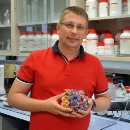 Polikanov Yury S合作论文数Dept Biol Sci, Univ Illinois10
 Ivan B Lomakin合作论文数Morse Institute for Molecular Genetics, State University of New York Health Science10J L Hansen合作论文数9Mark R. Sanderson合作论文数Cancer Research Campaign Biomolecular Structure Unit The Institute of Cancer Research, Sutton, Surrey SM2 5NG, U.K.9PA RICE合作论文数8J Hansen合作论文数8
 Robert Fletterick合作论文数Department of Biochemistry, University of California, San Francisco8Betty Freeborn合作论文数8
 Matthieu G. Gagnon合作论文数Dept Mol Biophys & Biochem, Yale Univ8Mark a Rould合作论文数8NDF GRINDLEY合作论文数7
 John Perona合作论文数Department of Biochemistry and Molecular Biology, Oregon Health & Science University;Department of Chemistry, Portland State University7
 Bin Liu合作论文数Department of Molecular Biophysics and Biochemistry,7David L. Ollis合作论文数Research School of Chemistry, Australian National University7
 Jinzhong Lin (林金钟)合作论文数School of Life Sciences, Fudan University7Yong Xiong合作论文数7
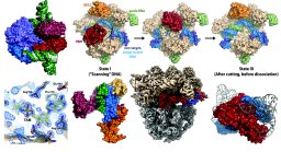 Miljan Simonovic合作论文数Department of Biochemistry and Molecular Biology, University of Illinois College of Medicine7Steve C. Schultz合作论文数6
 Jeffrey L. Hansen合作论文数6
 Jonathan Malcolm Friedman合作论文数6
 Scott Bailey合作论文数Chemistry-Biology Interface Program, Krieger School of Arts & Sciences, Johns Hopkins University6Yuhong Zuo合作论文数Department of Molecular, Cellular and Developmental Biology, Yale University6
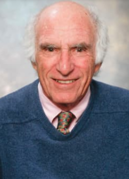 William Konigsberg合作论文数Biochemistry, Quantitative Biology, Biophysics, and Structural Biology, Biological & Biomedical Sciences, Yale University6
 Klein Daniel合作论文数6
 Phoebe A. Rice合作论文数Department of Biochemistry & Molecular Biology, The University of Chicago6
 Wayne Anderson合作论文数6C. Axel Innis合作论文数6
 Yousif Shamoo合作论文数Rice University6
 William Lipscomb合作论文数Harvard University6C. C. Correll合作论文数6Catherine M. Joyce合作论文数6Adrian Goldman合作论文数5Robin E. Stanley合作论文数5
 Chad Brautigam合作论文数Department of Biophysics, UT Southwestern Medical Center/Department of Microbiology, UT Southwestern Medical Center5McKay D B合作论文数5
 George C Shields合作论文数Department of Chemistry;Hamilton College;Department of Chemistry, Hamilton College5




