INTRODUCTION:Biallelic variants in Fanconi Anemia-associated Nuclease 1 (FAN1) cause karyomegalic tubulointerstitial nephropathy (KIN), a condition poorly characterized in terms of kidney survival, patient survival, and clinical characteristics. Therefore, we undertook a cross-sectional collaborative study to better characterize KIN-FAN1. METHODS:To gather data, we distributed a REDCap survey on clinical characteristics and genetic variants of KIN-FAN1 to colleagues and case report authors. RESULTS:Based on the survey, we identified 86 families affected (122 individuals) from 22 countries. There were 56 families (83 individuals) with a genetic diagnosis of KIN-FAN1, including 38 distinct FAN1 variants, and 30 families (39 individuals) with KIN with no predisposing risk factors and without molecular FAN1 testing. The median age at presentation was 38.5 years (interquartile range: 29-43), 62% male. Of the cohort, 46% had asymptomatic elevation of liver function tests, 39% had pulmonary complications, and 6% developed cancer. The median age of kidney failure was 45 years (95% confidence interval (CI): 38-56). Of the cohort, 27.1% died at a median age of 55 years (95% CI: 43-75). Pulmonary complications was/were the cause of death in 15.4% of patients on dialysis and 23.1% of kidney transplant recipients. Compared to other variants, patients with the p.W707X-FAN1 variant were at a significantly higher risk of pulmonary complications (adjusted odds ratio: 8.26 (95% CI: 1.7-40.1) and had a significantly shorter lifespan (hazard ratio: 3.24 (95% CI: 1.13-9.28). No genetic covariates were statistically associated with the progression to kidney failure. CONCLUSIONS:Patients with KIN-FAN1 develop kidney failure at a median age of 45 years. Survival is compromised with many dying of pulmonary disease.
FSGS is the most common primary glomerular lesion that causes kidney failure in the US. FSGS results from injury or loss of glomerular visceral epithelial cells (i.e. podocytes). Pathogenic variants in TRPC6 can cause FSGS through dysregulated calcium conductance and associated disturbances in podocyte physiology. Here, we describe a 5-generation kindred with FSGS caused by a novel compound C-terminal TRPC6 mutation. Analysis of patient-specific iPSC-derived podocytes and glomerular capillary wall-on-a-chip systems revealed that the compound variants disrupt TRPC6 protein structure, intermolecular interactions, and membrane localization. Combined therapy with Sildenafil and Losartan ameliorated these disturbances compared to conventional immunosuppressive treatment. ### Competing Interest Statement S. Musah is an inventor on a patent for stem cell differentiation into podocytes and establishment of a glomerulus chip. S. Musah is also an inventor on a pending patent for engineering in vivo-like models of kidney tissues.
Focal segmental glomerulosclerosis (FSGS) is a histologic lesion caused by a variety of injurious stimuli that lead to dysfunction/loss of glomerular visceral epithelial cells (i.e., podocytes). Pathogenic mutations in crumbs homolog-2 (CRB2), encoding the type 1 transmembrane protein crumbs homolog-2, have been shown to cause early-onset corticosteroid-resistant nephrotic syndrome (SRNS)/FSGS. Here, we identified a two-generation Indian kindred (DUK40595) with biopsy-proven SRNS/FSGS caused by a compound heterozygous mutation in CRB2 comprised of the previously described truncating mutation p.Gly1036_Alafs*43 and a rare 9-bp deletion mutation p.Leu1074_Asp1076del. Because compound heterozygous mutations involving the truncating p.Gly1036_Alafs*43 variant have been associated with reduced CRB2 expression in podocytes and autosomal recessive SRNS/FSGS, we sought to define the pathogenic effects of CRB2 deficiency in podocytes. We show that CRB2 knockdown induces yes-associated protein (YAP) activity and target gene expression in podocytes. It upregulates YAP-mediated mechanosignaling and increases the density of focal adhesion and F-actin. Using elastic resonator interference stress microscopy (ERISM), we demonstrate that CRB2 knockdown also enhances podocyte contractility in a substrate stiffness-dependent manner. The knockdown effect decreases with increasing substrate stiffness, indicating impaired mechanosensing in CRB2 knockdown cells at low substrate stiffness. Although the mechanical activation of CRB2 knockdown cells is associated with increased YAP activity, the enhanced cell contractility is not significantly reduced by the selective YAP inhibitors K-975 and verteporfin, suggesting that multiple pathways may be involved in mechanosignaling downstream of CRB2. Taken together, these studies provide the first evidence that CRB2 deficiency may impair podocyte mechanotransduction via disruption of YAP signaling in podocytes.NEW & NOTEWORTHY We identified a rare compound heterozygous CRB2 mutation as the cause of familial SRNS/FSGS in a two-generation East Asian kindred. Modeling the effect of the mutation, we show that CRB2 knockdown in podocytes induces YAP transcriptional activity and upregulates YAP-mediated mechanosignaling. Using elastic resonator interference stress microscopy (ERISM), we demonstrate that CRB2 knockdown enhances podocyte contractility in a substrate stiffness-dependent manner. The knockdown effect decreases with increasing substrate stiffness, indicating impaired mechanosensing in CRB2-deficient podocytes.
Rationale & Objective:Cardiovascular (CV) and thromboembolic (TE) events are known complications of glomerular disease (GD), but their incidence and risk factors are poorly characterized. This analysis describes CV and TE outcomes in the Cure GlomeruloNephropathy (CureGN) Network. Study Design:Prospective cohort study. Setting & Participants:CureGN is a prospective cohort study of children and adults with biopsy-proven minimal change disease (MCD), focal segmental glomerulosclerosis (FSGS), membranous nephropathy (MN), or IgA nephropathy (IgAN)/vasculitis (IgAV). Data from 2,545 children and adults (23% MCD, 23% MN, 25% FSGS, 29% IgAN/IgAV) was analyzed. Exposure:Estimated glomerular filtration rate (eGFR), proteinuria, serum albumin, tobacco use, body mass index, hypertension, renin-angiotensin-aldosterone system. Outcomes:CV and TE events. Analytic Approach:Kaplan-Meier curves were used to estimate cumulative incidence, and multivariable Cox proportional hazards models were fitted to estimate associations of histologic diagnosis, age, biological sex, and race. Laboratory and other clinical data were evaluated separately in models adjusted for base model covariates. Results:Median follow-up time was 4.6 years (IQR 2.7-6.1). The cumulative incidence of first CV and TE event postbiopsy was 3% and 2% in children and 10% and 5% in adults, respectively. No association between GD subtype and risk of CV or TE event was detected. Older age and Black race were associated with higher risk of first CV and TE event {hazard ratio (HR) (95% confidence interval {CI}) per 5 years, CV = 1.17 (1.12-1.23); TE = 1.11 (1.05-1.18); for Black race, CV = 1.62 (1.03-2.56), TE = 2.25 (1.27-4.01)}. Lower eGFR, higher urinary protein-creatinine ratio (UPCR), and lower serum albumin levels at enrollment were associated with higher risk of first CV and TE event (eGFR per 10 mL/min/1.73 m2, CV = 0.87 [0.81-0.93], TE = 0.80 [0.73-0.88]; UPCR per mg/mg, CV = 1.04 [1.02-1.07], TE = 1.03 [1.00-1.07]; serum albumin per g/dL, CV = 0.75 [0.59-0.95], TE = 0.71 [0.53-0.96]). Limitations:Age of cohort, duration of follow-up. Conclusions:In the CureGN cohort, elevated risk of incident CV and TE events is associated with severity of kidney disease rather than GD subtype.
The term atypical hemolytic uremic syndrome has been in use since the mid-1970s.It was initially used to describe the familial or sporadic form of hemolytic uremic syndrome as opposed to the epidemic, typical form of the disease.Over time, the atypical hemolytic uremic syndrome term has evolved into being used to refer to anything that is not Shiga toxin-associated hemolytic uremic syndrome.The term describes a heterogeneous group of diseases of disparate causes, a circumstance that makes defining disease-specific natural history and/or targeted treatment approaches challenging.A working group of specialty-specific experts in the thrombotic microangiopathies was convened to review the validity of this broad term in an era of swiftly advancing science and targeted therapeutics.A Delphi approach was used to define and interrogate some of the key issues related to the atypical hemolytic uremic syndrome nomenclature.
BACKGROUND:IgA nephropathy is the most common primary GN. Clinical features of IgA nephropathy include proteinuria, which is the strongest known surrogate of progression to kidney failure. Complement pathway activation is a critical driver of inflammation and tissue injury in IgA nephropathy. Cemdisiran is an investigational RNA interference therapeutic that suppresses hepatic production of complement component 5 (C5), thereby potentially reducing proteinuria in IgA nephropathy. We evaluated the efficacy and safety of cemdisiran in adult patients with IgA nephropathy at high risk of kidney disease progression. METHODS:In this phase 2, 36-week, double-blind study, adult patients with IgA nephropathy and urine protein ≥1 g/24 hours were randomized (2:1) to subcutaneous cemdisiran 600 mg or placebo every 4 weeks in combination with the standard of care. The primary end point was percentage change from baseline at week 32 in urine protein-to-creatinine ratio (UPCR) measured by 24-hour urine collection. Additional end points included change from baseline in UPCR measured by spot urine, serum C5 level, and safety assessments. RESULTS:Thirty-one patients were randomized (cemdisiran, N =22; placebo, N =9). Cemdisiran-treated patients had a placebo-adjusted geometric mean change in 24-hour UPCR of -37.4% (cemdisiran-adjusted geometric mean ratio to baseline [SEM], 0.69 [0.10]) at week 32. Spot UPCR was consistent with 24-hour UPCR placebo-adjusted change of -45.8% (cemdisiran-adjusted geometric mean ratio to baseline [SEM], 0.73 [0.11]). Mean (SD) change in serum C5 level from baseline at week 32 was -98.7% (1.2) with cemdisiran and 25.2% (57.7) with placebo. Over 36 weeks, most adverse events were mild or moderate and transient; the most common adverse event after cemdisiran treatment was injection-site reaction (41%). CONCLUSIONS:These findings indicate that treatment with cemdisiran resulted in a reduction of proteinuria at week 32 and was well tolerated.
ABSTRACT:Complement-mediated thrombotic microangiopathy (CM-TMA) or hemolytic uremic syndrome, previously identified as atypical hemolytic uremic syndrome, is a TMA characterized by germ line variants or acquired antibodies to complement proteins and regulators. Building upon our prior experience with the modified Ham (mHam) assay for ex vivo diagnosis of complementopathies, we have developed an array of cell-based complement "biosensors" by selective removal of complement regulatory proteins (CD55 and CD59, CD46, or a combination thereof) in an autonomously bioluminescent HEK293 cell line. These biosensors can be used as a sensitive method for diagnosing CM-TMA and monitoring therapeutic complement blockade. Using specific complement pathway inhibitors, this model identifies immunoglobulin M (IgM)-driven classical pathway stimulus during both acute disease and in many patients during clinical remission. This provides a potential explanation for ∼50% of patients with CM-TMA who lack an alternative pathway "driving" variant and suggests at least a subset of CM-TMA is characterized by a breakdown of IgM immunologic tolerance.
Complement-mediated hemolytic uremic syndrome (CM-HUS) is a thrombotic microangiopathy characterized by germline variants or acquired antibodies to complement proteins and regulators. Building upon our prior experience with the modified Ham (mHam) assay for ex vivo diagnosis of complementopathies, we have developed an array of cell-based complement "biosensors'' by selective removal of complement regulatory proteins (CD55 and CD59, CD46, or a combination thereof) in an autonomously bioluminescent HEK293 cell line. These biosensors can be used as a sensitive method for diagnosing CM-HUS and monitoring therapeutic complement blockade. Using specific complement pathway inhibitors, this model identifies IgM-driven classical pathway stimulus during both acute disease and in many patients during clinical remission. This provides a potential explanation for ~50% of CM-HUS patients who lack an alternative pathway "driving" variant and suggests at least a subset of CM-HUS is characterized by a breakdown of IgM immunologic tolerance. Key Points:CM-HUS has a CP stimulus driven by polyreactive IgM, addressing the mystery of why 40% of CM-HUS lack complement specific variantsComplement biosensors and the bioluminescent mHam can be used to aid in diagnosis of CM-HUS and monitor complement inhibitor therapy.
Familial hypokalemic periodic paralysis (HypoPP) is an uncommon genetic disorder characterized by recurrent episodes of muscle weakness and hypokalemia, typically starting in early adulthood. The existence of hyperthyroidism in the presence of HypoPP is more strongly associated with a diagnosis of thyrotoxic periodic paralysis (TPP), with most cases occurring in Asian males with pathogenic KCNJ2 or KCNJ18 variants and without a family history of the condition. This case is novel due to the combination of familial HypoPP and hyperthyroidism induced by Graves’ disease, a rare occurrence especially in non-Asian populations. A 40-year-old African American man presented with profound muscle weakness after consuming a high-salt meal. He had a significant family history of hyperthyroidism and hypokalemia. On examination, he showed profound weakness in all extremities. Laboratory tests confirmed hypokalemia and hyperthyroidism, and genetic testing identified a pathogenic variant in the CACNA1S gene (c.1583 G > A, p. R528H), with normal SCN4A, KCNJ2 and KCNJ18 sequencing. He was diagnosed with familial HypoPP and hyperthyroidism due to Graves’ disease. He was started on PO methimazole 10 mg three times a day and PO acetazolamide 250 mg twice a day. He was advised to follow a low carbohydrate and low salt diet. This case highlights the importance of considering a genetic basis for HypoPP in patients with a family history of the condition, even when hyperthyroidism is present. The combination of familial HypoPP and Graves’ disease is rare and emphasizes the need for careful genetic and clinical evaluation in similar cases. Management should focus on correcting hypokalemia, treating hyperthyroidism, and lifestyle modifications to prevent recurrence.
Malakoplakia is a rare inflammatory disorder believed to result from a defect in macrophage phagocytic function triggering a granulomatous reaction. It can present with genitourinary, gastrointestinal, or cutaneous manifestations in immunocompromised or, less commonly, immunocompetent hosts. We describe a case of renal malakoplakia in a young, otherwise healthy patient presenting with nephromegaly and sepsis following an E. coli urinary tract infection. We discuss diagnosis and management, including antibiotic selection and the decision to pursue nephrectomy. This case highlights the potential for kidney recovery with prolonged antibiotic therapy in conjunction with adjunct immunomodulatory therapies and source control.
In the clinical setting, routine identification of the main types of tissue amyloid deposits, light-chain amyloid (AL) and serum amyloid A (AA), is based on histochemical staining; rarer types of amyloid require mass spectrometry analysis. Raman spectroscopic imaging is an analytical tool, which can be used to chemically map, and thus characterize, the molecular composition of fluid and solid tissue. In this proof-of-concept study, we tested the feasibility of applying Raman spectroscopy combined with artificial intelligence to detect and characterize amyloid deposits in unstained frozen tissue sections from kidney biopsies with pathologic diagnosis of AL and AA amyloidosis and control biopsies with no amyloidosis (NA). Raman hyperspectral images, mapped in a 2D grid-like fashion over the tissue sections, were obtained. Three machine learning–assisted analysis models of the hyperspectral images could accurately distinguish AL (types λ and κ), AA, and NA 93–100% of the time. Although very preliminary, these findings illustrate the potential of Raman spectroscopy as a technique to identify, and possibly, subtype renal amyloidosis.
Acute kidney injury (AKI) in COVID-19 patients is associated with high mortality and morbidity. Critically ill COVID-19 patients are at twice the risk of in-hospital mortality compared to non-COVID AKI patients. We know little about the cell-specific mechanism in the kidney that contributes to worse clinical outcomes in these patients. New generation single cell technologies have the potential to provide insights into physiological states and molecular mechanisms in COVID-AKI. One of the key limitations is that these patients are severely ill posing significant risks in procuring additional biopsy tissue. We recently generated single nucleus RNA-sequencing data using COVID-AKI patient biopsy tissue as part of the human kidney atlas. Here we describe this approach in detail and report deeper comparative analysis of snRNAseq of 4 COVID-AKI, 4 reference, and 6 non-COVID-AKI biopsies. We also generated and analyzed urine transcriptomics data to find overlapping COVID-AKI-enriched genes and their corresponding cell types in the kidney from snRNA-seq data. We identified all major and minor cell types and states by using by using less than a few cubic millimeters of leftover tissue after pathological workup in our approach. Differential expression analysis of COVID-AKI biopsies showed pathways enriched in viral response, WNT signaling, kidney development, and cytokines in several nephron epithelial cells. COVID-AKI profiles showed a much higher proportion of altered TAL cells than non-COVID AKI and the reference samples. In addition to kidney injury and fibrosis markers indicating robust remodeling we found that, 17 genes overlap between urine cell COVID-AKI transcriptome and the snRNA-seq data from COVID-AKI biopsies. A key feature was that several of the distal nephron and collecting system cell types express these markers. Some of these markers have been previously observed in COVID-19 studies suggesting a common mechanism of injury and potentially the kidney as one of the sources of soluble factors with a potential role in disease progression. Translational Statement The manuscript describes innovation, application and discovery that impact clinical care in kidney disease. First, the approach to maximize use of remnant frozen clinical biopsies to inform on clinically relevant molecular features can augment existing pathological workflow for any frozen tissue without much change in the protocol. Second, this approach is transformational in medical crises such as pandemics where mechanistic insights are needed to evaluate organ injury, targets for drug therapy and diagnostic and prognostic markers. Third, the cell type specific and soluble markers identified and validated can be used for diagnoses or prognoses in AKI due to different etiologies and in multiorgan injury.
Introduction: The role of complement is increasingly recognized in disease pathogenesis, however diagnostic assays are limited by low predictive value, technical requirements, and inability to assess relative contributions of each pathway of complement (classical, alternative, or lectin). The traditional mHam was developed as a functional assay of complement-mediated eukaryotic cell death in atypical hemolytic uremic syndrome (aHUS). However, broader use of the assay has been limited by inter-operator dependence and technical requirements. We have developed a bioluminescent mHam that is robust and easily performed (no cell washes or exogenous reagent addition) and utilizes an autonomously bioluminescent HEK293 cell line to allow for real-time assessment of changes in metabolic health due to complement activation. Given both HEK293 and endothelial cells lack complement receptor 1 (CR1), a crucial regulator of the classical pathway (CP), this assay measures complement activity of greater relevance to disease pathogenesis. Methods: The bioluminescent mHam uses LiveLight (LL) HEK293 cells (490 BioTech) engineered to stably express the bacterial luciferase ( lux) and flavin reductase genes ( luxF) to create autonomous bioluminescence. A PIGA null clone (LL PIGA- HEK293) was created through proaerolysin selection (removing GPI-anchored complement regulators CD55 and CD59). To perform the assay, 40,000 cells were harvested with trypsin/EDTA and plated as triplicates in gelatin veronal buffer (GVB ++,80 µL). Serum (20 µL) was added after specified treatments (heat inactivation or inhibitor addition). Activity was monitored by serial luminescence measurements every 5 min at 37 °C. A relative luminescence of <25% at one hour is designated as positive based upon lowest replicate of any healthy control. Results: The bioluminescent mHam, unlike the original mHam, provides continuous measurement of complement activity (example tracing Figure 1). The complement specificity of the luminescent changes were demonstrated with heat inactivation, terminal complement blockade with eculizumab, and calcium/magnesium dependence. The relative contribution of CP to changes in luminescence was confirmed with the specific CP inhibitor sutimlimab as well as use of pathway specific buffers. Healthy control (HC) samples show a relative luminescence of 53.7% (95%CI 44.0-63.4) with no difference observed in acute thrombotic thrombocytopenic purpura (TTP) (mean 56.2%, 95%CI 41.2-71.2, p=>0.9999). We next utilized samples of aHUS patients known to be in remission, off complement inhibitor therapy. A large proportion of these patients have ongoing assay positivity even in the traditional mHam (9 of 13 in one unpublished cohort). The bioluminescent mHam shows that 5 of 6 aHUS patients in remission have ongoing positivity (mean 17.56%, 95%CI -10.5-45.6, p=0.0131). An acute aHUS sample showed similar results (17.8% after plasmapheresis). Unexpectedly, the relative luminescence is rescued by addition of either C1s or C5 inhibitors, but use of alternative pathway (AP) inhibitors has no significant rescuing effect ( Figure 1-2). Of aHUS patients tested while on complement inhibition, 91% (10/11) show greater than 88% blockade ( Figure 2 aHUS on therapy) . Finally, in one cohort, the traditional mHam was positive in 52% (14/27) of primary antiphospholipid syndrome (PAPS) cases and 15% (10/66) of secondary APS (SAPS) (unpublished data). Thus far, the bioluminescent mHam shows abnormal complement activity in 92% (12/13) of PAPS and 17% (2/12) of patients in SAPS ( Figure 2). Again, complement inhibition with C1s or C5 inhibitors abrogated the effect in a dose dependent manner. Conclusions: The bioluminescent mHam test is an exquisitely sensitive real-time measure of abnormal complement activity. It is easy to perform, more closely approximates the complement regulators on renal endothelium, can be utilized for therapeutic monitoring, and has already offered novel disease insights through the use of specific pathway inhibitors. Although aHUS is traditionally categorized as a disorder of the AP, these results suggest the “trigger” mediating disease pathology is the classical pathway. Also, we show that a large proportion of primary APS patients have abnormal CP activation responsive to C1s and C5 inhibition, suggesting the disease may be responsive to complement-based therapeutics.
Amyloids are proteins with characteristic beta-sheet secondary structures that display fibrillary ultrastructural configurations. They can result in pathologic lesions when deposited in human organs. Various types of amyloid protein can be routinely identified in human tissue specimens by special stains, immunolabeling, and electron microscopy, and, for certain forms of amyloidosis, mass spectrometry is required. In this study, we applied Raman spectroscopy to identify immunoglobulin light chain and amyloid A amyloidosis in human renal tissue biopsies and compared the results with a normal kidney biopsy as a control case. Raman spectra of amyloid fibrils within unstained, frozen, human kidney tissue demonstrated changes in conformation of protein secondary structures. By using t-distributed stochastic neighbor embedding (t-SNE) and density-based spatial clustering of applications with noise (DBSCAN), Raman spectroscopic data were accurately classified with respect to each amyloid type and deposition site. To the best of our knowledge, this is the first time Raman spectroscopy has been used for amyloid characterization of ex vivo human kidney tissue samples. Our approach, using Raman spectroscopy with machine learning algorithms, shows the potential for the identification of amyloid in pathologic lesions.
 Shruti Chaturvedi合作论文数Department of Otolaryngology-Head and Neck Surgery, School of Medicine, Johns Hopkins University11
 Mohamed G Atta合作论文数School of Medicine, Johns Hopkins University11
 Robert A. Brodsky合作论文数School of Medicine, Johns Hopkins University;Heart and Vascular Institute, Johns Hopkins University;Sidney Kimmel Comprehensive Cancer Center, Johns Hopkins University11
 Nester Carla M合作论文数9
 Bernard G. Jaar合作论文数Epidemiology, Bloomberg School of Public Health, Johns Hopkins University8
 Alison R. Moliterno合作论文数School of Medicine Ross Research, Johns Hopkins University8Michael J. Choi合作论文数7
 Xuan Yuan合作论文数7
 Serena M Bagnasco合作论文数Department of Otolaryngology-Head and Neck Surgery, School of Medicine, Johns Hopkins University7
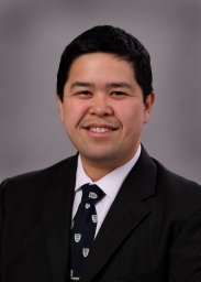 Menez Steven合作论文数1Johns Hopkins University School of Medicine7
 Rasheed Adebayo Gbadegesin合作论文数Department of Medicine, Duke University School of Medicine6
 Andrew Stephen Bomback合作论文数Department of Nephrology, Columbia University;Department of Surgery, Columbia University Irving Medical Center6
 Katherine Twombley合作论文数6
 Laura C. Plantinga合作论文数Department of Medicine, Johns Hopkins University School of Medicine6Gerald B. Appel合作论文数6
 Rulan Parekh合作论文数Division of Nephrology, The Hospital for Sick Children6
 Derek Fine合作论文数Department of Medicine, Johns Hopkins University School of Medicine6
 Aftab Chishti合作论文数5
 Myda Khalid合作论文数5
 Margaret E. Helmuth合作论文数Arbor Research Collaborative for Health5Sandeep Soman合作论文数5
 Raquel Greer合作论文数Department of Epidemiology, Johns Hopkins Bloomberg School of Public Health;Johns Hopkins University School of Medicine5
 Daniel Cattran合作论文数Toronto General Hospital, University of Toronto;Institute of Medical Science, Temerty Faculty of Medicine, University of Toronto5
 Jeffrey B. Kopp合作论文数Kidney Disease Section, Kidney Diseases Branch, National institute of Diabetes and Digestive and Kidney Diseases, National Institutes of Health5Braunstein Evan M合作论文数Department of Genetics, Albert Einstein College of Medicine5Kidd Jason M合作论文数5
 Larry Greenbaum合作论文数5
 David Thissen合作论文数Department of Psychology & Neuroscience, University of North Carolina at Chapel Hill5
 Michelle M O'Shaughnessy合作论文数Galway University Hospitals, National University of Ireland5
) Giuseppe Remuzzi合作论文数Mario Negri Institute for Pharmacological Research IRCCS5
Giuseppe Remuzzi合作论文数Mario Negri Institute for Pharmacological Research IRCCS5 Tarak Srivastava合作论文数Nephrology Research Laboratory, Children's Mercy Adele Hall;School of Medicine, University of Missouri-Kansas City5
 Kimberly J. Reidy合作论文数Department of Pediatrics, Albert Einstein College of Medicine, Montefiore Einstein;Montefiore Medical Center, Albert Einstein College of Medicine, Montefiore Einstein5
 Michelle Estrella合作论文数School of Medicine, University of California, San Francisco;San Francisco VA Health Care System;Kidney Health Research Collaborative;U.S. Department of Veterans Affairs5
 Bradley P Dixon合作论文数5Kara Rice合作论文数4Gloria F Gerber合作论文数4
 Brenda Gillespie合作论文数Department of Biostatistics, School of Public Health, University of Michigan4
 Craig B Langman合作论文数Kidney Diseases, Department of Pediatrics, Feinberg School of Medicine, Northwestern University4
 Debbie Gipson合作论文数George M. O'Brien Kidney Translational Core Center, Medical School, University of Michigan-Ann Arbor4
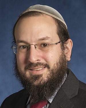 Avi Z Rosenberg合作论文数Department of Otolaryngology-Head and Neck Surgery, School of Medicine, Johns Hopkins University4
 Chirag R. Parikh合作论文数Bloomberg School of Public Health, Johns Hopkins University;Division of Nephrology, Johns Hopkins University School of Medicine4Lucas Gregory M合作论文数4
 Tetyana L Vasylyeva合作论文数4
 Mottl Amy K合作论文数School of Medicine, University of North Carolina at Chapel Hill4
 Terence Cook合作论文数Department of Immunology and Inflammation, Faculty of Medicine, Imperial College London4
 J. F. M. Wetzels (Jack)合作论文数Faculteit der Medische Wetenschappen, Radboud University Medical Center4
 Bruce Robinson合作论文数Northern Clinical School, University of Sydney;Sydney Southeast Asia Centre, University of Sydney;Kolling Institute of Medical Research4
 Lightstone Liz合作论文数4
 Erica Daina合作论文数Clinical Center, Istituto di Ricerche Farmacologiche Mario Negri IRCCS4




