OBJECTIVES:The aim is to investigate the proteomic profile of different molecular subtypes of pancreatic ductal adenocarcinoma (PDAC) and understand their impact on patient outcomes, particularly focusing on pathways involved in xenobiotic metabolism and drug resistance. MATERIALS AND METHODS:The study utilized the serum-free PACO cell culture model and a quantitative prefractionation-based MALDI/MS approach to establish the proteomic profiles of various PDAC subtypes. Differential protein regulation was analyzed to identify systematic alterations in metabolic and drug resistance pathways. Mechanistic studies involved the knockdown and overexpression of key proteins to assess their role in drug resistance. RESULTS:Proteomic analysis revealed subtype-specific alterations, particularly in pathways associated with xenobiotic metabolism and drug resistance. Notably, CYP2S1, a member of the CYP450 family, was upregulated in the HNF1A+ PDAC subtype. CYP2S1 levels were further inducible by polyaromatic hydrocarbons (PAHs) and SN38, the active metabolite of irinotecan via AHR. Mechanistic studies demonstrated that knockdown of AHR or CYP2S1 sensitized PDAC cells to SN38, whereas overexpression of CYP2S1 increased resistance to SN38. CONCLUSIONS:The findings highlight the significant role of CYP2S1 in mediating drug resistance in certain PDAC subtypes. Targeting CYP2S1 and its regulatory pathways could enhance the efficacy of chemotherapeutic agents like irinotecan in treating PDAC. These results provide new insights into the molecular mechanisms underlying PDAC subtype-specific drug resistance and suggest potential therapeutic targets.
Abstract Extrachromosomal DNA (ecDNA) is increasingly recognized as a driver of cellular plasticity, yet its role in therapy adaptation in pancreatic ductal adenocarcinoma (PDAC) remains poorly defined. In patient-derived PDAC models, we identify extrachromosomal ABCB1 amplification as a mechanism of resistance to paclitaxel and KRAS inhibitors and show that ABCB1 copy number dynamically adjusts to selective pressure. Notably, paclitaxel-resistant cells remain primed for rapid de novo ecDNA generation: after eliminating pre-existing ABCB1 ecDNA through single-cell cloning, new and structurally distinct ABCB1 ecDNA rapidly emerges, indicating treatment-induced molecular changes that prime these cells for ecDNA formation. In metastatic breast cancer, taxane-associated ABCB1 amplification is likewise observed and can be tracked in cell-free DNA (cfDNA), underscoring clinical relevance beyond PDAC. Finally, gemcitabine co-treatment suppresses ABCB1 ecDNA generation, suggesting potential strategies to counteract ecDNA-mediated resistance. Together, these findings demonstrate that dynamic ecDNA modulation and inducible ecDNA biogenesis enable rapid, reversible drug resistance, providing a rationale for ecDNA-targeted combination therapies and longitudinal monitoring. Citation Format: Tim Vorberg, Manuel Reitberger, Bernardo Rodriguez Martin, Maja Starostecka, Dominique Schulz, Arlou K. Angeles, Kate I. Glennon, Tasneem Cheytan, Roberto Würth, Vera Thiel, Paul Schwerd-Kleine, Verena Thewes, Laura Michel, Ewgenija Gutjahr, Simon J. Ogrodnik, Heike Conrad, Steffi O. Mehlhorn, Vanessa Vogel, Corinna Klein, Albrecht Stenzinger, Peter Lichter, Andreas Schneeweiss, Martin Granzow, Marc Zapatka, Anna Jauch, Holger Sültmann, Jan Korbel, Andreas Trumpp, Martin R. Sprick. Dynamic copy number changes and de novo generation of extrachromosomal DNA modulate therapy resistance [abstract]. In: Proceedings of the American Association for Cancer Research Annual Meeting 2026; Part 1 (Regular Abstracts); 2026 Apr 17-22; San Diego, CA. Philadelphia (PA): AACR; Cancer Res 2026;86(7 Suppl):Abstract nr 1803.
BACKGROUND:RNA-binding proteins (RBPs) are fundamental regulators of cellular biology that affect all steps in the generation and processing of RNA molecules. Recent evidence suggests that regulation of RBPs that modulate both RNA stability and translation may have a profound effect on the proteome. However, regulation of RBPs in clinically relevant experimental conditions has not been studied systematically.METHODS:We used RNA interactome capture, a method for the global identification of RBPs to characterize the global RNA-binding proteome (RBPome) associated with polyA-tailed RNA species in murine ciliated epithelial cells of the inner medullary collecting duct. To study regulation of RBPs in a clinically relevant condition, we analyzed hypoxia-associated changes of the RBPome.RESULTS:We identified >1000 RBPs that had been previously found using other systems. In addition, we found a number of novel RBPs not identified by previous screens using mouse or human cells, suggesting that these proteins may be specific RBPs in differentiated kidney epithelial cells. We also found quantitative differences in RBP-binding to mRNA that were associated with hypoxia versus normoxia.CONCLUSIONS:These findings demonstrate the regulation of RBPs through environmental stimuli and provide insight into the biology of hypoxia-response signaling in epithelial cells in the kidney. A repository of the RBPome and proteome in kidney tubular epithelial cells, derived from our findings, is freely accessible online, and may contribute to a better understanding of the role of RNA-protein interactions in kidney tubular epithelial cells, including the response of these cells to hypoxia.
Amyloid-β (Aβ) plaque deposits in the brain are a hallmark of Alzheimer’s disease (AD) neuropathology. Plaques consist of complex mixtures of peptides like Aβ1–42 and characteristic lipids such as gangliosides, and they are targeted by reactive microglia and astrocytes. Background: In pharmaceutical research and development, it is a formidable challenge to contextualize the different biomolecular classes and cell types of the Aβ plaque microenvironment in a coherent experimental workflow on a single tissue section and on a benchtop imaging reader. Methods: Here, we developed a workflow that combines lipid MALDI mass spectrometry imaging using a vacuum-stable matrix with histopathology stains and with the MALDI HiPLEX immunohistochemistry of plaques and multiple protein markers on a benchtop imaging mass spectrometer. The three data layers consisting of lipids, protein markers, and histology could be co-registered and evaluated together. Results: Multimodal data analysis suggested the extensive co-localization of Aβ plaques with the peptide precursor protein, with a defined subset of lipids and with reactive glia cells on a single brain section in APPPS1 mice. Plaque-associated lipids like ganglioside GM2 and phosphatidylinositol PI38:4 isoforms were readily identified using the tandem MS capabilities of the mass spectrometer. Conclusions: Altogether, our data suggests that complex pathology involving multiple lipids, proteins and cell types can be interrogated by this spatial multiomics workflow on a user-friendly benchtop mass spectrometer.
Circulating tumor cells (CTCs) drive metastasis, the leading cause of death in individuals with breast cancer. Due to their low abundance in the circulation, robust CTC expansion protocols are urgently needed to effectively study disease progression and therapy responses. Here we present the establishment of long-term CTC-derived organoids from female individuals with metastatic breast cancer. Multiomics analysis of CTC-derived organoids along with preclinical modeling with xenografts identified neuregulin 1 (NRG1)–ERBB2 receptor tyrosine kinase 3 (ERBB3/HER3) signaling as a key pathway required for CTC survival, growth and dissemination. Genome-wide CRISPR activation screens revealed that fibroblast growth factor receptor 1 (FGFR1) signaling serves a compensatory function to the NRG1–HER3 axis and rescues NRG1 deficiency in CTCs. Conversely, NRG1–HER3 activation induced resistance to FGFR1 inhibition, whereas combinatorial blockade impaired CTC growth. The dynamic interplay between NRG1–HER3 and FGFR1 signaling reveals the molecular basis of cancer cell plasticity and clinically relevant strategies to target it. Our CTC organoid platform enables the identification and validation of patient-specific vulnerabilities and represents an innovative tool for precision medicine. Trumpp and colleagues develop a method to obtain long-term circulating tumor cell-derived organoids from individuals with metastatic breast cancer and identify the neuregulin 1–HER3 axis as important for organoid growth and a promising therapeutic target.
The peripheral nervous system (PNS) orchestrates organ function in health and disease. Most cancers, including pancreatic ductal adenocarcinoma (PDAC), are infiltrated by PNS neurons, and this contributes to the complex tumour microenvironment (TME)1,2. However, neuronal cell bodies reside in various PNS ganglia, far from the tumour mass. Thus, cancer-innervating or healthy-organ-innervating neurons are lacking in current tissue-sequencing datasets. To molecularly characterize pancreas- and PDAC-innervating neurons at single-cell resolution, we developed Trace-n-Seq. This method uses retrograde tracing of axons from tissues to their respective ganglia, followed by single-cell isolation and transcriptomic analysis. By characterizing more than 5,000 individual sympathetic and sensory neurons, with about 4,000 innervating PDAC or healthy pancreas, we reveal novel neuronal cell types and molecular networks that are distinct to the pancreas, pancreatitis, PDAC or melanoma metastasis. We integrate single-cell datasets of innervating neurons and the TME to establish a neuron-cancer-microenvironment interactome, delineate cancer-driven neuronal reprogramming and generate a pancreatic-cancer nerve signature. Pharmacological denervation induces a pro-inflammatory TME and increases the effectiveness of immune-checkpoint inhibitors. The taxane nab-paclitaxel causes intratumoral neuropathy, which attenuates PDAC growth and, in combination with sympathetic denervation, results in synergistic tumour regression. Our multi-dimensional data provide insights into the networks and functions of PDAC-innervating neurons, and support the inclusion of denervation in future therapies.
Human microglia are critically involved in Alzheimer’s disease (AD) progression, as shown by genetic and molecular studies. However, their role in tau pathology progression in human brain has not been well described. Here, we characterized 32 human donors along progression of AD pathology, both in time—from early to late pathology—and in space—from entorhinal cortex (EC), inferior temporal gyrus (ITG), prefrontal cortex (PFC) to visual cortex (V2 and V1)—with biochemistry, immunohistochemistry, and single nuclei-RNA-sequencing, profiling a total of 337,512 brain myeloid cells, including microglia. While the majority of microglia are similar across brain regions, we identified a specific subset unique to EC which may contribute to the early tau pathology present in this region. We calculated conversion of microglia subtypes to diseased states and compared conversion patterns to those from AD animal models. Targeting genes implicated in this conversion, or their upstream/downstream pathways, could halt gene programs initiated by early tau progression. We used expression patterns of early tau progression to identify genes whose expression is reversed along spreading of spatial tau pathology (EC > ITG > PFC > V2 > V1) and identified their potential involvement in microglia subtype conversion to a diseased state. This study provides a data resource that builds on our knowledge of myeloid cell contribution to AD by defining the heterogeneity of microglia and brain macrophages during both temporal and regional pathology aspects of AD progression at an unprecedented resolution.
Abstract Breast cancer is a highly complex multifactorial disease, which can be driven by the aberrant regulation of different signaling pathways including receptor tyrosine kinase (RTK) signaling. Notably, while RTKs have been classically described as transmembrane receptors, they can also translocate to the nucleus, even though the functional importance of this process remains largely unexplored for most RTKs. Here, we report a novel role for the nuclear form of the RTK human epidermal growth factor receptor 3 (HER3) in driving primary breast cancer growth and metastasis. Firstly, using different patient-derived organoids (PDOs) established from metastatic breast cancer patients, we demonstrated the robust translocation of the activated phosphorylated HER3 receptor (pHER3) from the plasma membrane to the nucleus in response to its ligand Neuregulin 1 (NRG1). Interestingly, the nuclear translocation was observed not only in the HER2-positive breast cancer subtype but also in luminal and triple-negative breast cancer-derived cells. In order to decipher the functional role of nuclear HER3 (nHER3), we identified and mutated its nuclear localization signal, resulting in decreased nuclear translocation of the receptor while its membrane form remained intact. This reduction in nHER3 remarkably impeded primary tumor growth and metastatic spread in different patient-derived xenograft (PDX) models. Conversely, selective overexpression of HER3 in the nucleus led to increased primary tumor growth and metastatic burden in vivo. In order to decipher the mechanism of action of nHER3, we performed co-immunoprecipitation followed by global mass spectrometric analysis to identify the protein binding partners of the receptor in the nucleus. Specifically, nHER3 was found to interact with different transcription factors such as the AP-2 family of transcription factors and with major chromatin remodeling complexes like the nucleosome remodeling and deacetylase (NuRD) complex, implicating its role in transcriptional regulation. Furthermore, gene expression analysis of PDOs expressing the wild-type or the mutated form of HER3 revealed the modulation of several key growth regulators such as early growth response 3 (EGR3) by nHER3. The nHER3-EGR3 axis further controlled downstream effectors like the immune chemoattractants CXCL1 and CXCL8. Accordingly, PDX from breast cancer cells overexpressing nHER3 showed significantly increased immune infiltration, suggesting a possible role of nHER3 in regulating the tumor microenvironment. Altogether, we report here a novel non-canonical role of the nuclear form of HER3 receptor in driving breast tumorigenesis, with key implications for our understanding and targeting of RTK signaling in breast cancer. Citation Format: Tasneem Cheytan, Roberto Würth, Nina Hahnen, Elisa Donato, Corinna Klein, Rebecca Weber, Daniele Colombo, Jeroen Krijgsveld, Martin Sprick, Andreas Trumpp. Nuclear translocation of HER3 promotes breast cancer progression and dissemination recruiting immune cells via CXCL1 and CXCL8 [abstract]. In: Proceedings of the American Association for Cancer Research Annual Meeting 2024; Part 1 (Regular Abstracts); 2024 Apr 5-10; San Diego, CA. Philadelphia (PA): AACR; Cancer Res 2024;84(6_Suppl):Abstract nr 3940.
Ferroptosis is a form of cell death that has received considerable attention not only as a means to eradicate defined tumour entities but also because it provides unforeseen insights into the metabolic adaptation that tumours exploit to counteract phospholipid oxidation1,2. Here, we identify proferroptotic activity of 7-dehydrocholesterol reductase (DHCR7) and an unexpected prosurvival function of its substrate, 7-dehydrocholesterol (7-DHC). Although previous studies suggested that high concentrations of 7-DHC are cytotoxic to developing neurons by favouring lipid peroxidation3, we now show that 7-DHC accumulation confers a robust prosurvival function in cancer cells. Because of its far superior reactivity towards peroxyl radicals, 7-DHC effectively shields (phospho)lipids from autoxidation and subsequent fragmentation. We provide validation in neuroblastoma and Burkitt's lymphoma xenografts where we demonstrate that the accumulation of 7-DHC is capable of inducing a shift towards a ferroptosis-resistant state in these tumours ultimately resulting in a more aggressive phenotype. Conclusively, our findings provide compelling evidence of a yet-unrecognized antiferroptotic activity of 7-DHC as a cell-intrinsic mechanism that could be exploited by cancer cells to escape ferroptosis. Proferroptotic activity of 7-dehydrocholesterol reductase is shown along with an unexpected prosurvival function of its substrate, 7-dehydrocholesterol, indicating a cell-intrinsic mechanism that could be used by cancer cells to protect phospholipids from oxidative damage and escape ferroptosis.
Intraneuronal aggregates of the microtubule binding protein Tau are a hallmark of different neurodegenerative diseases including Alzheimer's disease (AD). In these aggregates, Tau is modified by posttranslational modifications such as phosphorylation as well as by proteolytic cleavage. Here we identify a novel Tau cleavage site at aspartate 65 (D65) that is specific for caspase-2. In addition, we show that the previously described cleavage site at D421 is also efficiently processed by caspase-2, and both sites are cleaved in human brain samples. Caspase-2-generated Tau fragments show increased aggregation potential in vitro, but do not accumulate in vivo after AAV-mediated overexpression in mouse hippocampus. Interestingly, we observe that steady-state protein levels of caspase-2 generated Tau fragments are low in our in vivo model despite strong RNA expression, suggesting efficient clearance. Consistent with this hypothesis, we find that caspase-2 cleavage significantly improves the recognition of Tau by the ubiquitin E3 ligase CHIP, leading to increased ubiquitination and faster degradation of Tau fragments. Taken together our data thus suggest that CHIP-induced ubiquitination is of particular importance for the clearance of caspase-2 generated Tau fragments in vitro and in vivo.
The acquisition of mesenchymal traits is considered a hallmark of breast cancer progression. However, the functional relevance of epithelial-to-mesenchymal transition (EMT) remains controversial and context dependent. Here, we isolate epithelial and mesenchymal populations from human breast cancer metastatic biopsies and assess their functional potential in vivo. Strikingly, progressively decreasing epithelial cell adhesion molecule (EPCAM) levels correlate with declining disease propagation. Mechanistically, we find that persistent EPCAM expression marks epithelial clones that resist EMT induction and propagate competitively. In contrast, loss of EPCAM defines clones arrested in a mesenchymal state, with concomitant suppression of tumorigenicity and metastatic potential. This dichotomy results from distinct clonal trajectories impacting global epigenetic programs that are determined by the interplay between human ZEB1 and its target GRHL2. Collectively, our results indicate that susceptibility to irreversible EMT restrains clonal propagation, whereas resistance to mesenchymal reprogramming sustains disease spread in multiple models of human metastatic breast cancer, including patient-derived cells in vivo.
Topic: 3. Acute myeloid leukemia - Biology & Translational Research Background: Acute Myeloid Leukemia (AML) is a highly aggressive adult leukemia with a high incidence of relapse and mortality. AML hijacked the hierarchical organization of normal hematopoiesis with Leukemia Stem Cells (LSCs) at the top of the differentiation tree and differentiated and highly proliferating myeloid blast at the bottom. Similar to normal hematopoietic stem cells (HSCs), LSCs have been originally identified as CD34+CD38- cells. However, this strategy to isolate LSCs is particularly challenging in NPM1 mutated AMLs, since expression of CD34 on leukemic cells is low or absent altogether. Aims: In this study, we aimed to establish a novel sorting strategy to isolate LSCs independently of CD34 surface marker in both CD34positive and CD34negative AMLs. Methods: Using a genetically highly homogeneous cohort of 46 AML patients carrying DNMT3A and NPM1 mutations, we established a novel isolation strategy to enrich for LSCs even in CD34negative AMLs by additionally including a positive selection for GPR56 and a negative selection for mature markers such as NKG2DLigandss. Results: By combining functional in vivo data, mutational analyses on xenografts and transcriptomic data, we found that CD34+GPR56+NKG2DL- subpopulations can contain along with LSCs also normal and/or pre-leukemic (HSCs). In contrast, no such residual normal or pre-leukemic HSCs were detected in CD34-GPR56+NKG2DL- subpopulations. Finally, we show that naïve AML patients who retain a relevant reserve of normal and/or preleukemic HSCs, able to reconstitute immunocompromised mice at time of diagnosis, have significant longer relapse-free and overall survival compared to patients without engrafting HSCs. Summary/Conclusion: These data suggest that the presence of normal and/or preleukemic HSCs with multilineage engrafting potential in the bone marrow of naïve patients is associated with good response to standard chemotherapy, providing a basis for the development of novel predictive biomarkers for standard chemotherapy regime. Keywords: DNMT3A, AML, Leukemic stem cell, Stem cell marker
Background: Despite typically high response rates to initial chemotherapy, the rate of mortality in acute myeloid leukemia (AML) remains high due to frequent and hard to control relapses. Persistence of therapy resistant Leukemic Stem Cells (LSCs) harboring clonal outgrowth capacity and present within Minimal Residual Disease (MRD) during complete remission stage are thought to be the origin of relapse. However, identifying the exact cellular composition of MRD in patients during remission has been notoriously difficult due to the exceedingly low number of resistant leukemia cells in MRD stage hiding within the vast majority of healthy blood cells. Thus, the molecular and cellular mechanisms responsible for the functional maintenance of LSCs in patient MRD remains poorly understood. Aims: Clinical MRD sample analysis is limited by the difficulty of accessing such samples in a longitudinal manner. Therefore, we established and characterized an MRD human xenograft mouse model to circumvent the difficulty to analyze the MRD fractions of AML patients. Based on the knowledge attained, precise strategies targeting or preventing the appearance of resistant persister leukemic cells including LSCs will be developed. Methods: A unique set of four longitudinally collected triplet samples (diagnosis, remission, and relapse) were used for this analysis. Established in vivo patient derived xenograft (PDX) models from the longitudinally triplet samples were treated with combination chemotherapy (Cytarabine and Daunorubicin) in order to mimic the MRD state in the clinical setting. Using flow cytometry and RNA-seq, we analysed MRD fractions in both primary samples and the corresponding PDX models. Results: First, we determined the in vivo regimen and the optimum sub-lethal dose of Cytarabine (AraC) + Daunorubicin. Administration of AraC 30mg/kg/day for 4 consecutive days combined with 2 days of Daunorubicine 2.5mg/kg/day was determined as the most efficient treatment to observe a significant reduction in total AML cell tumor burden in the bone marrow and spleen at day 8. Additionally, we determined week 2 as the optimal time point to observe tumor regrowth mimicking the clinical MRD state. Our data show that this combination treatment (4 + 2) is well tolerated in NSG mice at a dose and schedule analogous to the clinically used 7 + 3 AML patient treatment regime, and thus allowing us to study mechanisms involved in drug resistance. Importantly, PDX generated from diagnosis samples, but not from MRD or relapse samples, displayed sensitivity to chemotherapy. Moreover, the residual AML-MRD cells surviving chemotherapy in PDX mice from diagnostic samples displayed a higher OxPhos metabolism compared to untreated controls, as we observed a higher mitochondrial membrane potential and mitochondrial mass as assessed by FACS assays TMRE and MTG respectively. In addition, MRD cells expressed higher level of the myeloid chemokine receptors. Collectively, these data correlate with the clinical data of the analysed longitudinal triplet samples as both primary patient and PDX MRD samples were enriched on High OxPhos signature and presented a higher gene expression of the myeloid chemokine receptors compared to the respective diagnosis samples. Summary/Conclusion: Taken together, we have established PDX mouse models that recapitulates the metabolic properties and the phenotypic features that we identified at the clinical MRD state. Single-cell multi-omics technologies will now be applied to gain novel insights into the cellular identity, heterogeneity, and molecular mechanism of therapy-resistant LSCs in AML. Keywords: Acute myeloid leukemia, Minimal residual disease (MRD), Chemoresistance
Acute myeloid leukemia (AML) is a heterogeneous disease characterized by high rate of relapse and mortality. Current chemotherapies whilst successful in eradicating blasts, are less effective in eliminating relapse-causing leukemic stem cells (LSCs). Although LSCs are usually identified as CD34+CD38- cells, there is significant heterogeneity in surface marker expression, and CD34- LSCs exist particularly in NPM1mut AMLs. By analyzing diagnostic primary DNMT3AmutNPM1mut AML samples, we suggest a novel flow cytometry sorting strategy particularly useful for CD34neg AML subtypes. To enrich for LSCs independently of CD34 status, positive selection for GPR56 and negative selection for NKG2D ligands are used. We show that the functional reconstitution capacity of CD34- and CD34+ LSCs as well as their transcriptomes are very similar which support phenotypic plasticity. Furthermore, we show that although CD34+ subpopulations can contain next to LSCs also normal and/or preleukemic hematopoietic stem cells (HSCs), this is not the case in CD34-GPR56+NKG2DL- enriched LSCs which thus can be isolated with high purity. Finally, we show that patients with AML, who retain at the time of diagnosis a reserve of normal and/or preleukemic HSCs in their bone marrow able to reconstitute immunocompromised mice, have significantly longer relapse-free and overall survival than patients with AML in whom functional HSCs are no longer detectable.
Intracellular abnormal aggregation of hyperphosphorylated tau protein is a neuropathological hallmark associated with Alzheimer’s Disease (AD). The insoluble tau aggregates called neurofibrillary tangles lead to microtubule dysfunction and neurodegeneration with AD progression. The TG4510 transgenic mice overexpressing the P301L mutant human tau protein exhibit region-specific tau inclusions, brain atrophy, and behavioral deficits, thereby serving as a widely used animal model of tauopathy in AD. This study aims to characterize the tissue-specific transcriptomic and epigenomic signatures of progressive tau pathology in TG4510 mice as well as investigate the correlation of brain and blood expression and epigenomic changes. Using UMI-based bulk RNA-Sequencing and Reduced-representation Bisulfite Sequencing (RRBS), we profiled the gene expression and DNA-methylation changes respectively, in the cortex, hippocampus, cerebellum and pre-sacrifice and terminal whole blood between three age groups (2-3 months, 6-7 months, 8-9 months old; N=20/tissue/group) of male and female TG4510 mice. The cortex and hippocampus show the largest transcriptional differences between mice, recapitulating phenotype differences with loss of neurotransmission and increased inflammation in these tissues. Systems-level co-expression analysis reveals common gene modules in immune system, behaviour and cognition and ECM/synaptic pathways between cortex/hippocampus and blood. Furthermore, the genes correlated between brain regions and blood in these mice were prioritized as potential biomarkers of tau pathology, which can be further validated in preclinical animal models and ultimately human studies. This study serves as one of the first evidence of multiomic molecular underpinnings in a tauopathy mouse model providing potential novel targets and biomarkers of progressive tau pathology in AD. All authors are employees of AbbVie. The design, study conduct, and financial support for this research were provided by AbbVie. AbbVie participated in the interpretation of data, review, and approval of the publication.
 Andreas Trumpp合作论文数Division of Stem Cells and Cancer, The Heidelberg Institute for Stem Cell Technology and Experimental Medicine;German Cancer Research Center54
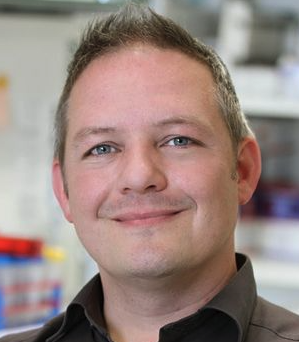 Martin Sprick合作论文数Heidelberg Institute for Stem Cell Technology and Experimental Medicine;German Cancer Research Center33
 Vogel Vanessa合作论文数24
 Wilko Weichert合作论文数Institute of Pathology, Technical University Munich;School of Medicine, Technical University Munich18
 Elisa Donato合作论文数HI-STEM gGmbH),, Heidelberg Institute for Stem Cell Technology and Experimental Medicine14Christian Eisen合作论文数13
 Wolf-K. Hofmann合作论文数Medizinische Fakulat Manheim, Universit Heidelberg;III. medical clinic, University Hospital Mannheim12
 Thomas Hofner合作论文数12
 Johann-Christoph Jann合作论文数Heidelberg Univ, Med Fac Mannheim, Dept Hematol & Oncol, Mannheim, Germany11
 Würth Roberto合作论文数Sez Farmacol, Univ Genoa11Rigo-Watermeier Teresa合作论文数11Florian Nolte合作论文数11Verena Nowak合作论文数11Fey Stephanie合作论文数10
 Andreas Schneeweiss合作论文数Universitätsklinikum Heidelberg10Maximilian Mossner合作论文数10
 Hind Medyouf合作论文数10
 Elisa Espinet合作论文数Institute for Research in Biomedicine (IRB)9Albrecht Stenzinger合作论文数Institute of Pathology, University Hospital9
 Claudia Haferlach合作论文数Leukämielabor für Leukämiediagnostik und Methoden;MLL MVZ GmbH9Zens Bettina合作论文数Division of Stem Cells and Cancer, Deutsches Krebsforschungszentrum (DKFZ),;Heidelberg Institute for Stem Cell Technology and Experimental Medicine (HI-STEM gGmbH), Im Neuenheimer Feld 280, 69120 Heidelberg, German.;Deutsches Krebsforschungszentrum (DKFZ),, Heidelberg Institute for Stem Cell Technology and Experimental Medicine (HI-STEM gGmbH),8N Giese合作论文数7
 Massimo Saini合作论文数Division of Stem Cells and Cancer, German Cancer Research Center (DKFZ), 69120 Heidelberg, Germany.7
 Correia Nádia C合作论文数Instituto de Medicina Molecular, Faculdade de Medicina da Universidade de Lisboa7Uwe Platzbecker合作论文数7J. Oblaender合作论文数6Manuel Reitberger合作论文数6
 Darja Karpova合作论文数6Adriana Przybylla合作论文数German Canc Res Ctr, Div Stem Cells & Canc, D-69120 Heidelberg, Germany6D. Nowak合作论文数6
 Nicole Pfarr合作论文数6
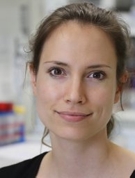 Baccelli Irène合作论文数Division of Stem Cells and Cancer, German Cancer Research Center (DKFZ), Heidelberg, Germany.6
 Falcone Mattia合作论文数Molecular Biotechnology Center, University of Torino6Zeisberger Petra合作论文数6
 Schlesner Matthias合作论文数Div Theoret Bioinformat, German Canc Res Ctr6
 Roland Eils合作论文数Center of Digital Health, Berlin Institute of Health;Charité Universitätsmedizin Berlin;Health Data Science Unit, Medical Faculty of Heidelberg University6Christian Thiede合作论文数6
 Daniel Nowak合作论文数6A. Fabarius合作论文数5Katja Muedder合作论文数5Markus Sohn合作论文数Heidelberg Inst Stem Cell Technol & Expt Med HI S, D-69120 Heidelberg, Germany5
 Daniel Huebschmann合作论文数German Canc Res Ctr, Div Theoret Bioinformat, Heidelberg, Germany5Carolin Andresen合作论文数5
 Saskia Spaich合作论文数Universitatsmedizin Mannheim5
 Strobel Oliver合作论文数5Eisen Christian合作论文数5Noll Elisa合作论文数5Dolnik Anna合作论文数5Obländer Julia合作论文数5




