Leiomyosarcoma (LMS) is a rare malignancy arising from the smooth muscle, which affects the inferior vena cava (IVC) in 30–40
BackgroundWe report on a series of consecutive patients with localized radiation-associated angiosarcoma (RAAS) of the breast region (BR) treated at two Italian sarcoma reference centers.Materials and methodsWe retrospectively reviewed all cases of primary, localized, resectable RAAS of the BR, treated at one of the two participating institutions from 2000 to 2019. Relapse-free survival (RFS) and overall survival (OS) were calculated. The prognostic role of several variables was investigated. A propensity score matched (PSM) analysis was carried out.ResultsEighty-four patients were retrospectively identified. Nineteen out of 84 patients (22.6%) were pretreated with an anthracycline-based regimen for previous cancer. All patients but one underwent surgery, with 37/84 (44.1%) receiving surgery alone and 46/84 (54.8%) a multimodal approach: 18/84 (21.4%) received radiation therapy (RT) and 46/84 (54.9%) received chemotherapy. An anthracycline-based regimen was used in 10/84 patients (11.9%), while a gemcitabine-based regimen was used in 33/84 (39.3%). With a median follow-up of 51 months (interquartile range: 30-126 months), 36/84 patients (42.9%) relapsed and 35/84 patients (41.7%) died (8/84, 9.5% in the lack of metastatic disease). Five-year OS and 5-year RFS were 57% [95% confidence interval (CI) 43% to 68%] and 52% (95% CI 39% to 63%), respectively. Both (neo)adjuvant RT and chemotherapy were associated with better RFS [hazard ratio (HR) 0.25, 95% CI 0.08-0.83; HR 0.45, 95% CI 0.23-0.89] with a trend towards a better OS (HR 0.51, 95% CI 0.18-1.46; HR 0.60, 95% CI 0.29-1.24). Gemcitabine-based regimens seemed to perform better (HR 4.28, 95% CI 1.29-14.14). PSM analysis retained the above results.ConclusionsThis retrospective study supports the use of (neo)adjuvant RT and chemotherapy, in primary, localized resectable RAAS of the BR. An effort to prospectively validate the role of (neo)adjuvant RT and chemotherapy is warranted.
Background: Retroperitoneal sarcoma (RPS) is a rare tumour that can reach exceptionally large size at diagnosis and affects body weight with its volume. We investigated the risk of nutritional status misclassification based on the Global Leadership Initiative on Malnutrition (GLIM) criteria in RPS patients. Methods: We retrospectively analyzed the baseline data of patients with primary RPS including anthropometry, Malnutrition Universal Screening Tool (MUST) score and skeletal muscle index (SMI) calculated on preoperative CT scan. Phenotypic GLIM criteria considered were non-volitional weight loss, low-BMI and low muscle mass. MUST score, being inclusive of weight loss, was chosen as its surrogate. Reduced muscle mass was defined with SMI cut-offs <38.5-52.4 cm2/m2 for female and male respectively. Results: From 2018 to 2020, 100 consecutive patients (male/female: 48/52) were included. Median age was 61 (48-68) years, median BMI 24.6 (21.5-27.4) kg/m2. Seven patients (7%) reported MUST score >2 and 80 (80%) score 0. Five patients (5%) were underweight, 44 (44%) overweight and obese. Sixty patients (60%) presented low SMI, of those 56 (93%) were normal or overweight, and 46 (77%) not at nutritional risk according to the MUST score. Both underweight BMI and MUST score >2 were not correlated with low SMI (p = 0.406, p = 0.612, respectively). No relevant discrepancy was found in the postoperative course of patients with low or normal muscle mass, although SMI was able to better characterize nutritional risk in the proportion of patents with high-grade liposarcoma and predicted worst overall survival. Conclusion: GLIM criteria are a useful tool for diagnosis of malnutrition. However, our analysis suggests that phenotypic criteria are not interchangeable with each other. An altered body composition is often hidden in patients with retroperitoneal sarcoma and the use of weight-based classification does not seem to be appropriate, enhancing the role of mass muscle measurement to prevent misdetections of malnourished patient. (c) 2022 European Society for Clinical Nutrition and Metabolism. Published by Elsevier Ltd. All rights reserved.
Epithelioid hemangioendothelioma (EHE) is an ultra-rare, translocated, vascular sarcoma. EHE clinical behavior is variable, ranging from that of a low-grade malignancy to that of a high-grade sarcoma and it is marked by a high propensity for systemic involvement. No active systemic agents are currently approved specifically for EHE, which is typically refractory to the antitumor drugs used in sarcomas. The degree of uncertainty in selecting the most appropriate therapy for EHE patients and the lack of guidelines on the clinical management of the disease make the adoption of new treatments inconsistent across the world, resulting in suboptimal outcomes for many EHE patients. To address the shortcoming, a global consensus meeting was organized in December 2020 under the umbrella of the European Society for Medical Oncology (ESMO) involving >80 experts from several disciplines from Europe, North America and Asia, together with a patient representative from the EHE Group, a global, disease-specific patient advocacy group, and Sarcoma Patient EuroNet (SPAEN). The meeting was aimed at defining, by consensus, evidence-based best practices for the optimal approach to primary and metastatic EHE. The consensus achieved during that meeting is the subject of the present publication.
Background: This multicentric, retrospective study conducted within the Italian Rare Cancer Network describes clinical features and explores their possible prognostic relevance in patients with advanced epithelioid haemangioendothelioma (EHE) started on surveillance. Patients and methods: We collected data on adult patients with molecularly confirmed, advanced EHE consecutively referred at five sarcoma reference centres between January 2010 and June 2018, with no evidence of progressive disease (PD) and started on surveillance. Overall survival (OS) and progression-free survival (PFS) univariable and multivariable Cox analyses were performed. In the latter, due to the low number of cases and events, penalized likelihood was applied, and variable selection was performed using a random forest model. Results: Sixty-seven patients were included. With a median follow-up of 50.2 months, 51 (76%) patients developed PD and 16 (24%) remained stable. PD at treatment start did not meet RECIST version 1.1 in 15/51 (29%) patients. The 3-year PFS and OS were 25.4% and 71.1%, respectively, in the whole population. Tumour-related pain (TRP) was the most common baseline symptom (32.8%), followed by temperature (20.9%), fatigue (17.9%), and weight loss (16.4%). Baseline TRP (P = 0.0002), development of TRP during follow-up (P = 0.005), baseline temperature (P = 0.002), and development of fatigue during follow-up (P = 0.007) were associated with a significantly worst PFS. An association between baseline TRP (P < 0.0001), development of TRP during follow-up (P = 0.0009), evidence of baseline serosal effusion (P = 0.121), and OS was recorded. Conclusion: Because of the poor outcome observed in EHE patients presenting with serosal effusion, TRP, temperature, or serosal effusion, upfront treatment in this subgroup could be considered.
Background The capacity of inducing tumor shrinkage in advanced soft tissue sarcomas (STS) is an unmet need beyond the first-line of systemic therapy. Pivotal studies of approved drugs reported RECIST response rates below 10%. Thus, it is challenging to relieve symptoms related to tumor volume in patients progressing to anthracycline-based schemes. Besides that, the overall response rate (ORR) is an appropriate surrogate for overall survival. This phase II trial explores the combination of trabectedin (T) and concurrent radiotherapy (RT) in the metastatic setting and is supported by preclinical experiments and previous phase I trial. Methods Patients received T 1.5 mg/m2 in 24-h infusion and started RT (30 Gy, 3 Gy/day) from the end of the first T perfusion. Most relevant inclusion criteria were metastatic progressing STS, a maximum of two previous systemic lines for advanced disease with at least one previous anthracycline-based line. Neither all the lesions nor all the sites were required to be irradiated. The main endpoint was ORR by RECIST 1.1. A Simon 2-stage was used to estimate 35% ORR of interest for further investigation (α = 0.1, power 90%; P0 = 0.10). Central pathology and radiological review were mandatory. Results From 10/2017 to 11/2018, 27 patients were enrolled. Histologies were: leiomyosarcoma 7 (26%), synovial sarcoma 6 (22%), liposarcoma 4 (15%) and other 10 (37%). The median of previous lines was 2, and ECOG distribution was 0: 19 (70%) and 1: 8 (30%). With a median follow-up of 6 months (1-12), there were 9 events of progression and 3 events of death. The 6-month PFSR was 75% and the 6-month OSR was 86%. From 26 evaluable patients by RECIST, there were 15 PR (57.7%), 7 SD (26.9%) and 4 PD (15.4%). One G3 and 3 G1 pneumonitis were observed. One toxic death occurred (sepsis) being G3-4 neutropenia observed of 7 (26%) while febrile neutropenia was reported in 2 (7.4%) cases. Conclusions T concurrent with RT showed a relevant activity in progressing metastatic setting in a wide range of STS types, giving options for tumor shrinkage beyond the first-line of advanced STS. The RECIST ORR of 57.7% and 6-m PFSR of 75% confirm the synergy of T+RT. Clinical trial identification NCT02275286. Legal entity responsible for the study GEIS (Spanish Group for Research on Sarcoma). Funding GEIS, ISG (Italian Sarcoma Group), FSG (French Sarcoma Group); PharmaMar. Disclosure J. Martin Broto: Research grant / Funding (institution): EISAI; Research grant / Funding (institution): Novartis; Advisory / Consultancy, Research grant / Funding (institution), Travel / Accommodation / Expenses: PharmaMar; Advisory / Consultancy: Lilly. A. Italiano: Honoraria (self), Advisory / Consultancy, Research grant / Funding (self): Roche; Honoraria (self), Advisory / Consultancy: Daiichi Sankyo; Advisory / Consultancy: Inmune Design; Honoraria (self), Advisory / Consultancy: Epizyme; Honoraria (self), Advisory / Consultancy, Research grant / Funding (self): Bayer; Honoraria (self), Advisory / Consultancy: Lilly; Honoraria (self): Novartis; Research grant / Funding (self): Merck Serono; Research grant / Funding (self): AstraZeneca/MedImmune; Research grant / Funding (self): MSD Oncology. R.M. Alvarez Alvarez: Honoraria (self), Advisory / Consultancy, Research grant / Funding (self), Travel / Accommodation / Expenses: PharmaMar; Advisory / Consultancy: Lilly; Honoraria (self), Advisory / Consultancy: Boehringer Ingelheim; Research grant / Funding (self), Travel / Accommodation / Expenses: Roche; Honoraria (self): Pierre Fabre; Honoraria (self): Pfizer; Honoraria (self), Research grant / Funding (institution): Novartis; Honoraria (self): Bayer; Honoraria (self): Bristol-Myers Squibb; Research grant / Funding (self): Roche; Research grant / Funding (self): AstraZeneca; Research grant / Funding (self): Peregrine Pharmaceuticals; Research grant / Funding (self): AbbVie; Research grant / Funding (self): Merrimack; Research grant / Funding (institution): Janssen. A. Lopez Pousa: Travel / Accommodation / Expenses: PharmaMar. A. Redondo: Advisory / Consultancy, Research grant / Funding (institution), Travel / Accommodation / Expenses: Roche; Advisory / Consultancy, Travel / Accommodation / Expenses: AstraZeneca; Advisory / Consultancy: Tesaro; Advisory / Consultancy, Research grant / Funding (institution), Travel / Accommodation / Expenses: PharmaMar; Advisory / Consultancy: Clovis Oncology; Research grant / Funding (institution): Eisai. J. Blay: Honoraria (self), Research grant / Funding (self): PharmaMar. A. Gronchi: Honoraria (self), Advisory / Consultancy: Novartis; Advisory / Consultancy: Bayer; Honoraria (self), Advisory / Consultancy: Lilly; Advisory / Consultancy, Travel / Accommodation / Expenses: Nanobiotix; Advisory / Consultancy, Research grant / Funding (institution), Travel / Accommodation / Expenses: PharmaMar; Honoraria (self): Pfizer. N. Hindi: Honoraria (self), Research grant / Funding (institution), Travel / Accommodation / Expenses: PharmaMar; Research grant / Funding (institution): Eisai; Research grant / Funding (institution): Novartis. All other authors have declared no conflicts of interest.
Percutaneous biopsy is recommended before surgery for suspected retroperitoneal sarcoma (RPS) to confirm the histological diagnosis and guide surgical strategy. The present study aimed to establish the diagnostic accuracy of percutaneous core biopsy with respect to histological diagnosis and tumour grade.Data on patients with suspected RPS who underwent percutaneous biopsy followed by surgical resection between 2005 and 2016 at one of two tertiary European sarcoma units were reviewed. Histological tumour type and tumour grade on biopsy were correlated with postoperative histology to evaluate diagnostic accuracy.A total of 239 patients underwent percutaneous core biopsy followed by surgical resection in Milan (163, 68·2 per cent) or Birmingham (76, 31·8 per cent). Diagnostic accuracy varied with histological diagnosis (P < 0·001), but demonstrated overall concordance with final pathology following resection in 67·2 per cent of biopsies (κ = 0·606). The majority of discrepancies occurred in dedifferentiated liposarcoma (DDLPS), owing to under-recognition of dedifferentiation in this group. Concordance between pathology on biopsy and resection improved to 81·1 per cent when DDLPS and well differentiated liposarcoma were grouped together as liposarcoma. Grade on biopsy was concordant with grade on resection specimen in 60·4 per cent of tumours (κ = 0·640). Diagnosis of high-grade tumours on biopsy had a high specificity (98 per cent), and moderate positive predictive value (85 per cent) and negative predictive value (78 per cent).A diagnosis of DDLPS or leiomyosarcoma on percutaneous biopsy is highly reliable. High-grade sarcomas can be identified with high specificity, which opens the door to a study on neoadjuvant therapy in these patients.
Background Perivascular epithelioid cell tumors (PEComa) are exceedingly rare mesenchymal neoplasms arising in a variety of anatomic sites. mTOR inhibitors are active in these neoplasms. However, no other effective treatments are available in those patients progressing to them. The PI3K–Akt–mTOR signaling pathway modulates neoplastic growth through signaling activation of ER and the EGF receptor family of receptor tyrosine kinases. ER drives PI3K/AKT activation in response to mTORC1 inhibition. This provides a rationale for combining anti-estrogens and mTORC1 inhibitors. Methods We retrospectively identified patients with advanced PEComa treated with mTOR inhibitors since January 2002 at Fondazione IRCCS Istituto Nazionale dei Tumori, Milan – Italy. In a subgroup of them, exemestane was added at the time of progression. Results Twenty-eight patients with advanced PEComa treated with mTOR inhibitors were identified. Twenty–six were evaluable for response. Twelve out 26 (46%) had a PR and seven (27%) a SD, with a median PFS of 7 months. At the time of progression to sirolimus, 5 patients received a combination of sirolimus and exemestane and one of sirolimus, exemestane and GnRH. Three patients out 6 had a PR, 2 out 6 had a SD, and 1 out 6 had a PD, with a median PFS of 6 months. In this subgroup of patients treated with the combination, previous PFS to mTOR inhibitors was 6.6 months. Conclusions In this small retrospective series, the combination of mTOR inhibitors and exemestane obtained a reversion of resistance to mTOR inhibitors in one half of patients. Legal entity responsible for the study The authors. Funding Has not received any funding. Disclosure R. Sanfilippo: Honoraria (self), Research grant / Funding (institution), Travel / Accommodation / Expenses: Lilly; Honoraria (self), Research grant / Funding (institution), Travel / Accommodation / Expenses: PharmaMar; Research grant / Funding (institution): Advanchen Laboratories; Research grant / Funding (institution): Amgen Dompe; Research grant / Funding (institution): AROG Pharmaceuticals; Research grant / Funding (institution): Bayer; Research grant / Funding (institution): Blueprint Medicines; Research grant / Funding (institution): Daiichi Sankyo; Research grant / Funding (institution): Deciphera; Research grant / Funding (institution): Eisai; Research grant / Funding (institution): Epizyme Inc; Research grant / Funding (institution): Galxo; Research grant / Funding (institution): Karyopharm; Research grant / Funding (institution): Novartis; Research grant / Funding (institution): Pfizer. C. Fabbroni: Research grant / Funding (institution): Advanchen Laboratories; Research grant / Funding (institution): Amgen Dompe’; Research grant / Funding (institution): AROG Pharmaceuticals; Research grant / Funding (institution): bayer; Research grant / Funding (institution): Blueprint Medicines; Research grant / Funding (institution): Daiichi Sankyo; Research grant / Funding (institution): Deciphera; Research grant / Funding (institution): Eisai; Research grant / Funding (institution): Lilly; Research grant / Funding (institution): Epizyme Inc; Research grant / Funding (institution): Glaxo; Research grant / Funding (institution): Karyopharm Pharmaceuticals; Research grant / Funding (institution): Novartis; Research grant / Funding (institution): Pfizer; Research grant / Funding (institution): PharmaMar. E. Fumagalli: Research grant / Funding (institution): Advenchen Laboratories; Research grant / Funding (institution): Amgen dompe’; Research grant / Funding (institution): AROG Pharmaceuticals; Research grant / Funding (institution): Bayer; Research grant / Funding (institution): Blueprint Medicines; Research grant / Funding (institution): Daiichi Sankyo; Research grant / Funding (institution): Deciphera; Research grant / Funding (institution): Eisai; Research grant / Funding (institution): Lilly; Research grant / Funding (institution): Epizyme; Research grant / Funding (institution): Novartis; Research grant / Funding (institution): PharmaMar; Research grant / Funding (institution): Glaxo; Research grant / Funding (institution): karyopharm Pharmaceuticals; Research grant / Funding (institution): Pfizer. R. Bertulli: Research grant / Funding (institution): Karyopharm Pharmaceuticals; Research grant / Funding (institution): Pfizer; Research grant / Funding (institution): Novartis; Research grant / Funding (institution): Eisai; Research grant / Funding (institution): Lilly; Research grant / Funding (institution): Advenchen Laboratories; Research grant / Funding (institution): Amgen dompe’; Research grant / Funding (institution): AROG Pharmaceuticals; Research grant / Funding (institution): Bayer; Research grant / Funding (institution): Blueprint Medicines; Research grant / Funding (institution): Daiichi Sankyo ; Research grant / Funding (institution): Deciphera; Research grant / Funding (institution): Epizyme; Research grant / Funding (institution): Glaxo; Research grant / Funding (institution): PharmaMar. S. Stacchiotti: Research grant / Funding (institution): Karyopharm Pharmaceuticals; Research grant / Funding (institution): Eisai; Research grant / Funding (institution): PharmaMar; Research grant / Funding (institution): Glaxo; Research grant / Funding (institution): Novartis; Research grant / Funding (institution): Lilly; Research grant / Funding (institution): Deciphera; Research grant / Funding (institution): Advenche Laboratories; Research grant / Funding (institution): Amgen; Research grant / Funding (institution): AROG Pharmaceuticals; Research grant / Funding (institution): Bayer; Research grant / Funding (institution): Blueprint Medicines; Research grant / Funding (institution): Daiichi Sankyo; Research grant / Funding (institution): Epizyme; Research grant / Funding (institution): Pfizer. P.G. Casali: Honoraria (self), Advisory / Consultancy, Research grant / Funding (institution): Bayer; Honoraria (self), Advisory / Consultancy, Research grant / Funding (institution): Deciphera; Honoraria (self), Advisory / Consultancy, Research grant / Funding (institution): Eisai; Honoraria (self), Advisory / Consultancy, Research grant / Funding (institution): Lilly; Honoraria (self), Advisory / Consultancy, Research grant / Funding (institution): Pfizer; Honoraria (self), Advisory / Consultancy, Research grant / Funding (institution): Nektar Therapeutics; Research grant / Funding (institution): Advenchen Laboratories; Research grant / Funding (institution): Novartis; Research grant / Funding (institution): Blueprint Midicines; Research grant / Funding (institution): AROG Pharmaceuticals; Research grant / Funding (institution): Amgen dompe’; Research grant / Funding (institution): Daiichi Sankyo; Research grant / Funding (institution): Epizyme; Research grant / Funding (institution): Glaxo; Research grant / Funding (institution): KaryopharmPharmaceuticals; Research grant / Funding (institution): PharmaMar. All other authors have declared no conflicts of interest.
Background: To evaluate the antitumor activity of imatinib in combination with everolimus in patients (pts) with advanced PDGFB- and/or PDGFRB-positive chordomas with evidence of mTOR and/or of its effectors (i.e. S6, 4EBP1) activation. Methods: Within an Italian academic prospective phase II clinical study carried out from January 2011 to March 2015, 45 patients with advanced PDGFB/PDGFRB and mTOR/S6/4EBP1 positive chordoma received imatinib 400 mg/day in combination with everolimus at the starting dose of 2.5 mg/day, until progression or limiting toxicity. Eligible pts had to have evidence of progression in the 6 months prior to study entry. The primary endpoint was overall tumor response rate (ORR), defined by the Choi criteria applied also to MRI. Secondary endpoints were RECIST response, progression-free survival (PFS), overall survival (OS). Results: Fifteen of 45 pts included in the study were pretreated with imatinib (as a single agent). All pts completed their treatment (22 progression; 16 toxicity; 7 other). Among 38/46 patients evaluable by Choi criteria, the best response was: 8 partial response (PR) (ORR, 21%), 23 stable disease (60%) and 7 progression. 42/46 pts were evaluable by RECIST with 1 PR (2%), 36 SD (85%) and 3. Median PFS by Choi criteria was 10 months (range 1-45), with 36% and 17% pts disease-free at 12 and 24 mos, respectively. Median PFS by RECIST was 13 months. At a median follow-up of 31 months, median OS was 47 months. Conclusions: Although formally negative (the planned target was a Choi ORR ≥60%), this study showed that imatinib + everolimus is active in a proportion of progressive advanced chordoma pts. Major dimensional responses were uncommon but disease stabilization was apparently longer than observed with imatinib as a single agent. Toxicity was not negligible. Clinical trial identification: EudraCT number: 2010-021755-34 Legal entity responsible for the study: Fondazione IRCCS Istituto Nazionale dei Tumori, Milan, Italy Funding: AIFA (Agenzia Italiana per il Farmaco) Disclosure: S. Stacchiotti, E. Palassini, A.M. Frezza: Novartis, research funding to my Institution for clinical trial in which I am involved. A. Gronchi: Novartis: Advisory Board (compensated), Honoraria. P.G. Casali: Novartis, Advisory Board (compensated), Honoraria, Research funding to my Institution for clinical trial in which I am involved. All other authors have declared no conflicts of interest.
Background: Preclinical models that mimic pathological and molecular features of solitary fibrous tumour (SFT) represent an important tool to select effective regimes and novel compounds to be tested in the clinic. This study was aimed at developing two preclinical models of SFT, assessing their predictive value in the clinic and selecting potential novel effective treatments.Material and methods: Two dedifferentiated-SFT (D-SFT) models obtained from patients' biopsies were grown in immunodeficient mice. The antitumour activity on these models of doxorubicin, dacarbazine (DTIC), ifosfamide (monotherapy or combination), trabectedin and eribulin was tested. Twelve SFT patients were treated with doxorubicin and DTIC. Response by RECIST, progression-free survival and overall survival were retrospectively evaluated, distinguishing malignant-SFT (M-SFT) and D-SFT.Results: Two D-SFT patient-derived xenografts (PDXs) that represent the first available pre clinical in vivo models of SFT were developed and characterised. Doxorubicin/DTIC, DTIC/ifosfamide, doxorubicin/ifosfamide combinations consistently induced better antitumour activity than the single-agents. Particularly, doxorubicin/DTIC combination caused a max tumour volume inhibition >80% in both models. Doxorubicin/DTIC combo showed activity also in the case-series. Best RECIST responses were: 6 responses (M-SFT = 2 of 7, D-SFT = 4 of 5), I stable disease, 5 progressions, with a 6-month median progression-free survival (M-SFT = 6, D-SFT = 10 months). The PDXs were very sensitive to trabectedin and eribulin.Conclusion: Doxorubicin plus DTIC combination was effective in our two D-SFT mice models and appeared to be active also in the clinic, especially in high-grade D-SFT patients. Among additional drugs tested in the PDXs, trabectedin and eribulin were highly effective, providing a rational to test these drugs in D-SFT patients. (C) 2017 Elsevier Ltd. All rights reserved.
Background: Pediatric oncology patients are more likely to develop venous thromboembolic events CVC–related, but the incidence of CVC-related thrombosis (CRT) is still under debate. First objective of this perspective monocentric study is to investigate the incidence of symptomatic and asymptomatic CRT using Doppler Ultrasound and evaluate the possible risk factors linked to this pathology. Methods: This study was performed on all paediatric patients with cancer, aged< 18, requiring CVC implantation for chemotherapy infusion, from October 2015 to December 2016 in Istituto Nazionale Tumori of Milan. All patients underwent US examination at 15, 30 and 90 days after implantation. Results: A total of 114 medium - long term CVC were inserted into 104 patients (median age; range: 9.5; 0.4-17 years). Patients have been followed up for an average of 269 (range: 11-503) days. Incidence of asymptomatic CRT was 0.11 (0.03-0.33) events for 1000 catheter-days. Only three cases of thrombosis were identified with Doppler Ultrasound screening. In no patient symptomatic DVT occurred. The only common risk factor in these cases was the presence of a previous catheter-related infection (p value= 0.0018). Conclusion: Despite the high risk of CRT associated with the considered sample, the incidence of symptomatic and asymptomatic thrombosis in our patients is very low, especially when compared to other studies. Following these results, the role of US surveillance for the prevention of CRT could be revaluated, perhaps limiting it to patients with previous CVC-related complications. Possible limitations of this study are the limited number of patients and the limited follow up.
Chordomas are rare, malignant bone tumors of the skull-base and axial skeleton. Until recently, there was no consensus among experts regarding appropriate clinical management of chordoma, resulting in inconsistent care and suboptimal outcomes for many patients. To address this shortcoming, the European Society of Medical Oncology (ESMO) and the Chordoma Foundation, the global chordoma patient advocacy group, convened a multi-disciplinary group of chordoma specialists to define by consensus evidence-based best practices for the optimal approach to chordoma. In January 2015, the first recommendations of this group were published, covering the management of primary and metastatic chordomas. Additional evidence and further discussion were needed to develop recommendations about the management of local-regional failures. Thus, ESMO and CF convened a second consensus group meeting in November 2015 to address the treatment of locally relapsed chordoma. This meeting involved over 60 specialists from Europe, the United States and Japan with expertise in treatment of patients with chordoma. The consensus achieved during that meeting is the subject of the present publication and complements the recommendations of the first position paper.
Although retroperitoneal sarcomas are rare tumours, they can be encountered by a wide variety of clinicians as they can be incidental findings on imaging or present with non specific symptoms and signs. Surgical resection can offer hope of cure and patient outcomes are improved when patients are managed in high-volume specialist centers. Failure to recognize retroperitoneal sarcomas on imaging can lead to inappropriate management in inexperienced centers. Therefore it is critical that a diagnosis of retroperitoneal sarcoma should be considered in the differential diagnosis of a retroperitoneal mass with prompt referral to a soft tissue sarcoma unit. In particular, the most common retroperitoneal sarcoma subtypes, liposarcoma and leiomyosarcoma, have characteristic imaging appearances which are discussed. This review therefore aims to set the context and guide clinicians through a diagnostic pathway for retroperitoneal masses in adults which arise extrinsic to the solid abdominal viscera.
 Paolo Giovanni Casali合作论文数Medical Oncology Unit 2 at the Istituto Nazionale Tumori;Department of Oncology and Hematology-Oncology, Università degli Studi di Milano46
 Alessandro Gronchi合作论文数Fondazione IRCCS Istituto Nazionale dei Tumori di Milano45
 Silvia Stacchiotti合作论文数Dipartimento di Oncologia Medica, Istituto Tumori di Milano36
 Silvana Pilotti合作论文数Laboratory of Experimental Molecular Pathology, Department of Pathology, Fondazione IRCCS Istituto Nazionale dei Tumori;Department of Diagnostic Pathology and Laboratory Medicine, Fondazione IRCCS Istituto Nazionale dei Tumori di Milano32
 Alfonso Vittorio Marchianò合作论文数Dipartimento Diagnostica Per Immagini e Radioterapia, Fondazione IRCCS Istituto Nazionale dei Tumori;Università degli Studi di Milano20
 Elena Palassini合作论文数20
 Roberta Sanfilippo合作论文数18F Crippa合作论文数18
 Ettore Seregni合作论文数Laboratory Nuclear Medicine Division Fondazione IRCCS, Istituto Nazionale dei Tumori Radioisotopes 18Carlo Spreafico合作论文数17
 Carlo Chiesa合作论文数17
 Marco Maccauro合作论文数Nuclear Medicine Department, Fondazione IRCCS Istituto Nazionale dei Tumori di Milano17
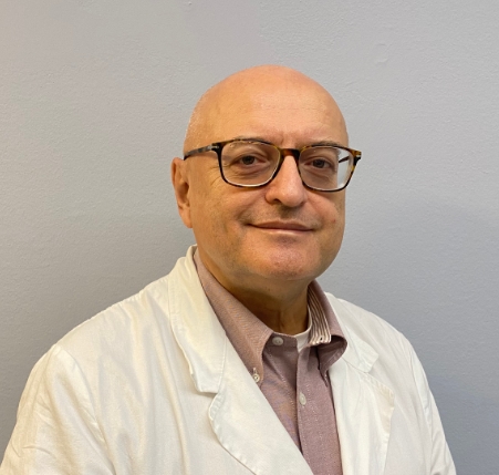 Mazzaferro Vincenzo Maria合作论文数Department of Oncology and Hematology-Oncology, Università degli Studi di Milano17R Romito合作论文数16R Lanocita合作论文数14Tiziana Negri合作论文数14Messina Antonella合作论文数14
 Marco Fiore合作论文数Department of Surgery, Istituto Nazionale Tumori14
 Carlo Sposito合作论文数14Paola Collini合作论文数13Nadia Zaffaroni合作论文数13E Civelli合作论文数12Rossella Bertulli合作论文数12Elena Tamborini合作论文数11Emilio Bombardieri合作论文数10Bhoori Sherrie合作论文数9Dei Tos Angelo Paolo合作论文数9
 Radaelli Stefano合作论文数Department of Surgery, Fondazione IRCCS Istituto Nazionale dei Tumori9Gianluca Aliberti合作论文数9tiziana camerini合作论文数8M. Mira合作论文数8Elena Fumagalli合作论文数8
 Federica Grosso合作论文数Azienda Ospedaliera SS Antonio e Biagio e Cesare Arrigo di Alessandria;Presidio Ospedaliero “Santo Spirito” di Casale Monferrato8Roberto Salvioni合作论文数8Cristina Colombo合作论文数8
 Nicola Nicolai合作论文数Fondazione IRCCS Istituto Nazionale dei Tumori di Milano8
 Maurizio D’Incalci合作论文数Department of Oncology, Istituto di Ricerche Farmacologiche Mario Negri IRCCS;Department of Biomedical Sciences, Humanitas University;Laboratory of Cancer Pharmacology, Humanitas Research Hospital7
 Andrea Necchi合作论文数Urological Research Institute, IRCCS San Raffaele Hospital and Scientific Institute;Department of Medical Oncology, Università Vita-Salute San Raffaele;Ospedale San Raffaele7P. Dileo合作论文数7E. Virdis合作论文数6Andrea Marrari合作论文数6
 Claudia Sangalli合作论文数6J. Martín Broto合作论文数5M. Massimino合作论文数5
 Jean-Yves Blay合作论文数Département De Médecine, Centre Léon Bérard;Université Claude Bernard Lyon5
 Luigi Mariani合作论文数Istituto Nazionale Tumori5Juan Carlos Tercero合作论文数5
 Monica Terenziani合作论文数Pediatric Unit, Fondazione IRCCS Istituto Nazionale Tumori5Cosimo Sacco合作论文数5




